


Article Figures & Data
Figures
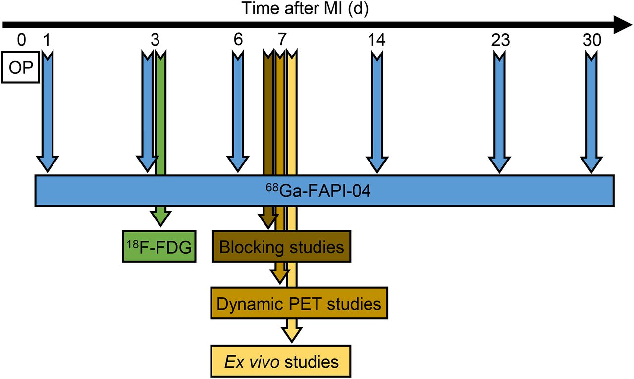
- FIGURE 1.
Experimental timeline. OP = operation.
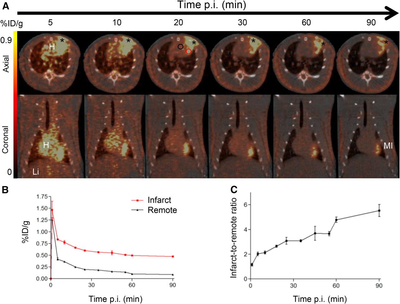
- FIGURE 2.
In vivo dynamic imaging of 68Ga-FAPI-04 uptake. (A) Serial PET/CT images (axial and coronal sections) from 90-min dynamic scan of MI rat at 7 d after coronary ligation. Representative regions of interest (2-dimensional) drawn over infarct border zone and remote myocardium are illustrated as red and black circles, respectively. Regions of interest in infarcts were placed relatively far from surgical wounds. 68Ga-FAPI-04 exhibited elevated uptake in scars from operation (asterisk). (B) Corresponding time–activity curves for infarcted and noninfarcted heart tissue (average and SD, n = 3). (C) Infarct-to-noninfarct ratio over time (average and SD, n = 3). H = heart; Li = liver; p.i. = after injection.
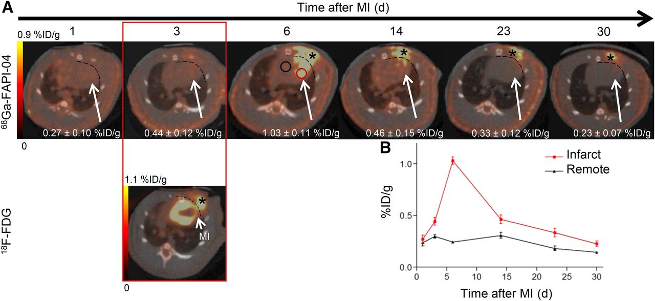
- FIGURE 3.
In vivo imaging of 68Ga-FAPI-04 uptake in longitudinal study. (A) Static PET/CT matched axial slices in same rat subjected to coronary ligation and scanned 1 h after injection of 68Ga-FAPI-04 (1, 3, 6, 14, 23, and 30 d after MI) and 18F-FDG (3 d after MI). Dashed lines separate tracer uptake in myocardium from uptake in surgical wounds. In 6 d after MI image, representative regions of interest (2-dimensional) drawn over infarct border zone and remote myocardium are illustrated as red and black circles, respectively. 68Ga-FAPI-04 uptake in regions of interest of infarcts is demonstrated. (B) Corresponding time–activity curves for infarcted and noninfarcted heart tissue (average and SD, n = 3). 68Ga-FAPI-04 and 18F-FDG exhibited elevated uptake in scars from operation (asterisk).
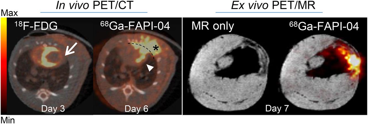
- FIGURE 4.
Axial sections of in vivo PET/CT imaging with 18F-FDG (day 3 after MI) and 68Ga-FAPI-04 (day 6 after MI) and corresponding ex vivo PET/MRI with 68Ga-FAPI-04 (day 7 after MI). 18F-FDG image was used to identify areas of infarcted myocardium (arrow), where increased uptake of 68Ga-FAPI-04 was apparent (arrowhead). 68Ga-FAPI-04 exhibited elevated uptake in postsurgical scar (asterisk). Dashed line separates 68Ga-FAPI-04 uptake in myocardium from surgical wound. High-resolution MR and PET/MR data confirmed infarcted area, where 68Ga-FAPI-04 uptake was increased.
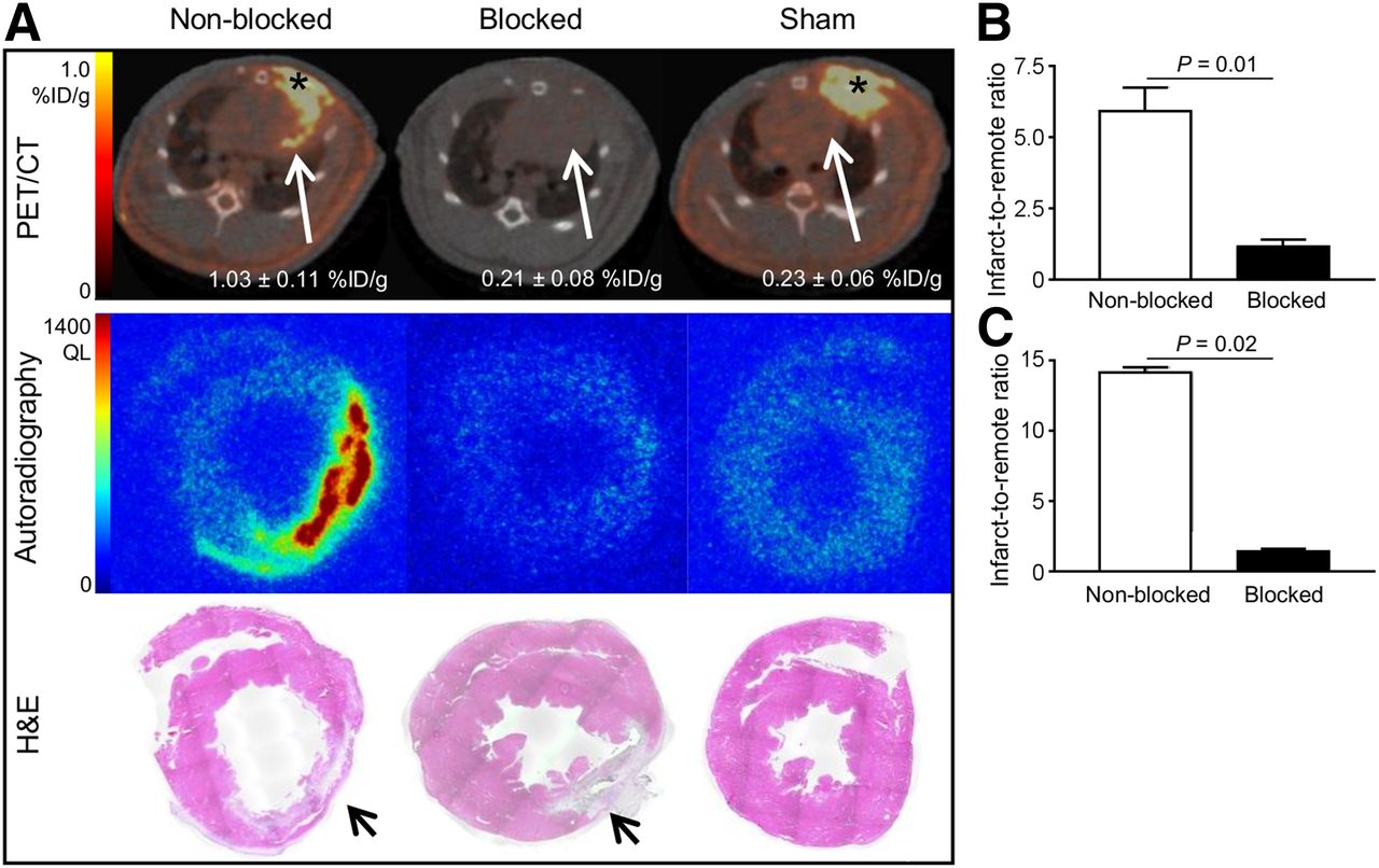
- FIGURE 5.
Binding specificity test. (A) PET/CT axial views, autoradiographs, and corresponding H&E stainings of 10-μm cross-sections prepared from MI nonblocked, blocked, and sham-operated rats. Autoradiographs and H&E stainings from nonblocked hearts show increased 68Ga-FAPI-04 uptake in infarcted area at 7 d after MI, whereas uptake is negligible after sham operation or injection of nonlabeled FAPI-04 (blocked). Infarcted areas in H&E stainings are identified with arrows. (B) PET image–derived infarct-to-noninfarct uptake ratio (derived from 6 nonblocked and 3 blocked rat hearts subjected to coronary ligation). (C) Autoradiography image–derived infarct-to-noninfarct uptake ratio (derived from 3 nonblocked and 3 blocked MI hearts). QL = quantum level.
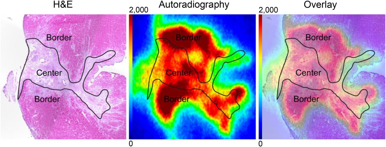
- FIGURE 6.
Ex vivo evaluation of 68Ga-FAPI-04 uptake in infarct at 7 d after MI. Representative autoradiograph and corresponding H&E staining from rat heart show elevated and heterogeneous uptake of 68Ga-FAPI-04 in infarct border compared with infarct center.
- FIGURE 7.
(A) Tile scan of entire infarcted heart section showing location of FAP+ fibroblasts. Insets 1, 2, and 3 show higher magnification from infarct border, infarct center, and noninfarcted remote myocardium, respectively. (B) Enumeration of FAP+ fibroblast density in MI hearts (n = 3) showing higher FAP+ fibroblast percentage in border zone than in infarct center or remote zone. (C) H&E-stained parallel section. (D) Photomicrographs of FAP, prolyl-4-hydroxylase β (P4H), α-smooth muscle actin, and vimentin-stained infiltrated fibroblasts in periinfarct border zone. Abundant colocalization of FAP, prolyl-4-hydroxylase β, and vimentin shows that accumulated fibroblasts are activated phenotypes, whereas small portion have differentiated into α-smooth muscle actin–positive mature myofibroblasts.


Additional Files
Supplemental Data
Files in this Data Supplement:


Jump to section
Related Articles
Cited By...
- Toward Quantitative Multisite Preclinical Imaging Studies in Acute Myocardial Infarction: Evaluation of the Immune-Fibrosis Axis
- Molecular Imaging Biomarkers in Cardiooncology: A View on Established Technologies and Future Perspectives
- Myocardial Fibrosis: Emerging Target for Cardiac Molecular Imaging and Opportunity for Image-Guided Therapy
- [68Ga]Ga-FAPI-46 PET for Visualization of Postinfarction Renal Fibrosis
- PET imaging of fibroblast activation protein alpha (FAP) detects incipient cardiotoxicity due to anthracycline chemotherapy
- Molecular Imaging of Myocardial Fibroblast Activation in Patients with Advanced Aortic Stenosis Before Transcatheter Aortic Valve Replacement: A Pilot Study
- [68Ga]Ga-FAPI-46 PET for Visualization of Postinfarction Renal Fibrosis
- Molecular Imaging of Myocardial Fibroblast Activation in Patients with Advanced Aortic Stenosis Before Transcatheter Aortic Valve Replacement: A Pilot Study
- Cardiac Fibroblast Activation in Patients Early After Acute Myocardial Infarction: Integration with MR Tissue Characterization and Subsequent Functional Outcome
- Radiotracers to Address Unmet Clinical Needs in Cardiovascular Imaging, Part 2: Inflammation, Fibrosis, Thrombosis, Calcification, and Amyloidosis Imaging
- Pitfalls and Common Findings in 68Ga-FAPI PET: A Pictorial Analysis
- PET/MRI, Part 4: Clinical Applications
- Feasibility, Biodistribution, and Preliminary Dosimetry in Peptide-Targeted Radionuclide Therapy of Diverse Adenocarcinomas Using 177Lu-FAP-2286: First-in-Humans Results
- The Latest Advances in Imaging Crosstalk Between the Immune System and Fibrosis in Cardiovascular Disease
- Combination of Forced Diuresis with Additional Late Imaging in 68Ga-PSMA-11 PET/CT: Effects on Lesion Visibility and Radiotracer Uptake
- The Latest Developments in Imaging of Fibroblast Activation Protein
- FAPI-74 PET/CT Using Either 18F-AlF or Cold-Kit 68Ga Labeling: Biodistribution, Radiation Dosimetry, and Tumor Delineation in Lung Cancer Patients
- Mars Shot for Nuclear Medicine, Molecular Imaging, and Molecularly Targeted Radiopharmaceutical Therapy
- Targeted PET Imaging of Chemokine Receptor 2-Positive Monocytes and Macrophages in the Injured Heart
- The Future of Nuclear Medicine Depends on the Quality of Its Research: Researchers at the University of Heidelberg Receive the Award for Best Article of the Year
- The Changing Face of Nuclear Cardiology: Guiding Cardiovascular Care Toward Molecular Medicine
- CARTing Away Cardiac Fibrosis
- FAP: The Next Billion Dollar Nuclear Theranostics Target?