
Main menu

  • Home
  • Content
    • Current
    • Ahead of print
    • Past Issues
    • JNM Supplement
    • SNMMI Annual Meeting Abstracts
    • Continuing Education
    • JNM Podcasts
  • Subscriptions
    • Subscribers
    • Institutional and Non-member
    • Rates
    • Journal Claims
    • Corporate & Special Sales
  • Authors
    • Submit to JNM
    • Information for Authors
    • Assignment of Copyright
    • AQARA requirements
  • Info
    • Reviewers
    • Permissions
    • Advertisers
  • About
    • About Us
    • Editorial Board
    • Contact Information
  • More
    • Alerts
    • Feedback
    • Help
    • SNMMI Journals
  • SNMMI
    • JNM
    • JNMT
    • SNMMI Journals
    • SNMMI

User menu

  • Subscribe
  • My alerts
  • Log in
  • My Cart

Search

  • Advanced search
Journal of Nuclear Medicine
  • SNMMI
    • JNM
    • JNMT
    • SNMMI Journals
    • SNMMI
  • Subscribe
  • My alerts
  • Log in
  • My Cart
Journal of Nuclear Medicine

Advanced Search

  • Home
  • Content
    • Current
    • Ahead of print
    • Past Issues
    • JNM Supplement
    • SNMMI Annual Meeting Abstracts
    • Continuing Education
    • JNM Podcasts
  • Subscriptions
    • Subscribers
    • Institutional and Non-member
    • Rates
    • Journal Claims
    • Corporate & Special Sales
  • Authors
    • Submit to JNM
    • Information for Authors
    • Assignment of Copyright
    • AQARA requirements
  • Info
    • Reviewers
    • Permissions
    • Advertisers
  • About
    • About Us
    • Editorial Board
    • Contact Information
  • More
    • Alerts
    • Feedback
    • Help
    • SNMMI Journals
  • View or Listen to JNM Podcast
  • Visit JNM on Facebook
  • Join JNM on LinkedIn
  • Follow JNM on Twitter
  • Subscribe to our RSS feeds
Research ArticleOncology

Improved Detection of Transosseous Meningiomas Using 68Ga-DOTATATE PET/CT Compared with Contrast-Enhanced MRI

Wolfgang G. Kunz, Lisa M. Jungblut, Philipp M. Kazmierczak, Franziska J. Vettermann, Andreas Bollenbacher, Jörg C. Tonn, Christian Schichor, Axel Rominger, Nathalie L. Albert, Peter Bartenstein, Maximilian F. Reiser and Clemens C. Cyran
Journal of Nuclear Medicine October 2017, 58 (10) 1580-1587; DOI: https://doi.org/10.2967/jnumed.117.191932
Wolfgang G. Kunz
1Institute for Clinical Radiology, Ludwig-Maximilians-University Hospital, Munich, Germany
  • Find this author on Google Scholar
  • Find this author on PubMed
  • Search for this author on this site
Lisa M. Jungblut
1Institute for Clinical Radiology, Ludwig-Maximilians-University Hospital, Munich, Germany
  • Find this author on Google Scholar
  • Find this author on PubMed
  • Search for this author on this site
Philipp M. Kazmierczak
1Institute for Clinical Radiology, Ludwig-Maximilians-University Hospital, Munich, Germany
  • Find this author on Google Scholar
  • Find this author on PubMed
  • Search for this author on this site
Franziska J. Vettermann
2Department of Nuclear Medicine, Ludwig-Maximilians-University Hospital, Munich, Germany; and
  • Find this author on Google Scholar
  • Find this author on PubMed
  • Search for this author on this site
Andreas Bollenbacher
2Department of Nuclear Medicine, Ludwig-Maximilians-University Hospital, Munich, Germany; and
  • Find this author on Google Scholar
  • Find this author on PubMed
  • Search for this author on this site
Jörg C. Tonn
3Department of Neurosurgery, Ludwig-Maximilians-University Hospital, Munich, Germany
  • Find this author on Google Scholar
  • Find this author on PubMed
  • Search for this author on this site
Christian Schichor
3Department of Neurosurgery, Ludwig-Maximilians-University Hospital, Munich, Germany
  • Find this author on Google Scholar
  • Find this author on PubMed
  • Search for this author on this site
Axel Rominger
2Department of Nuclear Medicine, Ludwig-Maximilians-University Hospital, Munich, Germany; and
  • Find this author on Google Scholar
  • Find this author on PubMed
  • Search for this author on this site
Nathalie L. Albert
2Department of Nuclear Medicine, Ludwig-Maximilians-University Hospital, Munich, Germany; and
  • Find this author on Google Scholar
  • Find this author on PubMed
  • Search for this author on this site
Peter Bartenstein
2Department of Nuclear Medicine, Ludwig-Maximilians-University Hospital, Munich, Germany; and
  • Find this author on Google Scholar
  • Find this author on PubMed
  • Search for this author on this site
Maximilian F. Reiser
1Institute for Clinical Radiology, Ludwig-Maximilians-University Hospital, Munich, Germany
  • Find this author on Google Scholar
  • Find this author on PubMed
  • Search for this author on this site
Clemens C. Cyran
1Institute for Clinical Radiology, Ludwig-Maximilians-University Hospital, Munich, Germany
  • Find this author on Google Scholar
  • Find this author on PubMed
  • Search for this author on this site
  • Article
  • Figures & Data
  • Supplemental
  • Info & Metrics
  • PDF
Loading

Article Figures & Data

Figures

  • Tables
  • Additional Files
  • FIGURE 1.
    • Download figure
    • Open in new tab
    • Download powerpoint
    FIGURE 1.

    Example of meningioma with transosseous extension. A 56-y-old woman presented with worsening headache. CE-MRI was performed and revealed large, homogeneously enhancing mass in left temporopolar region, consistent with meningioma (lower row). Before surgical resection, patient was examined using 68Ga-DOTATATE PET/CT, which additionally demonstrated transosseous extension as assessed by strong tracer uptake extending into sphenoid wing. This was not evident on nonenhanced T2- or T1-weighted images or on T1-weighted CE-MR images. Extension of meningioma into sphenoid wing was found during surgical resection and later confirmed by pathologic evaluation of transosseous secretory meningioma (World Health Organization grade I). w = weighted.

  • FIGURE 2.
    • Download figure
    • Open in new tab
    • Download powerpoint
    FIGURE 2.

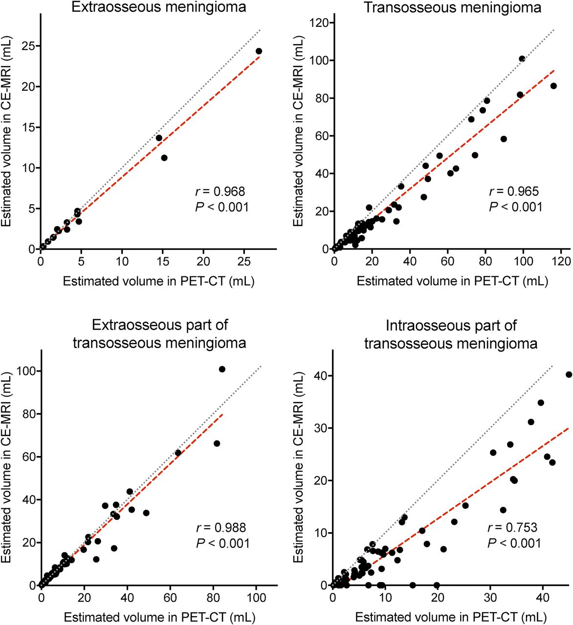
    Comparison of estimated volumes in 68Ga-DOTATATE PET/CT and CE-MRI. Correlation scatterplots depict estimated volumes using 68Ga-DOTATATE PET/CT (x-axis) against estimated volumes using CE-MRI (y-axis). Spearman correlation coefficients (r) were calculated. Gray line represents perfect positive correlation. Red line is calculated using least-squares fit of data. Data are based on 82 meningioma patients with pathologic exclusion or inclusion of osseous involvement. In cases of imaging-based false-negative osseous involvement, intraosseous part was attributed no volume (0 mL). Wilcoxon signed rank test demonstrated significant differences between PET and CE-MRI for volume estimation of total and intraosseous part of transosseous meningiomas (each with P < 0.001).
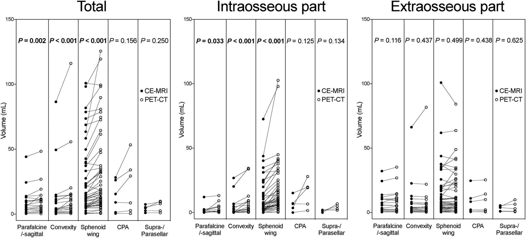
  • FIGURE 3.
    • Download figure
    • Open in new tab
    • Download powerpoint
    FIGURE 3.

    Comparison of estimated volumes stratified for meningioma location. Plots depict estimated volumes for total meningioma, intraosseous part, and extraosseous part using 68Ga-DOTATATE PET/CT (open circles) and CE-MRI (solid circles). Data are based on 105 meningioma patients, among whom 90 had imaging-based diagnosis of transosseous extension. Statistical comparison was performed using Wilcoxon signed rank test. Bold P values indicate statistical significance. CPA = cerebellopontine angle.
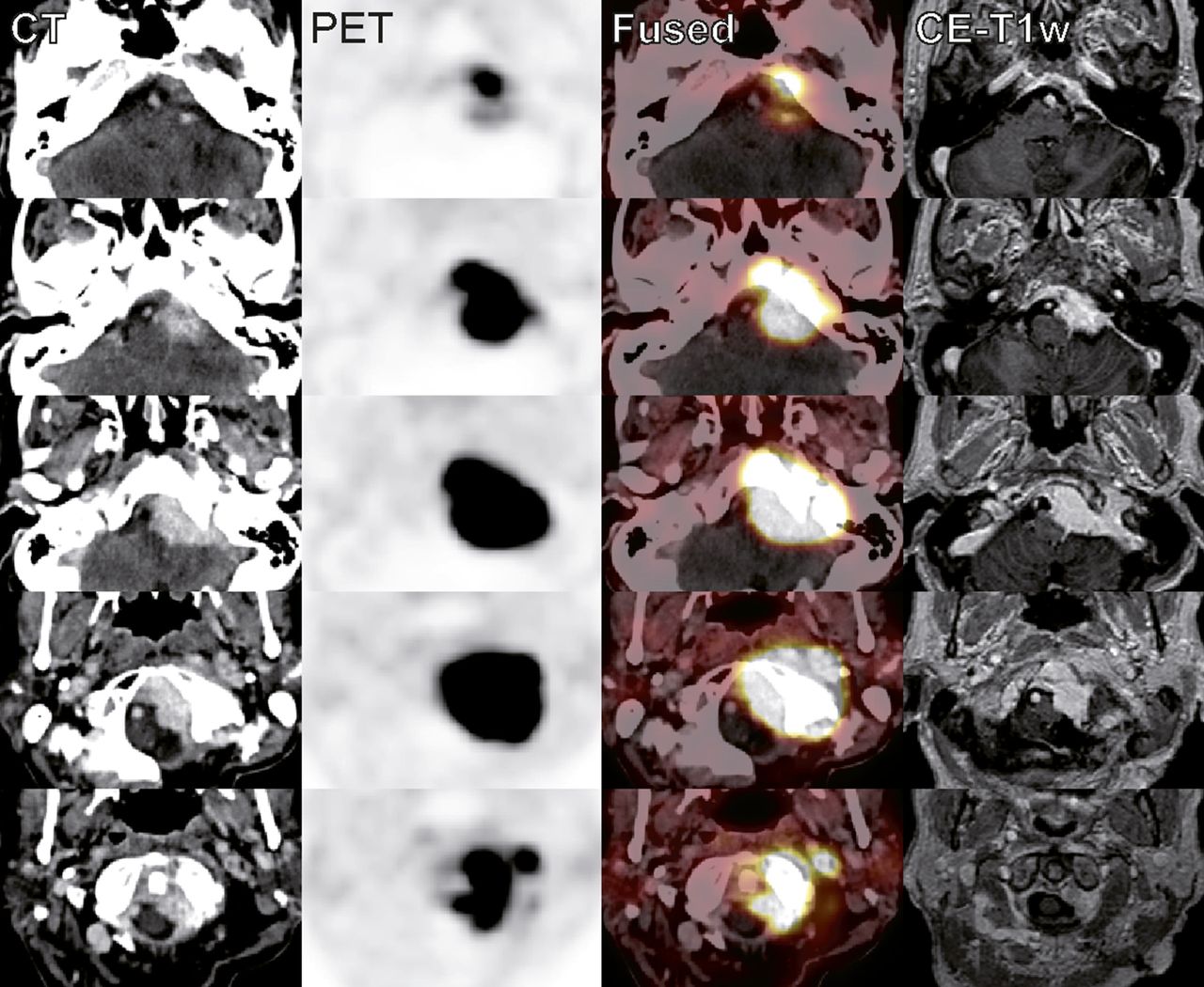
  • FIGURE 4.
    • Download figure
    • Open in new tab
    • Download powerpoint
    FIGURE 4.

    Example of estimated intraosseous volume discrepancy between 68Ga-DOTATATE PET/CT and CE-MRI in patient with skull base meningioma. A 75-y-old woman was followed up with 68Ga-DOTATATE PET/CT and CE-MRI after partial resection of transosseous transitional meningioma (World Health Organization grade I) in left cerebellopontine angle. Because of worsening and radiating pain in occipital region and hypoglossal nerve palsy, she was evaluated for stereotactic radiosurgery. Estimated volumes of transosseous extension were significantly larger on 68Ga-DOTATATE PET/CT than on CE-MRI (28.0 vs. 7.5 mL), whereas volumes of extraosseous part agreed well (25.3 vs. 22.6 mL). w = weighted.

Tables

  • Figures
  • Additional Files
    • View popup
    TABLE 1

    Characteristics of Extraosseous and Transosseous Meningiomas

    ParameterVariableExtraosseous (n = 15)Transosseous (n = 67)P
    Age (y)55 (51–66)49 (47–61)0.073
    Male sex6 (40%)13 (19%)0.087
    Pathology
     WHO gradeI9 (60%)56 (84%)NA
    II6 (40%)9 (13%)
    III0 (0%)2 (3%)
     Histologic subtypeTransitional5 (38%)22 (38%)NA
    Meningothelial2 (15%)15 (26%)
    Microcystic0 (0%)4 (7%)
    Fibroblastic0 (0%)1 (2%)
    Secretory0 (0%)5 (9%)
    Atypical6 (46%)9 (16%)
    Anaplastic0 (0%)2 (3%)
    Qualitative imaging
     Meningiomatosis9 (60%)26 (39%)0.134
     LocationParasagittal/parafalcine5 (33%)10 (15%)NA
    Convexity6 (40%)12 (18%)
    Sphenoid wing3 (20%)42 (63%)
    Cerebellopontine0 (0%)2 (3%)
    Suprasellar/parasellar1 (7%)1 (1%)
     PET/CT for osseous involvementNo signs of13 (87%)0 (0%)NA
    Suggestive of0 (0%)1 (1%)
    Consistent with2 (13%)66 (99%)
     CE-MRI for osseous involvementNo signs of9 (60%)12 (18%)NA
    Suggestive of5 (33%)19 (28%)
    Consistent with1 (7%)36 (54%)
     Peritumoral edemaNone8 (53%)33 (49%)0.858
    Mild3 (20%)18 (27%)
    Extensive4 (27%)16 (24%)
    Quantitative imaging
     Meningioma volume in PET/CT (mL)3.3 (1.6–4.7)12.8 (6.8–32.9)<0.001*
     Meningioma volume in CE-MRI (mL)2.5 (1.4–4.7)10.6 (4.6–22.0)0.001*
     Meningioma SUVmax7.6 (4.3–13.9)14.2 (10.0–22.4)0.011*
     Meningioma SUVmean2.7 (1.9–3.0)4.3 (3.1–5.9)0.001*
     Pituitary SUVmax15.8 (12.9–20.0)15.7 (11.6–19.3)0.852
     Pituitary SUVmean3.7 (3.5–4.2)4.0 (3.3–4.7)0.587
    • ↵* Statistically significant.

    • WHO = World Health Organization; NA = not applicable.

    • Data are count followed by percentage for categoric variables and median followed by interquartile range for continuous variables. Proportion analysis was tested using χ2 test. Nonparametric testing for continuous variables was performed using Mann–Whitney U test.

    • View popup
    TABLE 2

    Diagnostic Performance of 68Ga-DOTATATE PET/CT and CE-MRI for Detection of Osseous Involvement in Intracranial Meningiomas

    MethodSensitivitySpecificityPositive LRNegative LRPrevalence
    Overall (n = 82)
     PET/CT98.5 (92.0–99.9)86.7 (60.0–98.3)7.39 (2.03–26.9)0.02 (0.00–0.12)81.7 (71.6–89.4)
     CE-MRI53.7 (41.1–66.0)93.3 (68.1–99.8)8.06 (1.20–54.2)0.50 (0.37–0.66)81.7 (71.6–89.4)
    Preoperative (n = 39)
     PET/CT100 (88.4–100)77.8 (40.0–97.2)4.50 (1.33–15.3)0.00 (NA)76.9 (60.7–88.9)
     CE-MRI53.3 (34.3–71.7)100 (66.4–100)∞ (NA)0.47 (0.32–0.68)76.9 (60.7–88.9)
    Postoperative (n = 43)
     PET/CT97.3 (85.8–99.9)100 (54.1–100)∞ (NA)0.03 (0.00–0.19)86.0 (72.1–94.7)
     CE-MRI54.1 (36.9–70.5)83.3 (35.9–99.6)3.24 (0.53–19.9)0.55 (0.33–0.91)86.0 (72.1–94.7)
    • LR = likelihood ratio; NA = not applicable.

    • Data are percentages (or ratios) followed by 95% confidence interval. Imaging diagnosis of “consistent with” was set as test-positive. McNemar test showed significant differences in diagnostic performance of PET/CT and CE-MRI (P < 0.001).

    • View popup
    TABLE 3

    Receiver-Operating-Characteristic Analysis of Imaging Parameters for Osseous Involvement

    ParameterArea under curveP
    PET/CT qualitative assessment0.932 (0.830–1.000)<0.001*
    PET/CT meningioma volume0.803 (0.681–0.925)<0.001*
    PET/CT meningioma SUVmean0.778 (0.640–0.916)0.001*
    CE-MRI qualitative assessment0.773 (0.637–0.909)0.001*
    CE-MRI meningioma volume0.768 (0.641–0.895)0.001*
    PET/CT meningioma SUVmax0.710 (0.557–0.864)0.011*
    PET/CT peritumoral edema0.508 (0.342–0.675)0.919
    CE-MRI peritumoral edema0.477 (0.307–0.646)0.778
    • ↵* Statistically significant.

    • 95% confidence intervals are in parentheses.

    • View popup
    TABLE 4

    PET/CT Measurements in Transosseous Meningiomas

    ParameterOverall (n = 67)Parafalcine/parasagittal (n = 10)Convexity (n = 12)Sphenoid wing (n = 42)Cerebellopontine (n = 2)Suprasellar/parasellar (n = 1)
    SUVmax total14.2 (10–22)10.8 (7–12)15.6 (10–31)14.8 (11–26)7.3 (6–9)12.6 (13–13)
    SUVmax intraosseous13.4 (10–22)10.0 (7–11)15.6 (10–31)14.2 (11–26)7.3 (6–9)12.6 (13–13)
    SUVmean total4.3 (3–6)3.7 (3–4)4.7 (3–7)4.5 (4–6)2.6 (3–3)4.5 (4–4)
    SUVmean intraosseous4.6 (3–7)3.4 (3–4)5.7 (3–8)5.0 (4–8)2.7 (3–3)4.8 (5–5)
    Volume total (mL)12.8 (7–33)6.4 (4–15)13.3 (5–18)17.7 (10–47)5.6 (3–9)9.9 (10–10)
    Volume intraosseous (mL)6.8 (3–15)2.7 (1–5)5.1 (2–11)9.3 (6–21)4.1 (2–7)6.8 (7–7)
    • Data are median followed by interquartile range. SUVmean in total meningioma compared with intraosseous part of meningioma showed statistically significant differences on Wilcoxon signed rank testing (P < 0.001).

Additional Files

  • Figures
  • Tables
  • Supplemental Data

    Files in this Data Supplement:

    • Supplemental Data
PreviousNext
Back to top

In this issue

Journal of Nuclear Medicine: 58 (10)
Journal of Nuclear Medicine
Vol. 58, Issue 10
October 1, 2017
  • Table of Contents
  • Table of Contents (PDF)
  • About the Cover
  • Index by author
Print
Download PDF
Article Alerts
Sign In to Email Alerts with your Email Address
Email Article

Thank you for your interest in spreading the word on Journal of Nuclear Medicine.

NOTE: We only request your email address so that the person you are recommending the page to knows that you wanted them to see it, and that it is not junk mail. We do not capture any email address.

Enter multiple addresses on separate lines or separate them with commas.
Improved Detection of Transosseous Meningiomas Using 68Ga-DOTATATE PET/CT Compared with Contrast-Enhanced MRI
(Your Name) has sent you a message from Journal of Nuclear Medicine
(Your Name) thought you would like to see the Journal of Nuclear Medicine web site.
Citation Tools
Improved Detection of Transosseous Meningiomas Using 68Ga-DOTATATE PET/CT Compared with Contrast-Enhanced MRI
Wolfgang G. Kunz, Lisa M. Jungblut, Philipp M. Kazmierczak, Franziska J. Vettermann, Andreas Bollenbacher, Jörg C. Tonn, Christian Schichor, Axel Rominger, Nathalie L. Albert, Peter Bartenstein, Maximilian F. Reiser, Clemens C. Cyran
Journal of Nuclear Medicine Oct 2017, 58 (10) 1580-1587; DOI: 10.2967/jnumed.117.191932

Citation Manager Formats

  • BibTeX
  • Bookends
  • EasyBib
  • EndNote (tagged)
  • EndNote 8 (xml)
  • Medlars
  • Mendeley
  • Papers
  • RefWorks Tagged
  • Ref Manager
  • RIS
  • Zotero
Share
Improved Detection of Transosseous Meningiomas Using 68Ga-DOTATATE PET/CT Compared with Contrast-Enhanced MRI
Wolfgang G. Kunz, Lisa M. Jungblut, Philipp M. Kazmierczak, Franziska J. Vettermann, Andreas Bollenbacher, Jörg C. Tonn, Christian Schichor, Axel Rominger, Nathalie L. Albert, Peter Bartenstein, Maximilian F. Reiser, Clemens C. Cyran
Journal of Nuclear Medicine Oct 2017, 58 (10) 1580-1587; DOI: 10.2967/jnumed.117.191932
Twitter logo Facebook logo LinkedIn logo Mendeley logo
  • Tweet Widget
  • Facebook Like
  • Google Plus One
Bookmark this article

Jump to section

  • Article
    • Abstract
    • MATERIALS AND METHODS
    • RESULTS
    • DISCUSSION
    • CONCLUSION
    • DISCLOSURE
    • Footnotes
    • REFERENCES
  • Figures & Data
  • Supplemental
  • Info & Metrics
  • PDF

Related Articles

  • This Month in JNM
  • PubMed
  • Google Scholar

Cited By...

  • Meningioma: Molecular Updates from the 2021 World Health Organization Classification of CNS Tumors and Imaging Correlates
  • Meningioma Revisited: Should Whole-Body Staging with [68Ga]Ga-DOTATOC PET/CT of High-Grade Meningiomas Become Standard Practice?
  • DOTATATE PET/MR Imaging Differentiates Secondary-Progressive from de Novo World Health Organization Grade 3 Meningiomas
  • Evaluation of [68Ga]-DOTATOC PET/MRI in Patients with Meningioma of the Subcranial and Intraorbital Space
  • Cost-Effectiveness Analysis of 68Ga-DOTATATE PET/MRI in Radiotherapy Planning in Patients with Intermediate-Risk Meningioma
  • The Complementary Role of 68Ga-DOTATATE PET/CT in Diagnosis of Recurrent Meningioma
  • Improved Detection of Postoperative Residual Meningioma with [68Ga]Ga-DOTA-TOC PET Imaging Using a High-resolution Research Tomograph PET Scanner
  • Google Scholar

More in this TOC Section

Oncology

  • 18F-FDG PET/CT manifestations of three Cases of Female Desmoplastic Small Round Cell Tumor
  • 18F-FDG PET/CT imaging features of parotid lesions; Case based pictorial review and its multi-modality correlation.
  • Utility of Fluorine-18-fluorodeoxyglucose PET/CT in Uterine sarcoma patients.
Show more Oncology

Clinical

  • TauIQ: A Canonical Image Based Algorithm to Quantify Tau PET Scans
  • Dual PET Imaging in Bronchial Neuroendocrine Neoplasms: The NETPET Score as a Prognostic Biomarker
  • Addition of 131I-MIBG to PRRT (90Y-DOTATOC) for Personalized Treatment of Selected Patients with Neuroendocrine Tumors
Show more Clinical

Similar Articles

Keywords

  • meningioma
  • Osseous involvement
  • PET-CT
  • 68Ga-DOTATATE
  • MRI
SNMMI

© 2025 SNMMI

Powered by HighWire