
Main menu

  • Home
  • Content
    • Current
    • Ahead of print
    • Past Issues
    • JNM Supplement
    • SNMMI Annual Meeting Abstracts
    • Continuing Education
    • JNM Podcasts
  • Subscriptions
    • Subscribers
    • Institutional and Non-member
    • Rates
    • Journal Claims
    • Corporate & Special Sales
  • Authors
    • Submit to JNM
    • Information for Authors
    • Assignment of Copyright
    • AQARA requirements
  • Info
    • Reviewers
    • Permissions
    • Advertisers
  • About
    • About Us
    • Editorial Board
    • Contact Information
  • More
    • Alerts
    • Feedback
    • Help
    • SNMMI Journals
  • SNMMI
    • JNM
    • JNMT
    • SNMMI Journals
    • SNMMI

User menu

  • Subscribe
  • My alerts
  • Log in
  • Log out
  • My Cart

Search

  • Advanced search
Journal of Nuclear Medicine
  • SNMMI
    • JNM
    • JNMT
    • SNMMI Journals
    • SNMMI
  • Subscribe
  • My alerts
  • Log in
  • Log out
  • My Cart
Journal of Nuclear Medicine

Advanced Search

  • Home
  • Content
    • Current
    • Ahead of print
    • Past Issues
    • JNM Supplement
    • SNMMI Annual Meeting Abstracts
    • Continuing Education
    • JNM Podcasts
  • Subscriptions
    • Subscribers
    • Institutional and Non-member
    • Rates
    • Journal Claims
    • Corporate & Special Sales
  • Authors
    • Submit to JNM
    • Information for Authors
    • Assignment of Copyright
    • AQARA requirements
  • Info
    • Reviewers
    • Permissions
    • Advertisers
  • About
    • About Us
    • Editorial Board
    • Contact Information
  • More
    • Alerts
    • Feedback
    • Help
    • SNMMI Journals
  • View or Listen to JNM Podcast
  • Visit JNM on Facebook
  • Join JNM on LinkedIn
  • Follow JNM on Twitter
  • Subscribe to our RSS feeds
Meeting ReportInstrumentation & Data Analysis Track

4D Multimodal Atlas of the Swine Heart from PET-CT Images

Jonghye Woo, Marc Normandin, Nicolas Guehl, Dustin Wooten, Thomas Brady, Remi Baghdady, Timothy Shoup, Jinsong Ouyang, Georges El Fakhri and Nathaniel Alpert
Journal of Nuclear Medicine May 2016, 57 (supplement 2) 1898;
Jonghye Woo
1Gordon Center for Medical Imaging, NMMI Massachusetts General Hospital & Harvard Medical School Boston MA United States
  • Find this author on Google Scholar
  • Find this author on PubMed
  • Search for this author on this site
Marc Normandin
1Gordon Center for Medical Imaging, NMMI Massachusetts General Hospital & Harvard Medical School Boston MA United States
  • Find this author on Google Scholar
  • Find this author on PubMed
  • Search for this author on this site
Nicolas Guehl
1Gordon Center for Medical Imaging, NMMI Massachusetts General Hospital & Harvard Medical School Boston MA United States
  • Find this author on Google Scholar
  • Find this author on PubMed
  • Search for this author on this site
Dustin Wooten
1Gordon Center for Medical Imaging, NMMI Massachusetts General Hospital & Harvard Medical School Boston MA United States
  • Find this author on Google Scholar
  • Find this author on PubMed
  • Search for this author on this site
Thomas Brady
1Gordon Center for Medical Imaging, NMMI Massachusetts General Hospital & Harvard Medical School Boston MA United States
  • Find this author on Google Scholar
  • Find this author on PubMed
  • Search for this author on this site
Remi Baghdady
1Gordon Center for Medical Imaging, NMMI Massachusetts General Hospital & Harvard Medical School Boston MA United States
  • Find this author on Google Scholar
  • Find this author on PubMed
  • Search for this author on this site
Timothy Shoup
1Gordon Center for Medical Imaging, NMMI Massachusetts General Hospital & Harvard Medical School Boston MA United States
  • Find this author on Google Scholar
  • Find this author on PubMed
  • Search for this author on this site
Jinsong Ouyang
1Gordon Center for Medical Imaging, NMMI Massachusetts General Hospital & Harvard Medical School Boston MA United States
  • Find this author on Google Scholar
  • Find this author on PubMed
  • Search for this author on this site
Georges El Fakhri
1Gordon Center for Medical Imaging, NMMI Massachusetts General Hospital & Harvard Medical School Boston MA United States
  • Find this author on Google Scholar
  • Find this author on PubMed
  • Search for this author on this site
Nathaniel Alpert
1Gordon Center for Medical Imaging, NMMI Massachusetts General Hospital & Harvard Medical School Boston MA United States
  • Find this author on Google Scholar
  • Find this author on PubMed
  • Search for this author on this site
  • Article
  • Figures & Data
  • Info & Metrics
Loading

Abstract

1898

Objectives Multimodal imaging systems, such as hybrid PET-CT, capture the dynamics of cardiac anatomy and function. Yet, the quantitative characterization and subsequent comparison of anatomy and function of the heart is a challenging task due to the variations in both anatomy and function across different subjects. The goal of this work is to develop a 4D (3D space with time) probabilistic multimodal atlas of the swine heart for study of methods of automated analysis that can potentially be translated to clinical application.

Methods We acquired fifteen healthy pigs with weights ranging from 30-92 Kg, using a hybrid PET-CT scanner yielding three data sets per study, including CT (0.97mm×0.97mm×3mm), CTA (0.33mm×0.33mm×1mm), and PET (2.14mm×2.14 mm×3mm). The 15 volume sets were used to compute mean and variance of anatomic, vascular, and physiological maps of flow in a standard orientation and consistent physical size—i.e., the multimodal atlas. PET volumes were acquired dynamically with the blood flow tracer [18F]Flurpiridaz and parametric perfusion maps were computed with a kinetic model. The approach is based on a hierarchical method, constructing the atlas transformations from the CTA volumes to the PET volumes. First, we created a common spatial space from CTA volumes using a diffeomorphic groupwise registration method to define a basic structural layer in which the other modalities can be mapped. Second, we transported all other modalities including CT, dynamic PET, and gated PET volumes of all subjects into this common space via the deformations learnt above. Third, we constructed a gated 4D PET atlas from deformed gated PET volumes using a frame-by-frame atlas construction scheme. In addition, we determined the best matching phase of gated PET atlas volumes, corresponding to the phase of CTA, followed by creating a “motion-frozen” volume (MFV) from gated PET atlas volumes for each subject using consecutive diffeomorphic registrations between the best matching phase and the remaining phases. For the atlas construction, widely used similarity measures including mutual information, sum-of-squared-differences (SSD), and cross correlation (CC) were used to evaluate the CTA atlas. For the quantitative comparison, two sharpness measures, including the intensity variance measure (M1) and the energy of image gradient measure (M2) were used. In addition, to measure the accuracy of the registration during the atlas building, two expert observers manually selected anatomical landmarks and measured target registration error (TRE).

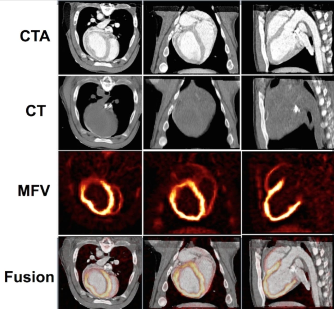
Results Our mean multimodal atlas was successfully constructed as illustrated in Fig. 1, where the CTA atlas, CT atlas, and MFV volumes, and the fusion of all the modalities are depicted. We evaluated image quality using the two sharpness measures as listed in Table 1. The data suggest that the CC similarity measure provided the best quality. The TRE obtained from the two anatomical landmarks was 2.47±1.56mm.

Conclusions This atlas provides myocardial images of a standard size and orientation combining anatomic and physiological data for pigs weighing from 30 to 92 Kg. To our knowledge, this is the first probabilistic multimodal atlas combining anatomic and functional images in a standard space. This method can potentially be extended to clinical applications involving automated image interpretation. $$graphic_8B12ADD6-084C-4099-8656-A8E7E8107FA6$$

Figure
  • Download figure
  • Open in new tab
  • Download powerpoint
View this table:
  • View inline
  • View popup

Comparison of Similarity Measures

Previous
Back to top

In this issue

Journal of Nuclear Medicine
Vol. 57, Issue supplement 2
May 1, 2016
  • Table of Contents
  • Index by author
Article Alerts
Sign In to Email Alerts with your Email Address
Email Article

Thank you for your interest in spreading the word on Journal of Nuclear Medicine.

NOTE: We only request your email address so that the person you are recommending the page to knows that you wanted them to see it, and that it is not junk mail. We do not capture any email address.

Enter multiple addresses on separate lines or separate them with commas.
4D Multimodal Atlas of the Swine Heart from PET-CT Images
(Your Name) has sent you a message from Journal of Nuclear Medicine
(Your Name) thought you would like to see the Journal of Nuclear Medicine web site.
Citation Tools
4D Multimodal Atlas of the Swine Heart from PET-CT Images
Jonghye Woo, Marc Normandin, Nicolas Guehl, Dustin Wooten, Thomas Brady, Remi Baghdady, Timothy Shoup, Jinsong Ouyang, Georges El Fakhri, Nathaniel Alpert
Journal of Nuclear Medicine May 2016, 57 (supplement 2) 1898;

Citation Manager Formats

  • BibTeX
  • Bookends
  • EasyBib
  • EndNote (tagged)
  • EndNote 8 (xml)
  • Medlars
  • Mendeley
  • Papers
  • RefWorks Tagged
  • Ref Manager
  • RIS
  • Zotero
Share
4D Multimodal Atlas of the Swine Heart from PET-CT Images
Jonghye Woo, Marc Normandin, Nicolas Guehl, Dustin Wooten, Thomas Brady, Remi Baghdady, Timothy Shoup, Jinsong Ouyang, Georges El Fakhri, Nathaniel Alpert
Journal of Nuclear Medicine May 2016, 57 (supplement 2) 1898;
Twitter logo Facebook logo LinkedIn logo Mendeley logo
  • Tweet Widget
  • Facebook Like
  • Google Plus One
Bookmark this article

Jump to section

  • Article
  • Figures & Data
  • Info & Metrics

Related Articles

  • No related articles found.
  • Google Scholar

Cited By...

  • No citing articles found.
  • Google Scholar

More in this TOC Section

Instrumentation & Data Analysis Track

  • Deep Learning Based Kidney Segmentation for Glomerular Filtration Rate Measurement Using Quantitative SPECT/CT
  • Comparison of 22 partial volume correction methods for amyloid PET imaging with 11C-PiB
  • The Benefit of Time-of-Flight in Digital Photon Counting PET Imaging: Physics and Clinical Evaluation
Show more Instrumentation & Data Analysis Track

MTA II: Data Analysis & Management Posters

  • An adaptive motion correction method for PET/CT Brain Imaging
  • Iterative factor analysis : Strategy for estimating input function in dynamic 18F-FDG brain PET
  • Quantitative image validation of PET-MR against stand-alone PET - A dynamic 11C-PE2I PET study.
Show more MTA II: Data Analysis & Management Posters

Similar Articles

SNMMI

© 2025 SNMMI

Powered by HighWire