
Main menu

  • Home
  • Content
    • Current
    • Ahead of print
    • Past Issues
    • JNM Supplement
    • SNMMI Annual Meeting Abstracts
    • Continuing Education
    • JNM Podcasts
  • Subscriptions
    • Subscribers
    • Institutional and Non-member
    • Rates
    • Journal Claims
    • Corporate & Special Sales
  • Authors
    • Submit to JNM
    • Information for Authors
    • Assignment of Copyright
    • AQARA requirements
  • Info
    • Reviewers
    • Permissions
    • Advertisers
  • About
    • About Us
    • Editorial Board
    • Contact Information
  • More
    • Alerts
    • Feedback
    • Help
    • SNMMI Journals
  • SNMMI
    • JNM
    • JNMT
    • SNMMI Journals
    • SNMMI

User menu

  • Subscribe
  • My alerts
  • Log in
  • My Cart

Search

  • Advanced search
Journal of Nuclear Medicine
  • SNMMI
    • JNM
    • JNMT
    • SNMMI Journals
    • SNMMI
  • Subscribe
  • My alerts
  • Log in
  • My Cart
Journal of Nuclear Medicine

Advanced Search

  • Home
  • Content
    • Current
    • Ahead of print
    • Past Issues
    • JNM Supplement
    • SNMMI Annual Meeting Abstracts
    • Continuing Education
    • JNM Podcasts
  • Subscriptions
    • Subscribers
    • Institutional and Non-member
    • Rates
    • Journal Claims
    • Corporate & Special Sales
  • Authors
    • Submit to JNM
    • Information for Authors
    • Assignment of Copyright
    • AQARA requirements
  • Info
    • Reviewers
    • Permissions
    • Advertisers
  • About
    • About Us
    • Editorial Board
    • Contact Information
  • More
    • Alerts
    • Feedback
    • Help
    • SNMMI Journals
  • View or Listen to JNM Podcast
  • Visit JNM on Facebook
  • Join JNM on LinkedIn
  • Follow JNM on Twitter
  • Subscribe to our RSS feeds
Meeting ReportInstrumentation & Data Analysis Track

Quantitative image validation of PET-MR against stand-alone PET - A dynamic 11C-PE2I PET study.

Joao Sousa, Lieuwe Appel, Stergios Papadimitriou, Dag Nyholm, Jens Sorensen, Torsten Danfors, Elna-Marie Larsson, Hakan Ahlstrom and Mark Lubberink
Journal of Nuclear Medicine May 2016, 57 (supplement 2) 1935;
Joao Sousa
1Department of Surgical Sciences Uppsala University Uppsala Sweden
  • Find this author on Google Scholar
  • Find this author on PubMed
  • Search for this author on this site
Lieuwe Appel
1Department of Surgical Sciences Uppsala University Uppsala Sweden
  • Find this author on Google Scholar
  • Find this author on PubMed
  • Search for this author on this site
Stergios Papadimitriou
2Neurosciences Uppsala University Uppsala Sweden
  • Find this author on Google Scholar
  • Find this author on PubMed
  • Search for this author on this site
Dag Nyholm
2Neurosciences Uppsala University Uppsala Sweden
  • Find this author on Google Scholar
  • Find this author on PubMed
  • Search for this author on this site
Jens Sorensen
1Department of Surgical Sciences Uppsala University Uppsala Sweden
  • Find this author on Google Scholar
  • Find this author on PubMed
  • Search for this author on this site
Torsten Danfors
1Department of Surgical Sciences Uppsala University Uppsala Sweden
  • Find this author on Google Scholar
  • Find this author on PubMed
  • Search for this author on this site
Elna-Marie Larsson
1Department of Surgical Sciences Uppsala University Uppsala Sweden
  • Find this author on Google Scholar
  • Find this author on PubMed
  • Search for this author on this site
Hakan Ahlstrom
1Department of Surgical Sciences Uppsala University Uppsala Sweden
  • Find this author on Google Scholar
  • Find this author on PubMed
  • Search for this author on this site
Mark Lubberink
1Department of Surgical Sciences Uppsala University Uppsala Sweden
  • Find this author on Google Scholar
  • Find this author on PubMed
  • Search for this author on this site
  • Article
  • Figures & Data
  • Info & Metrics
Loading

Abstract

1935

Objectives For PET-MR, challenges incorporating bone in attenuation maps remain, which may affect quantitative accuracy of PET data. The stand-alone PET ECAT HR+ scanner, using rotating 68Ge rod sources for attenuation correction, is often considered the gold standard for quantitative brain PET imaging. The aim of the present work was to compare quantitative measures of dopamine transporter availability and relative cerebral blood flow based on dynamic 11C-PE2I-PET data acquired on a time-of-flight integrated PET-MR and a stand-alone PET scanner.

Methods Five patients evaluated for Parkinsonism underwent two 80 min dynamic PET scans using the specific dopamine transporter radioligand 11C-PE2I (about 5 MBq/kg ). First they were investigated on a stand-alone PET scanner (ECAT HR+) and within 6 months they underwent a second investigation on a time-of-flight integrated PET-MR scanner (Signa PET-MR). ECAT images were reconstructed using OSEM (6 iterations, 8 subsets, 4 mm Hanning post-filter). PET-MR images were reconstructed using a resolution-matched protocol (OSEM; 2 iterations, 28 subsets, 5 mm Gaussian filter). PET-MR attenuation correction was based on a head atlas method. Both dynamic PET series were co-registered to T1-MRI images using SPM8. Volumes of interest were defined using a probabilistic template (PVElab) on the T1- MRI images and transferred to both sets of PET images. Binding potential (BPND), reflecting dopamine transporter availability, and relative cerebral blood flow (R1) were derived from the PET images for a number of clinically relevant tissues using the simplified reference tissue model, with cerebellar grey matter as reference tissue. BPND in putamen, and R1 in striatum as well as anterior cortical, posterior cortical, and limbic regions, were compared between both modalities using regression and Bland-Altman analysis.

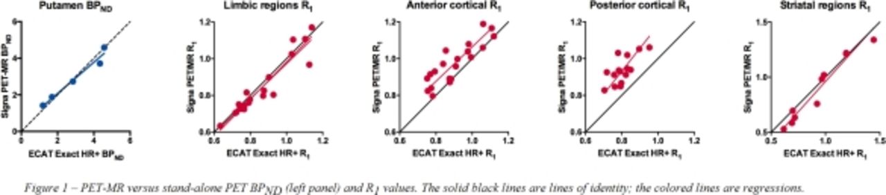
Results PET-MR BPND in putamen agreed well with stand-alone PET BPND (R2 0.97), with a slope of 0.84 (CI 0.55-1.13) and intercept of 0.40 (CI -0.54 - 1.34), and no significant bias. PET-MR R1 correlated best with stand-alone PET in striatum and limbic regions (R2 0.94 and 0.89, respectively), with lower values in anterior and posterior cortex (R2 0.76 and 0.50). A significant positive bias of PET-MR R1 values was found in posterior cortex (bias 0.13, CI 0.03-0.24). Correlation plots are shown in Figure 1.

Conclusions This study showed a good agreement between the PET-MR and stand-alone PET for BPND in putamen and for R1 especially in striatal and limbic regions. Cortical regions presented relatively low correlations between both modalities,which might be explained by the chosen attenuation correction method. Besides extension of the number of patients, additional investigations assessing, other attenuation correction methods for further improvement of the quantitative accuracy of the dynamic PET-MR data are required.

Figure
  • Download figure
  • Open in new tab
  • Download powerpoint
Previous
Back to top

In this issue

Journal of Nuclear Medicine
Vol. 57, Issue supplement 2
May 1, 2016
  • Table of Contents
  • Index by author
Article Alerts
Sign In to Email Alerts with your Email Address
Email Article

Thank you for your interest in spreading the word on Journal of Nuclear Medicine.

NOTE: We only request your email address so that the person you are recommending the page to knows that you wanted them to see it, and that it is not junk mail. We do not capture any email address.

Enter multiple addresses on separate lines or separate them with commas.
Quantitative image validation of PET-MR against stand-alone PET - A dynamic 11C-PE2I PET study.
(Your Name) has sent you a message from Journal of Nuclear Medicine
(Your Name) thought you would like to see the Journal of Nuclear Medicine web site.
Citation Tools
Quantitative image validation of PET-MR against stand-alone PET - A dynamic 11C-PE2I PET study.
Joao Sousa, Lieuwe Appel, Stergios Papadimitriou, Dag Nyholm, Jens Sorensen, Torsten Danfors, Elna-Marie Larsson, Hakan Ahlstrom, Mark Lubberink
Journal of Nuclear Medicine May 2016, 57 (supplement 2) 1935;

Citation Manager Formats

  • BibTeX
  • Bookends
  • EasyBib
  • EndNote (tagged)
  • EndNote 8 (xml)
  • Medlars
  • Mendeley
  • Papers
  • RefWorks Tagged
  • Ref Manager
  • RIS
  • Zotero
Share
Quantitative image validation of PET-MR against stand-alone PET - A dynamic 11C-PE2I PET study.
Joao Sousa, Lieuwe Appel, Stergios Papadimitriou, Dag Nyholm, Jens Sorensen, Torsten Danfors, Elna-Marie Larsson, Hakan Ahlstrom, Mark Lubberink
Journal of Nuclear Medicine May 2016, 57 (supplement 2) 1935;
Twitter logo Facebook logo LinkedIn logo Mendeley logo
  • Tweet Widget
  • Facebook Like
  • Google Plus One
Bookmark this article

Jump to section

  • Article
  • Figures & Data
  • Info & Metrics

Related Articles

  • No related articles found.
  • Google Scholar

Cited By...

  • No citing articles found.
  • Google Scholar

More in this TOC Section

Instrumentation & Data Analysis Track

  • Deep Learning Based Kidney Segmentation for Glomerular Filtration Rate Measurement Using Quantitative SPECT/CT
  • The Benefit of Time-of-Flight in Digital Photon Counting PET Imaging: Physics and Clinical Evaluation
  • Preclinical validation of a single-scan rest/stress imaging technique for 13NH3 cardiac perfusion studies
Show more Instrumentation & Data Analysis Track

MTA II: Data Analysis & Management Posters

  • Molecular Imaging And Kinetic Analysis Toolbox (MIAKAT) - A Quantitative Software Package for the Analysis of PET Neuroimaging Data
  • Detection of dementia-related hypometabolism using two different age-adjusted reference FDG- PET databases
  • Localized Quantitative Analysis of Positron Emission Tomography (PET) for Temporal Lobe Epilepsy Lateralization and Surgical Intervention
Show more MTA II: Data Analysis & Management Posters

Similar Articles

SNMMI

© 2025 SNMMI

Powered by HighWire