
Main menu

  • Home
  • Content
    • Current
    • Ahead of print
    • Past Issues
    • JNM Supplement
    • SNMMI Annual Meeting Abstracts
    • Continuing Education
    • JNM Podcasts
  • Subscriptions
    • Subscribers
    • Institutional and Non-member
    • Rates
    • Journal Claims
    • Corporate & Special Sales
  • Authors
    • Submit to JNM
    • Information for Authors
    • Assignment of Copyright
    • AQARA requirements
  • Info
    • Reviewers
    • Permissions
    • Advertisers
  • About
    • About Us
    • Editorial Board
    • Contact Information
  • More
    • Alerts
    • Feedback
    • Help
    • SNMMI Journals
  • SNMMI
    • JNM
    • JNMT
    • SNMMI Journals
    • SNMMI

User menu

  • Subscribe
  • My alerts
  • Log in
  • My Cart

Search

  • Advanced search
Journal of Nuclear Medicine
  • SNMMI
    • JNM
    • JNMT
    • SNMMI Journals
    • SNMMI
  • Subscribe
  • My alerts
  • Log in
  • My Cart
Journal of Nuclear Medicine

Advanced Search

  • Home
  • Content
    • Current
    • Ahead of print
    • Past Issues
    • JNM Supplement
    • SNMMI Annual Meeting Abstracts
    • Continuing Education
    • JNM Podcasts
  • Subscriptions
    • Subscribers
    • Institutional and Non-member
    • Rates
    • Journal Claims
    • Corporate & Special Sales
  • Authors
    • Submit to JNM
    • Information for Authors
    • Assignment of Copyright
    • AQARA requirements
  • Info
    • Reviewers
    • Permissions
    • Advertisers
  • About
    • About Us
    • Editorial Board
    • Contact Information
  • More
    • Alerts
    • Feedback
    • Help
    • SNMMI Journals
  • View or Listen to JNM Podcast
  • Visit JNM on Facebook
  • Join JNM on LinkedIn
  • Follow JNM on Twitter
  • Subscribe to our RSS feeds
Meeting ReportInstrumentation & Data Analysis Track

Molecular Imaging And Kinetic Analysis Toolbox (MIAKAT) - A Quantitative Software Package for the Analysis of PET Neuroimaging Data

Roger Gunn, Christopher Coello and Graham Searle
Journal of Nuclear Medicine May 2016, 57 (supplement 2) 1928;
Roger Gunn
1Imanova Ltd London United Kingdom
2Imanova Ltd London United Kingdom
3Imperial College London London United Kingdom
  • Find this author on Google Scholar
  • Find this author on PubMed
  • Search for this author on this site
Christopher Coello
1Imanova Ltd London United Kingdom
2Imanova Ltd London United Kingdom
  • Find this author on Google Scholar
  • Find this author on PubMed
  • Search for this author on this site
Graham Searle
1Imanova Ltd London United Kingdom
2Imanova Ltd London United Kingdom
  • Find this author on Google Scholar
  • Find this author on PubMed
  • Search for this author on this site
  • Article
  • Figures & Data
  • Info & Metrics
Loading

Abstract

1928

Objectives Quantitative analysis of clinical PET neuroimaging data involves a sequence of steps or processes that transform the primary data, from a number of sources, into parameters with biological significance. These outcome measures are used routinely in human studies of normo- and pathophysiology, disease progression and drug development. Until relatively recently, different components of the analysis workflow have been performed using separate applications, with significant human interaction and thus inefficiency with the potential for the introduction of errors and lack of reproducibility. MIAKAT addresses these issues and is being shared freely with the academic community to provide accessible software tools that will enable a broad set of users to conduct high quality, state of the art analysis of PET neuroimaging research data.

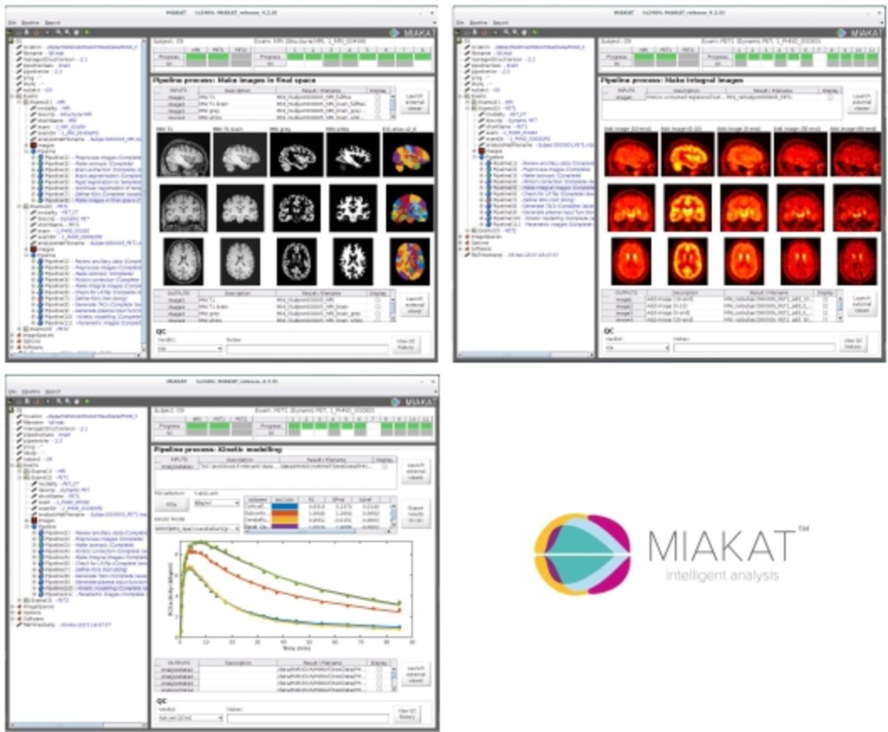
Methods MIAKAT (www.miakat.org) is a suite of analysis tools for PET neuroimaging data integrating state of the art tools in a single user friendly software environment making it easy for novice or advanced imaging scientists to efficiently and accurately analyse their data in a fully quantitative manner. The software includes functionality for motion correction of dynamic images, neuoranatomical parcellation of regions of interest (ROIs), blood/plasma input function modelling and tracer kinetic modelling for regional or parametric image analyses. MIAKAT is implemented in MATLAB[1], and has a central GUI that facilitates “point and click” operation. The standard brain pipeline configurations implemented in MIAKAT take the primary experimental data (dynamic PET and structural MR images, along with ancillary data such as arterial blood measurements) and perform a sequence of processes which ultimately produce results in the form of regional (or voxel-wise) parameters extracted via a comprehensive set of modelling techniques. MIAKAT combines in-house code with wrappers for SPM[2] and FSL[3] commands in order to provide start of the art functionality, including inbuilt audit trails, within a coherent framework.

Results Users can run preconfigured standard analysis workflows, or bespoke pipelines and processes to suit their needs. The software is distributed as MATLAB m-code, enabling users to understand and interact with the software at any level. The MIAKAT distribution consists of around 60,000 lines of Matlab code. MIAKAT implements a set of default image analysis workflows, as well as providing the ability to easily develop variations or new workflows as required. The standard PET neuroimaging analysis pipeline demonstrated here starts with a subject’s dynamic PET data and associated structural MRI in the NIfTI file format, along with associated ancillary data (such as frame times, injected radioactivity data, measure arterial blood, plasma and metabolite activity data) in a well-defined text-based .anc file format. The full analysis pipeline typically takes less than one hour per PET scan to perform all spatial and kinetic analysis processes, and has been used to analyse over 500 clinical PET scans.

Conclusions MIAKAT brings together state of the art tools for the quantitative analysis of PET molecular imaging data in a single user friendly software environment enabling high quality quantitative and reproducible analysis of PET neuroimaging studies. The inbuilt audit trails ensure quality and consistency. The modular nature of the software design easily permits the incorporation of additional algorithms as they become available, and the efficient implementation of alternative workflows. This will be a valuable resource for the academic community that currently does not have free access to such tools. [1] Matlab R2012b, The MathWorks Inc., Natick, MA, USA (www.mathworks.com) [2] SPM, Wellcome Trust Centre for Neuroimaging, London, UK (http://www.fil.ion.ucl.ac.uk/spm/) [3] FSL, FMRIB, Oxford, UK (http://fsl.fmrib.ox.ac.uk) $$graphic_8B78BF3C-E78E-4266-9AC1-356D0D95ED55$$

Figure
  • Download figure
  • Open in new tab
  • Download powerpoint
Previous
Back to top

In this issue

Journal of Nuclear Medicine
Vol. 57, Issue supplement 2
May 1, 2016
  • Table of Contents
  • Index by author
Article Alerts
Sign In to Email Alerts with your Email Address
Email Article

Thank you for your interest in spreading the word on Journal of Nuclear Medicine.

NOTE: We only request your email address so that the person you are recommending the page to knows that you wanted them to see it, and that it is not junk mail. We do not capture any email address.

Enter multiple addresses on separate lines or separate them with commas.
Molecular Imaging And Kinetic Analysis Toolbox (MIAKAT) - A Quantitative Software Package for the Analysis of PET Neuroimaging Data
(Your Name) has sent you a message from Journal of Nuclear Medicine
(Your Name) thought you would like to see the Journal of Nuclear Medicine web site.
Citation Tools
Molecular Imaging And Kinetic Analysis Toolbox (MIAKAT) - A Quantitative Software Package for the Analysis of PET Neuroimaging Data
Roger Gunn, Christopher Coello, Graham Searle
Journal of Nuclear Medicine May 2016, 57 (supplement 2) 1928;

Citation Manager Formats

  • BibTeX
  • Bookends
  • EasyBib
  • EndNote (tagged)
  • EndNote 8 (xml)
  • Medlars
  • Mendeley
  • Papers
  • RefWorks Tagged
  • Ref Manager
  • RIS
  • Zotero
Share
Molecular Imaging And Kinetic Analysis Toolbox (MIAKAT) - A Quantitative Software Package for the Analysis of PET Neuroimaging Data
Roger Gunn, Christopher Coello, Graham Searle
Journal of Nuclear Medicine May 2016, 57 (supplement 2) 1928;
Twitter logo Facebook logo LinkedIn logo Mendeley logo
  • Tweet Widget
  • Facebook Like
  • Google Plus One
Bookmark this article

Jump to section

  • Article
  • Figures & Data
  • Info & Metrics

Related Articles

  • No related articles found.
  • Google Scholar

Cited By...

  • No citing articles found.
  • Google Scholar

More in this TOC Section

Instrumentation & Data Analysis Track

  • Deep Learning Based Kidney Segmentation for Glomerular Filtration Rate Measurement Using Quantitative SPECT/CT
  • The Benefit of Time-of-Flight in Digital Photon Counting PET Imaging: Physics and Clinical Evaluation
  • Preclinical validation of a single-scan rest/stress imaging technique for 13NH3 cardiac perfusion studies
Show more Instrumentation & Data Analysis Track

MTA II: Data Analysis & Management Posters

  • Detection of dementia-related hypometabolism using two different age-adjusted reference FDG- PET databases
  • Localized Quantitative Analysis of Positron Emission Tomography (PET) for Temporal Lobe Epilepsy Lateralization and Surgical Intervention
  • An adaptive motion correction method for PET/CT Brain Imaging
Show more MTA II: Data Analysis & Management Posters

Similar Articles

SNMMI

© 2025 SNMMI

Powered by HighWire