
Main menu

  • Home
  • Content
    • Current
    • Ahead of print
    • Past Issues
    • JNM Supplement
    • SNMMI Annual Meeting Abstracts
    • Continuing Education
    • JNM Podcasts
  • Subscriptions
    • Subscribers
    • Institutional and Non-member
    • Rates
    • Journal Claims
    • Corporate & Special Sales
  • Authors
    • Submit to JNM
    • Information for Authors
    • Assignment of Copyright
    • AQARA requirements
  • Info
    • Reviewers
    • Permissions
    • Advertisers
  • About
    • About Us
    • Editorial Board
    • Contact Information
  • More
    • Alerts
    • Feedback
    • Help
    • SNMMI Journals
  • SNMMI
    • JNM
    • JNMT
    • SNMMI Journals
    • SNMMI

User menu

  • Subscribe
  • My alerts
  • Log in
  • Log out
  • My Cart

Search

  • Advanced search
Journal of Nuclear Medicine
  • SNMMI
    • JNM
    • JNMT
    • SNMMI Journals
    • SNMMI
  • Subscribe
  • My alerts
  • Log in
  • Log out
  • My Cart
Journal of Nuclear Medicine

Advanced Search

  • Home
  • Content
    • Current
    • Ahead of print
    • Past Issues
    • JNM Supplement
    • SNMMI Annual Meeting Abstracts
    • Continuing Education
    • JNM Podcasts
  • Subscriptions
    • Subscribers
    • Institutional and Non-member
    • Rates
    • Journal Claims
    • Corporate & Special Sales
  • Authors
    • Submit to JNM
    • Information for Authors
    • Assignment of Copyright
    • AQARA requirements
  • Info
    • Reviewers
    • Permissions
    • Advertisers
  • About
    • About Us
    • Editorial Board
    • Contact Information
  • More
    • Alerts
    • Feedback
    • Help
    • SNMMI Journals
  • View or Listen to JNM Podcast
  • Visit JNM on Facebook
  • Join JNM on LinkedIn
  • Follow JNM on Twitter
  • Subscribe to our RSS feeds
Meeting ReportInstrumentation & Data Analysis Track

Preclinical validation of a single-scan rest/stress imaging technique for 13NH3 cardiac perfusion studies

Nicolas Guehl, Matthieu Pelletier-Galarneau, Dustin Wooten, J. Guerrero, Julia-Ann Scotton, Eric McDonald, Brittan Morris, Aurelie Kas, Marc Normandin, Georges El Fakhri and Nathaniel Alpert
Journal of Nuclear Medicine May 2018, 59 (supplement 1) 1711;
Nicolas Guehl
1Gordon Center for Medical Imaging Massachusetts General Hospital Boston MA United States
  • Find this author on Google Scholar
  • Find this author on PubMed
  • Search for this author on this site
Matthieu Pelletier-Galarneau
1Gordon Center for Medical Imaging Massachusetts General Hospital Boston MA United States
  • Find this author on Google Scholar
  • Find this author on PubMed
  • Search for this author on this site
Dustin Wooten
1Gordon Center for Medical Imaging Massachusetts General Hospital Boston MA United States
  • Find this author on Google Scholar
  • Find this author on PubMed
  • Search for this author on this site
J. Guerrero
1Gordon Center for Medical Imaging Massachusetts General Hospital Boston MA United States
  • Find this author on Google Scholar
  • Find this author on PubMed
  • Search for this author on this site
Julia-Ann Scotton
1Gordon Center for Medical Imaging Massachusetts General Hospital Boston MA United States
  • Find this author on Google Scholar
  • Find this author on PubMed
  • Search for this author on this site
Eric McDonald
1Gordon Center for Medical Imaging Massachusetts General Hospital Boston MA United States
  • Find this author on Google Scholar
  • Find this author on PubMed
  • Search for this author on this site
Brittan Morris
1Gordon Center for Medical Imaging Massachusetts General Hospital Boston MA United States
  • Find this author on Google Scholar
  • Find this author on PubMed
  • Search for this author on this site
Aurelie Kas
2Pitié-Salpêtrière Hospital Paris France
  • Find this author on Google Scholar
  • Find this author on PubMed
  • Search for this author on this site
Marc Normandin
1Gordon Center for Medical Imaging Massachusetts General Hospital Boston MA United States
  • Find this author on Google Scholar
  • Find this author on PubMed
  • Search for this author on this site
Georges El Fakhri
1Gordon Center for Medical Imaging Massachusetts General Hospital Boston MA United States
  • Find this author on Google Scholar
  • Find this author on PubMed
  • Search for this author on this site
Nathaniel Alpert
1Gordon Center for Medical Imaging Massachusetts General Hospital Boston MA United States
  • Find this author on Google Scholar
  • Find this author on PubMed
  • Search for this author on this site
  • Article
  • Figures & Data
  • Info & Metrics
Loading

Abstract

1711

Objectives: Due to the 10 min half-life of 13N, two separate syntheses of 13NH3 are required for rest/stress imaging, which is performed in two scans that are separated by 50 min. This work validates a single-scan, two-injection method which takes less than 15 min and requires one radiosynthesis. The new method is based on previous work with 18F-Flurpiridaz (Alpert et al. 2012, Guehl et al. 2017a) and a more computationally efficient algorithm for the generation of rest and stress kinetic parameters (Guehl et al. 2017b). In this work we validated our methods for 13NH3 myocardial perfusion imaging (MPI) using experimental data acquired in a pig model. We compared our single-scan technique with separate rest and stress PET acquisitions, and with microsphere flow measurements. Methods: 9 studies were performed in 6 pigs for a total of 27 scans. 5 studies were done in the control state and 4 after infarction of the left anterior descending artery. Each study consisted of 3 scans (Supplemental Figure, A): a two-injection rest/rest single-scan (scan 1), a two-injection rest/stress single-scan (scan 2), and a conventional one-injection stress scan (scan 3). Variable doses of adenosine (140-300 μg/min/kg) combined with dobutamine (0-15 μg/min/kg) were administered to cover a wide range of myocardial blood flow (MBF). In 4 studies, 13NH3 injections were paired with microsphere injections. The two-injection single-scan measurements were fitted with our non-stationary kinetic model (MGH2). MBF estimates were compared to those obtained with the standard method that analyzes rest ans stress separately with a stationary kinetic model (Hutchins et al. 1990) and with microsphere flows. We used a model-based method to generate rest and stress perfusion images, as if these images were acquired separately: the rest and stress kinetic parameter maps, computed with our rapid algorithm, were used, with the resting part of the input function, to solve the equations describing a stationary kinetic model.

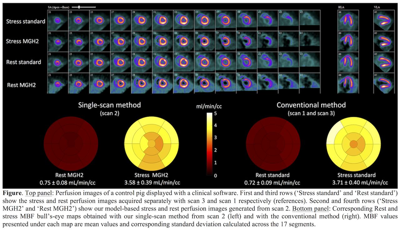
Results: MBF was investigated over the range 0.1 to ~8 mL/min/cc. For all datasets, the MGH2 kinetic model fitted the dynamic 13NH3 data in detail (Supplemental Figure, B). In the absence of stressor administration (scan 1), the estimated MBF values were nearly the same for the two radiotracer injections (mean difference: 0.067±0.070 ml/min/cc), demonstrating the basic repeatability of the method. Our simultaneous rest-stress method (scan 2) was in very good agreement with the conventional method for both rest (mean difference: -0.034±0.035 ml/min/cc; slope= 0.922±0.009; intercept= 0.024±0.007; R2= 0.97) and stress (mean difference: -0.011±0.291 ml/min/cc; slope= 1.050±0.025; intercept= -0.121±0.060; R2= 0.93) MBF measurements, which led in turn to very good agreement in myocardial flow reserve (MFR) values (mean difference: 0.123±0.498 ml/min/cc; slope= 1.110±0.036; intercept= -0.175±0.105; R2= 0.88). PET and microsphere MBF measurements correlated closely (slope= 1.111±0.071; intercept= -0.255±0.097; R2=0.94). In addition, our model-based method provided very good quality rest and stress perfusion images (Figure). Summed rest score (SRS) and summed stress score (SSS) were concordant with those obtained with the conventional method. Conclusions: 13NH3 rest/stress MPI procedure, that is normally acquired as two separate scans, can be compressed into a single ~15 min PET scan session while still obtaining accurate rest ans stress MBF measurements and good quality model-based perfusion images. Our method could significantly reduce 13NH3 MPI imaging cost through optimization of camera time and cyclotron usage, as only one 13N production is required. Support: 2017 Bradley-Alavi Student Fellowship; R01HL137230

Figure
  • Download figure
  • Open in new tab
  • Download powerpoint
Previous
Back to top

In this issue

Journal of Nuclear Medicine
Vol. 59, Issue supplement 1
May 1, 2018
  • Table of Contents
  • Index by author
Article Alerts
Sign In to Email Alerts with your Email Address
Email Article

Thank you for your interest in spreading the word on Journal of Nuclear Medicine.

NOTE: We only request your email address so that the person you are recommending the page to knows that you wanted them to see it, and that it is not junk mail. We do not capture any email address.

Enter multiple addresses on separate lines or separate them with commas.
Preclinical validation of a single-scan rest/stress imaging technique for 13NH3 cardiac perfusion studies
(Your Name) has sent you a message from Journal of Nuclear Medicine
(Your Name) thought you would like to see the Journal of Nuclear Medicine web site.
Citation Tools
Preclinical validation of a single-scan rest/stress imaging technique for 13NH3 cardiac perfusion studies
Nicolas Guehl, Matthieu Pelletier-Galarneau, Dustin Wooten, J. Guerrero, Julia-Ann Scotton, Eric McDonald, Brittan Morris, Aurelie Kas, Marc Normandin, Georges El Fakhri, Nathaniel Alpert
Journal of Nuclear Medicine May 2018, 59 (supplement 1) 1711;

Citation Manager Formats

  • BibTeX
  • Bookends
  • EasyBib
  • EndNote (tagged)
  • EndNote 8 (xml)
  • Medlars
  • Mendeley
  • Papers
  • RefWorks Tagged
  • Ref Manager
  • RIS
  • Zotero
Share
Preclinical validation of a single-scan rest/stress imaging technique for 13NH3 cardiac perfusion studies
Nicolas Guehl, Matthieu Pelletier-Galarneau, Dustin Wooten, J. Guerrero, Julia-Ann Scotton, Eric McDonald, Brittan Morris, Aurelie Kas, Marc Normandin, Georges El Fakhri, Nathaniel Alpert
Journal of Nuclear Medicine May 2018, 59 (supplement 1) 1711;
Twitter logo Facebook logo LinkedIn logo Mendeley logo
  • Tweet Widget
  • Facebook Like
  • Google Plus One
Bookmark this article

Jump to section

  • Article
  • Figures & Data
  • Info & Metrics

Related Articles

  • No related articles found.
  • Google Scholar

Cited By...

  • No citing articles found.
  • Google Scholar

More in this TOC Section

Instrumentation & Data Analysis Track

  • Deep Learning Based Kidney Segmentation for Glomerular Filtration Rate Measurement Using Quantitative SPECT/CT
  • The Benefit of Time-of-Flight in Digital Photon Counting PET Imaging: Physics and Clinical Evaluation
Show more Instrumentation & Data Analysis Track

Data Analysis & Management Posters

  • Evaluation of Quantitative Whole Body Dynamic FDG-PET Using Block Sequence Regularized Expectation Maximization (BSREM) Reconstruction
  • Feasibility of 18F-THK5351 PET quantitation using the Centiloid scale
  • NaF-PET/CT for assessment of temporomandibular joint osseous activity in rheumatoid arthritis patients and healthy subjects
Show more Data Analysis & Management Posters

Similar Articles

SNMMI

© 2025 SNMMI

Powered by HighWire