
Main menu

  • Home
  • Content
    • Current
    • Ahead of print
    • Past Issues
    • JNM Supplement
    • SNMMI Annual Meeting Abstracts
    • Continuing Education
    • JNM Podcasts
  • Subscriptions
    • Subscribers
    • Institutional and Non-member
    • Rates
    • Journal Claims
    • Corporate & Special Sales
  • Authors
    • Submit to JNM
    • Information for Authors
    • Assignment of Copyright
    • AQARA requirements
  • Info
    • Reviewers
    • Permissions
    • Advertisers
  • About
    • About Us
    • Editorial Board
    • Contact Information
  • More
    • Alerts
    • Feedback
    • Help
    • SNMMI Journals
  • SNMMI
    • JNM
    • JNMT
    • SNMMI Journals
    • SNMMI

User menu

  • Subscribe
  • My alerts
  • Log in
  • My Cart

Search

  • Advanced search
Journal of Nuclear Medicine
  • SNMMI
    • JNM
    • JNMT
    • SNMMI Journals
    • SNMMI
  • Subscribe
  • My alerts
  • Log in
  • My Cart
Journal of Nuclear Medicine

Advanced Search

  • Home
  • Content
    • Current
    • Ahead of print
    • Past Issues
    • JNM Supplement
    • SNMMI Annual Meeting Abstracts
    • Continuing Education
    • JNM Podcasts
  • Subscriptions
    • Subscribers
    • Institutional and Non-member
    • Rates
    • Journal Claims
    • Corporate & Special Sales
  • Authors
    • Submit to JNM
    • Information for Authors
    • Assignment of Copyright
    • AQARA requirements
  • Info
    • Reviewers
    • Permissions
    • Advertisers
  • About
    • About Us
    • Editorial Board
    • Contact Information
  • More
    • Alerts
    • Feedback
    • Help
    • SNMMI Journals
  • View or Listen to JNM Podcast
  • Visit JNM on Facebook
  • Join JNM on LinkedIn
  • Follow JNM on Twitter
  • Subscribe to our RSS feeds
Meeting ReportInstrumentation & Data Analysis Track

SPECT/CT and MR image registration for 123I-FP-CIT imaging

Takeshi Hara, Yuta Takeda, Xiangrong Zhou, Masaki Matsusako, Yuki Tanaka, Tetsuro Katafuchi and Hiroshi Fujita
Journal of Nuclear Medicine May 2016, 57 (supplement 2) 1936;
Takeshi Hara
1GIfu University Graduate School of Medicine Gifu Japan
  • Find this author on Google Scholar
  • Find this author on PubMed
  • Search for this author on this site
Yuta Takeda
1GIfu University Graduate School of Medicine Gifu Japan
  • Find this author on Google Scholar
  • Find this author on PubMed
  • Search for this author on this site
Xiangrong Zhou
1GIfu University Graduate School of Medicine Gifu Japan
  • Find this author on Google Scholar
  • Find this author on PubMed
  • Search for this author on this site
Masaki Matsusako
3St. Luke's International Hospital Tokyo Japan
  • Find this author on Google Scholar
  • Find this author on PubMed
  • Search for this author on this site
Yuki Tanaka
3St. Luke's International Hospital Tokyo Japan
  • Find this author on Google Scholar
  • Find this author on PubMed
  • Search for this author on this site
Tetsuro Katafuchi
2GIfu University of Medical Sciences Seki Japan
  • Find this author on Google Scholar
  • Find this author on PubMed
  • Search for this author on this site
Hiroshi Fujita
1GIfu University Graduate School of Medicine Gifu Japan
  • Find this author on Google Scholar
  • Find this author on PubMed
  • Search for this author on this site
  • Article
  • Figures & Data
  • Info & Metrics
Loading

Abstract

1936

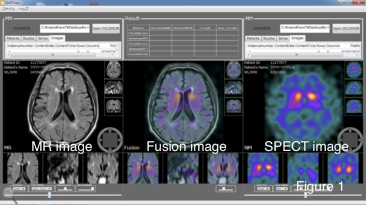
Objectives 123I-FP-CIT is used for the diagnosis of Parkinson's disease (PD) and Dementia with Lewy Bodies (DLB). It can visualize the distribution by binding to the dopamine neurons. In negative cases, the dopamine neurons are accumulated in form of comma, and in positive cases, the dopamine neurons are accumulated in form of dot. SPECT images can provide physiological information of a brain, but it is not possible to read anatomical information such as shape of corpus striatum. Therefore, it is possible to improve of diagnosis by presenting fusion image of SPECT image and MR image to readers as shown on Fig. 1.

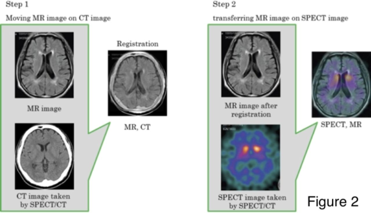
Methods Since image information represented by SPECT and MR images are different each other, an accurate image fusion of the two images is not easy. In this study, we propose a method fusion of SPECT image and MR image by intervening CT image taken by SPECT/CT devices (Fig. 2). Once the image fusion between CT and MR images was performed, the locations of MR images were transferred on SPECT images. SPECT/CT devices are general equipments integrated with gamma camera and multi-slice CT. Mostly, the CT images on SPECT/CT device were obtained with very low dose and were used for the absorption correction to generate SPECT images with high image quality. The CT images were utilized for the image registration with MR images. The location of CT and SPECT images were mechanically registered because the images were obtained at the almost same time. For the image registration between CT and MR images, we used a mutual information (MI) between the two images. The MI is the amount that represents a measure of the mutual dependence of two random variables. Before the MI was calculated, spatial resolutions of CT and MR images were unified, and the resolution of CT images were changed from 0.4297 mm to 2.9454 mm. After the resolutions were unified, rough alignments and angles of rotation were determined by calculating maximum mutual information. It is possible to limit search ranges and processing times. The maximum MI was obtained with fine steps of locations and angles to determine the final parameter of the location and the angles. Parallel computing method using CUDA was applied to obtain the parameter.

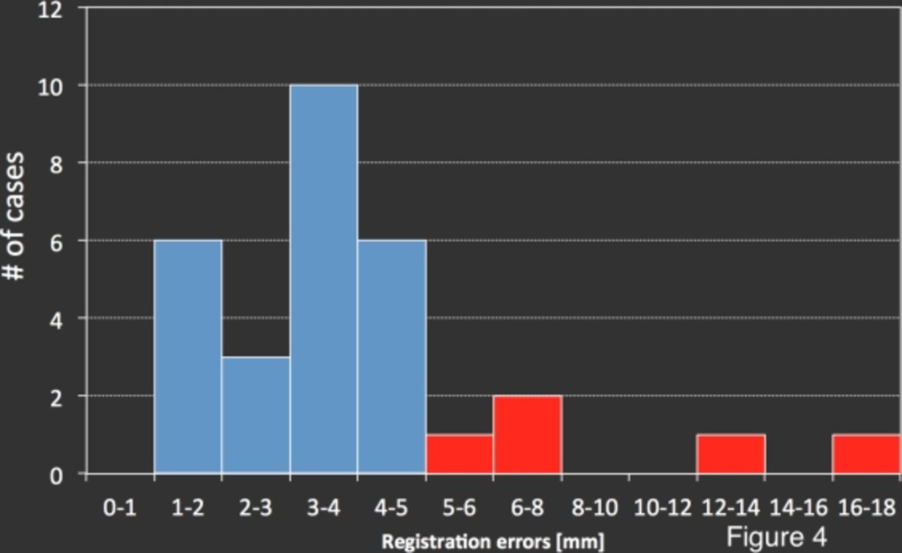
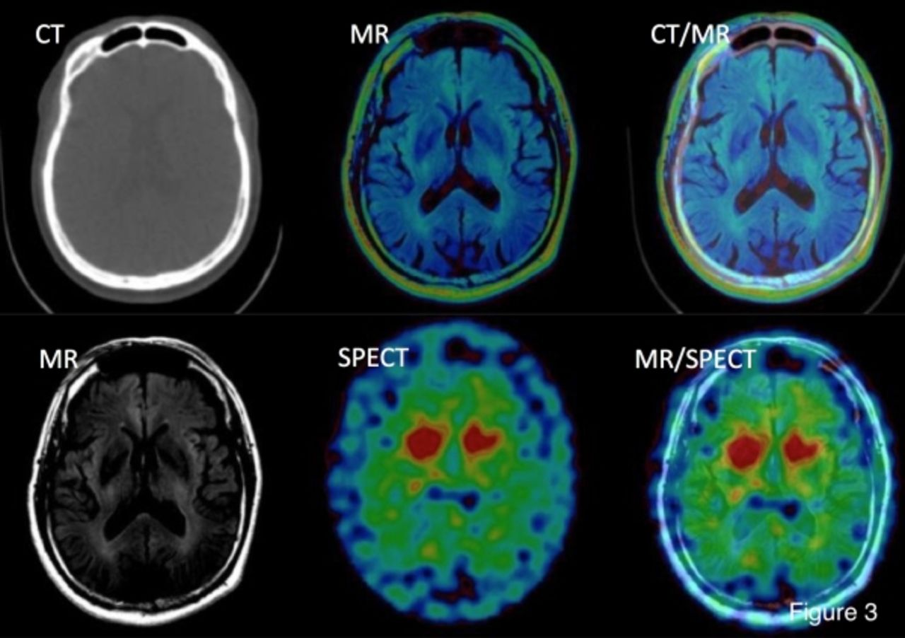
Results IRB approved 30 clinical cases of SPECT/CT and MR examinations were used in this study. 25 of 30 cases were registered correctly with registration errors up to 2mm. The other 5 cases were registered with registration errors from 7 to 16mm. Figure 3 shows an example for the image registration. The registration errors of 2mm are acceptable mis-registrations on SPECT images because the image resolution of SPECT is almost 3mm. The error can be ignored as mis-registration within a pixel size. The cause of errors in the other 5 cases of clinical results are in the configuration of initial ranges of deformation parameters during the global registration because the patient positioning at the MR examinations were quite different from SPECT/CT imaging. Figure 4 shows the distribution of registration errors.

Conclusions Image registration between brain SPECT and MR images can be performed by calculating MI between low dose CT images taken by SPECT/CT devices and the same patient’s MR images. Based on our registration scheme, the shape information determined on MR images could be superimposed on SPECT images to obtain quantitative counts.

Figure
  • Download figure
  • Open in new tab
  • Download powerpoint
Figure
  • Download figure
  • Open in new tab
  • Download powerpoint
Figure
  • Download figure
  • Open in new tab
  • Download powerpoint
Figure
  • Download figure
  • Open in new tab
  • Download powerpoint
Previous
Back to top

In this issue

Journal of Nuclear Medicine
Vol. 57, Issue supplement 2
May 1, 2016
  • Table of Contents
  • Index by author
Article Alerts
Sign In to Email Alerts with your Email Address
Email Article

Thank you for your interest in spreading the word on Journal of Nuclear Medicine.

NOTE: We only request your email address so that the person you are recommending the page to knows that you wanted them to see it, and that it is not junk mail. We do not capture any email address.

Enter multiple addresses on separate lines or separate them with commas.
SPECT/CT and MR image registration for 123I-FP-CIT imaging
(Your Name) has sent you a message from Journal of Nuclear Medicine
(Your Name) thought you would like to see the Journal of Nuclear Medicine web site.
Citation Tools
SPECT/CT and MR image registration for 123I-FP-CIT imaging
Takeshi Hara, Yuta Takeda, Xiangrong Zhou, Masaki Matsusako, Yuki Tanaka, Tetsuro Katafuchi, Hiroshi Fujita
Journal of Nuclear Medicine May 2016, 57 (supplement 2) 1936;

Citation Manager Formats

  • BibTeX
  • Bookends
  • EasyBib
  • EndNote (tagged)
  • EndNote 8 (xml)
  • Medlars
  • Mendeley
  • Papers
  • RefWorks Tagged
  • Ref Manager
  • RIS
  • Zotero
Share
SPECT/CT and MR image registration for 123I-FP-CIT imaging
Takeshi Hara, Yuta Takeda, Xiangrong Zhou, Masaki Matsusako, Yuki Tanaka, Tetsuro Katafuchi, Hiroshi Fujita
Journal of Nuclear Medicine May 2016, 57 (supplement 2) 1936;
Twitter logo Facebook logo LinkedIn logo Mendeley logo
  • Tweet Widget
  • Facebook Like
  • Google Plus One
Bookmark this article

Jump to section

  • Article
  • Figures & Data
  • Info & Metrics

Related Articles

  • No related articles found.
  • Google Scholar

Cited By...

  • No citing articles found.
  • Google Scholar

More in this TOC Section

Instrumentation & Data Analysis Track

  • Deep Learning Based Kidney Segmentation for Glomerular Filtration Rate Measurement Using Quantitative SPECT/CT
  • The Benefit of Time-of-Flight in Digital Photon Counting PET Imaging: Physics and Clinical Evaluation
  • Preclinical validation of a single-scan rest/stress imaging technique for 13NH3 cardiac perfusion studies
Show more Instrumentation & Data Analysis Track

MTA II: Data Analysis & Management Posters

  • Comparison of radiation dosimetry estimates for [11C]L-Deprenyl and [11C]Clorgyline across species and with D for H isotopic substitution.
  • Direct VOI-dedicated voxelwise Patlak estimation for quantitative dynamic imaging
Show more MTA II: Data Analysis & Management Posters

Similar Articles

SNMMI

© 2025 SNMMI

Powered by HighWire