
Main menu

  • Home
  • Content
    • Current
    • Ahead of print
    • Past Issues
    • JNM Supplement
    • SNMMI Annual Meeting Abstracts
    • Continuing Education
    • JNM Podcasts
  • Subscriptions
    • Subscribers
    • Institutional and Non-member
    • Rates
    • Journal Claims
    • Corporate & Special Sales
  • Authors
    • Submit to JNM
    • Information for Authors
    • Assignment of Copyright
    • AQARA requirements
  • Info
    • Reviewers
    • Permissions
    • Advertisers
  • About
    • About Us
    • Editorial Board
    • Contact Information
  • More
    • Alerts
    • Feedback
    • Help
    • SNMMI Journals
  • SNMMI
    • JNM
    • JNMT
    • SNMMI Journals
    • SNMMI

User menu

  • Subscribe
  • My alerts
  • Log in
  • Log out
  • My Cart

Search

  • Advanced search
Journal of Nuclear Medicine
  • SNMMI
    • JNM
    • JNMT
    • SNMMI Journals
    • SNMMI
  • Subscribe
  • My alerts
  • Log in
  • Log out
  • My Cart
Journal of Nuclear Medicine

Advanced Search

  • Home
  • Content
    • Current
    • Ahead of print
    • Past Issues
    • JNM Supplement
    • SNMMI Annual Meeting Abstracts
    • Continuing Education
    • JNM Podcasts
  • Subscriptions
    • Subscribers
    • Institutional and Non-member
    • Rates
    • Journal Claims
    • Corporate & Special Sales
  • Authors
    • Submit to JNM
    • Information for Authors
    • Assignment of Copyright
    • AQARA requirements
  • Info
    • Reviewers
    • Permissions
    • Advertisers
  • About
    • About Us
    • Editorial Board
    • Contact Information
  • More
    • Alerts
    • Feedback
    • Help
    • SNMMI Journals
  • View or Listen to JNM Podcast
  • Visit JNM on Facebook
  • Join JNM on LinkedIn
  • Follow JNM on Twitter
  • Subscribe to our RSS feeds
Meeting ReportInstrumentation & Data Analysis Track

Comparison of 22 partial volume correction methods for amyloid PET imaging with 11C-PiB

Tensho Yamao, Kenta Miwa, Kei Wagatsuma, Keisuke Matsubara, Reo Nemoto and Yuto Kamitaka
Journal of Nuclear Medicine May 2018, 59 (supplement 1) 1732;
Tensho Yamao
1Department of Radiological Sciences International University of Health and Welfare Ohtawara Japan
2Department of Radiological Sciences International University of Health and Welfare Ohtawara Japan
  • Find this author on Google Scholar
  • Find this author on PubMed
  • Search for this author on this site
Kenta Miwa
1Department of Radiological Sciences International University of Health and Welfare Ohtawara Japan
2Department of Radiological Sciences International University of Health and Welfare Ohtawara Japan
  • Find this author on Google Scholar
  • Find this author on PubMed
  • Search for this author on this site
Kei Wagatsuma
5Research Team for Neuroimaging Tokyo Metropolitan Institute of Gerontology Tokyo Japan
  • Find this author on Google Scholar
  • Find this author on PubMed
  • Search for this author on this site
Keisuke Matsubara
4Research Institute for Brain and Blood Vessels Aki Akita Japan
  • Find this author on Google Scholar
  • Find this author on PubMed
  • Search for this author on this site
Reo Nemoto
3Radiology International University of Health and Welfare Hos Nasushiobara Japan
  • Find this author on Google Scholar
  • Find this author on PubMed
  • Search for this author on this site
Yuto Kamitaka
1Department of Radiological Sciences International University of Health and Welfare Ohtawara Japan
2Department of Radiological Sciences International University of Health and Welfare Ohtawara Japan
  • Find this author on Google Scholar
  • Find this author on PubMed
  • Search for this author on this site
  • Article
  • Figures & Data
  • Info & Metrics
Loading

Abstract

1732

Objectives: Various strategies have been proposed to correct partial volume effects (PVE) in brain PET images. The present study aimed to determine the correction abilities of 22 partial volume correction (PVC) methods for amyloid 11C-PiB PET.

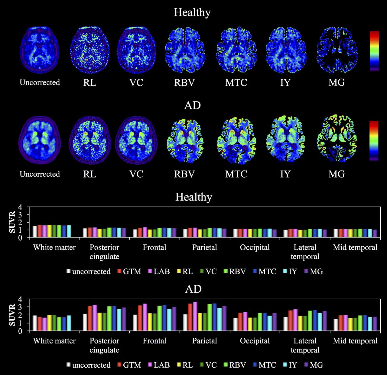
Methods: We retrospectively analyzed 11C-PiB PET/CT and 3T MRI data derived from nine healthy individuals (controls) and nine patients with Alzheimer disease (AD). Magnetic resonance images were parcellated using FreeSurfer and merged into 20 anatomical regions. We compared PVC by geometric transfer matrix (GTM), Labbe (LAB), Richardson-Lucy (RL), Van-Cittert (VC), region-based voxel-wise correction (RBV), multi target correction (MTC), iterative Yang (IY), Muller-Gartner (MG) and 14 combinations of these methods using PETPVC toolbox. Both uncorrected and PVC images were normalized with averaged ROI values of cerebellar grey matter to calculate standardized uptake value ratios (SUVR). The inter-subject coefficient of variation (COV) and intra-regional variability (COVr) were calculated based on data from all patients.

Results: The SUVR was almost identical in white matter regions with and without PVC. The SUVR and COV after PVC in images of grey matter regions increased by 8% and 22%, respectively, among the controls, and by 43% and 69%, respectively, among patients with AD. The RBV increased SUVR by 45% and decreased COV by 9% in these patients. The COVr values were more prominent when PVC methods included RL. Conclusions: The SUVR and image quality obviously differed according to the PVC

Methods: Thus, PVC should be cautiously applied to amyloid 11C-PiB PET images. Our results suggest that RBV or RBV+LAB was the most reliable means of qualifying Aβ plaque deposition and thus diagnosing AD. The present study provides useful information about how to select PVC methods for amyloid 11C-PiB PET images.

Figure
  • Download figure
  • Open in new tab
  • Download powerpoint
Previous
Back to top

In this issue

Journal of Nuclear Medicine
Vol. 59, Issue supplement 1
May 1, 2018
  • Table of Contents
  • Index by author
Article Alerts
Sign In to Email Alerts with your Email Address
Email Article

Thank you for your interest in spreading the word on Journal of Nuclear Medicine.

NOTE: We only request your email address so that the person you are recommending the page to knows that you wanted them to see it, and that it is not junk mail. We do not capture any email address.

Enter multiple addresses on separate lines or separate them with commas.
Comparison of 22 partial volume correction methods for amyloid PET imaging with 11C-PiB
(Your Name) has sent you a message from Journal of Nuclear Medicine
(Your Name) thought you would like to see the Journal of Nuclear Medicine web site.
Citation Tools
Comparison of 22 partial volume correction methods for amyloid PET imaging with 11C-PiB
Tensho Yamao, Kenta Miwa, Kei Wagatsuma, Keisuke Matsubara, Reo Nemoto, Yuto Kamitaka
Journal of Nuclear Medicine May 2018, 59 (supplement 1) 1732;

Citation Manager Formats

  • BibTeX
  • Bookends
  • EasyBib
  • EndNote (tagged)
  • EndNote 8 (xml)
  • Medlars
  • Mendeley
  • Papers
  • RefWorks Tagged
  • Ref Manager
  • RIS
  • Zotero
Share
Comparison of 22 partial volume correction methods for amyloid PET imaging with 11C-PiB
Tensho Yamao, Kenta Miwa, Kei Wagatsuma, Keisuke Matsubara, Reo Nemoto, Yuto Kamitaka
Journal of Nuclear Medicine May 2018, 59 (supplement 1) 1732;
Twitter logo Facebook logo LinkedIn logo Mendeley logo
  • Tweet Widget
  • Facebook Like
  • Google Plus One
Bookmark this article

Jump to section

  • Article
  • Figures & Data
  • Info & Metrics

Related Articles

  • No related articles found.
  • Google Scholar

Cited By...

  • No citing articles found.
  • Google Scholar

More in this TOC Section

Instrumentation & Data Analysis Track

  • Deep Learning Based Kidney Segmentation for Glomerular Filtration Rate Measurement Using Quantitative SPECT/CT
  • The Benefit of Time-of-Flight in Digital Photon Counting PET Imaging: Physics and Clinical Evaluation
  • Preclinical validation of a single-scan rest/stress imaging technique for 13NH3 cardiac perfusion studies
Show more Instrumentation & Data Analysis Track

Data Analysis & Management Posters

  • Evaluation of Quantitative Whole Body Dynamic FDG-PET Using Block Sequence Regularized Expectation Maximization (BSREM) Reconstruction
  • Feasibility of 18F-THK5351 PET quantitation using the Centiloid scale
  • NaF-PET/CT for assessment of temporomandibular joint osseous activity in rheumatoid arthritis patients and healthy subjects
Show more Data Analysis & Management Posters

Similar Articles

SNMMI

© 2025 SNMMI

Powered by HighWire