
Main menu

  • Home
  • Content
    • Current
    • Ahead of print
    • Past Issues
    • JNM Supplement
    • SNMMI Annual Meeting Abstracts
    • Continuing Education
    • JNM Podcasts
  • Subscriptions
    • Subscribers
    • Institutional and Non-member
    • Rates
    • Journal Claims
    • Corporate & Special Sales
  • Authors
    • Submit to JNM
    • Information for Authors
    • Assignment of Copyright
    • AQARA requirements
  • Info
    • Reviewers
    • Permissions
    • Advertisers
  • About
    • About Us
    • Editorial Board
    • Contact Information
  • More
    • Alerts
    • Feedback
    • Help
    • SNMMI Journals
  • SNMMI
    • JNM
    • JNMT
    • SNMMI Journals
    • SNMMI

User menu

  • Subscribe
  • My alerts
  • Log in
  • My Cart

Search

  • Advanced search
Journal of Nuclear Medicine
  • SNMMI
    • JNM
    • JNMT
    • SNMMI Journals
    • SNMMI
  • Subscribe
  • My alerts
  • Log in
  • My Cart
Journal of Nuclear Medicine

Advanced Search

  • Home
  • Content
    • Current
    • Ahead of print
    • Past Issues
    • JNM Supplement
    • SNMMI Annual Meeting Abstracts
    • Continuing Education
    • JNM Podcasts
  • Subscriptions
    • Subscribers
    • Institutional and Non-member
    • Rates
    • Journal Claims
    • Corporate & Special Sales
  • Authors
    • Submit to JNM
    • Information for Authors
    • Assignment of Copyright
    • AQARA requirements
  • Info
    • Reviewers
    • Permissions
    • Advertisers
  • About
    • About Us
    • Editorial Board
    • Contact Information
  • More
    • Alerts
    • Feedback
    • Help
    • SNMMI Journals
  • View or Listen to JNM Podcast
  • Visit JNM on Facebook
  • Join JNM on LinkedIn
  • Follow JNM on Twitter
  • Subscribe to our RSS feeds
Meeting ReportInstrumentation & Data Analysis Track

An adaptive motion correction method for PET/CT Brain Imaging

Man Wang, Yusheng Su, Jie Lu, Zhigang Liang, Defu Yang, Yang Lv, Yun Dong, Mu Chen and Lin An
Journal of Nuclear Medicine May 2016, 57 (supplement 2) 1934;
Man Wang
5XuanWu Hospital of Capital Medical University Beijing China
  • Find this author on Google Scholar
  • Find this author on PubMed
  • Search for this author on this site
Yusheng Su
5XuanWu Hospital of Capital Medical University Beijing China
  • Find this author on Google Scholar
  • Find this author on PubMed
  • Search for this author on this site
Jie Lu
5XuanWu Hospital of Capital Medical University Beijing China
  • Find this author on Google Scholar
  • Find this author on PubMed
  • Search for this author on this site
Zhigang Liang
5XuanWu Hospital of Capital Medical University Beijing China
  • Find this author on Google Scholar
  • Find this author on PubMed
  • Search for this author on this site
Defu Yang
3United Imaging Healthcare Shanghai China
4United Imaging Healthcare Shanghai China
  • Find this author on Google Scholar
  • Find this author on PubMed
  • Search for this author on this site
Yang Lv
3United Imaging Healthcare Shanghai China
4United Imaging Healthcare Shanghai China
  • Find this author on Google Scholar
  • Find this author on PubMed
  • Search for this author on this site
Yun Dong
3United Imaging Healthcare Shanghai China
4United Imaging Healthcare Shanghai China
  • Find this author on Google Scholar
  • Find this author on PubMed
  • Search for this author on this site
Mu Chen
3United Imaging Healthcare Shanghai China
4United Imaging Healthcare Shanghai China
1Duke Kunshan University Kunshan China
  • Find this author on Google Scholar
  • Find this author on PubMed
  • Search for this author on this site
Lin An
2Jining NO.1 People's Hospital Jining,Shandong China
  • Find this author on Google Scholar
  • Find this author on PubMed
  • Search for this author on this site
  • Article
  • Figures & Data
  • Info & Metrics
Loading

Abstract

1934

Objectives Head motion was commonly happened during the period of PET scanning, which would lead to severe artifacts in PET image reconstruction. In this study, a line of response (LOR) based adaptive motion correction method was presented to solve this problem.

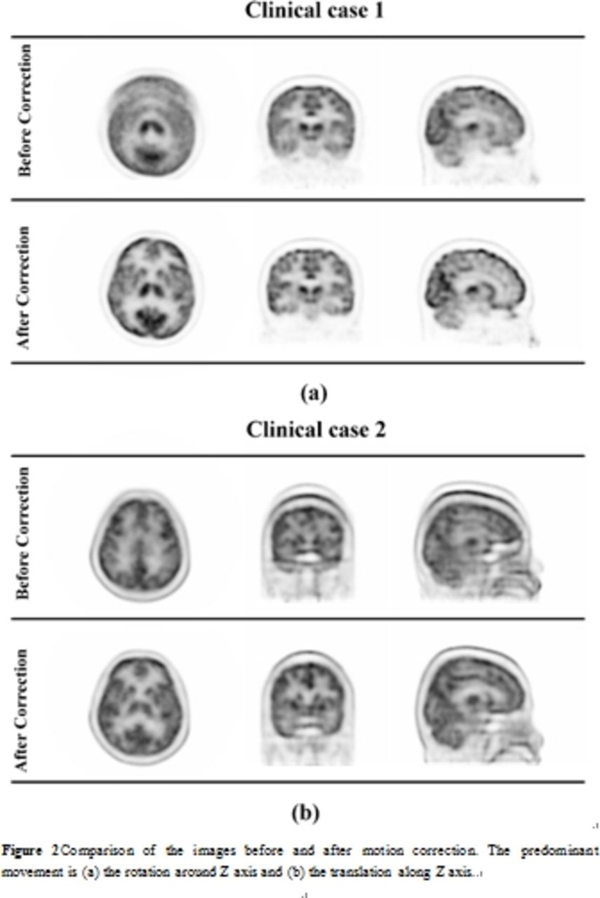
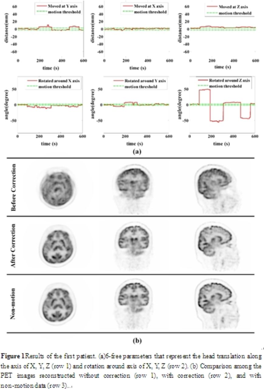
Methods The method mainly contains three important steps. First, without introducing additional motion tracking devices, the acquired data was dynamically reconstructed with very short time frames to find out the time intervals during which the motion happened. Second, from the multiple dynamic frames, head motion model could be established by mutual information based three-dimensional rigid registration method. Third, an event-by-event motion correction was performed on the list-mode data. The performance of the method was evaluated by several clinical datasets. All the datasets were obtained from uMI510 produced by United Imaging Healthcare. In the first experiment, two scans of the same patient were acquired. The patient was with frequent head motion in the first scan and kept steady in the second scan. Then the misplaced LOR was corrected into the right position to reconstruct the image. To further demonstrate the feasibly of the method, another two datasets were used. In one scan the patient was frequently shaking, and in the other scan the patient was frequently nodding. Then we compared their data before and after correction with this method.

Results Results of the first experiment showed that the proposed method could well detect the time intervals during which the motion happened. Then, moved LORs were corrected into their right position and the images were reconstructed. After motion correction, the resultant images were comparable to those from non-motion data. There were a lot of motion artifacts before correction. The images were blurry and the details cannot be showed. After correction the brain tissue structure of the images displayed clearly. There was no significant difference between the corrected images and non-motion images in the second experiment. Both results showed that our method could effectively detect the motion and eliminate the motion artifacts.

Conclusions In this paper, a motion correction method was proposed for PET/CT brain imaging. This method is adaptive, robust and effective, and has been validated by clinical datasets.

Figure
  • Download figure
  • Open in new tab
  • Download powerpoint
Figure
  • Download figure
  • Open in new tab
  • Download powerpoint
Previous
Back to top

In this issue

Journal of Nuclear Medicine
Vol. 57, Issue supplement 2
May 1, 2016
  • Table of Contents
  • Index by author
Article Alerts
Sign In to Email Alerts with your Email Address
Email Article

Thank you for your interest in spreading the word on Journal of Nuclear Medicine.

NOTE: We only request your email address so that the person you are recommending the page to knows that you wanted them to see it, and that it is not junk mail. We do not capture any email address.

Enter multiple addresses on separate lines or separate them with commas.
An adaptive motion correction method for PET/CT Brain Imaging
(Your Name) has sent you a message from Journal of Nuclear Medicine
(Your Name) thought you would like to see the Journal of Nuclear Medicine web site.
Citation Tools
An adaptive motion correction method for PET/CT Brain Imaging
Man Wang, Yusheng Su, Jie Lu, Zhigang Liang, Defu Yang, Yang Lv, Yun Dong, Mu Chen, Lin An
Journal of Nuclear Medicine May 2016, 57 (supplement 2) 1934;

Citation Manager Formats

  • BibTeX
  • Bookends
  • EasyBib
  • EndNote (tagged)
  • EndNote 8 (xml)
  • Medlars
  • Mendeley
  • Papers
  • RefWorks Tagged
  • Ref Manager
  • RIS
  • Zotero
Share
An adaptive motion correction method for PET/CT Brain Imaging
Man Wang, Yusheng Su, Jie Lu, Zhigang Liang, Defu Yang, Yang Lv, Yun Dong, Mu Chen, Lin An
Journal of Nuclear Medicine May 2016, 57 (supplement 2) 1934;
Twitter logo Facebook logo LinkedIn logo Mendeley logo
  • Tweet Widget
  • Facebook Like
  • Google Plus One
Bookmark this article

Jump to section

  • Article
  • Figures & Data
  • Info & Metrics

Related Articles

  • No related articles found.
  • Google Scholar

Cited By...

  • No citing articles found.
  • Google Scholar

More in this TOC Section

Instrumentation & Data Analysis Track

  • Deep Learning Based Kidney Segmentation for Glomerular Filtration Rate Measurement Using Quantitative SPECT/CT
  • The Benefit of Time-of-Flight in Digital Photon Counting PET Imaging: Physics and Clinical Evaluation
  • Preclinical validation of a single-scan rest/stress imaging technique for 13NH3 cardiac perfusion studies
Show more Instrumentation & Data Analysis Track

MTA II: Data Analysis & Management Posters

  • Detection of dementia-related hypometabolism using two different age-adjusted reference FDG- PET databases
  • Localized Quantitative Analysis of Positron Emission Tomography (PET) for Temporal Lobe Epilepsy Lateralization and Surgical Intervention
Show more MTA II: Data Analysis & Management Posters

Similar Articles

SNMMI

© 2025 SNMMI

Powered by HighWire