


Abstract
Imaging of neurofibrillary pathology in the brain helps in diagnosing dementia, tracking disease progression, and evaluating the therapeutic efficacy of antidementia drugs. The radiotracers used in this imaging must be highly sensitive and specific for tau protein fibrils in the human brain. We developed a novel tau PET tracer, 18F-THK5351, through compound optimization of arylquinoline derivatives. Methods: The in vitro binding properties, pharmacokinetics, and safety of 18F-THK5351 were investigated, and a clinical study on Alzheimer disease (AD) patients was performed. Results: 18F-THK5351 demonstrated higher binding affinity for hippocampal homogenates from AD brains and faster dissociation from white-matter tissue than did 18F-THK5117. The THK5351 binding amount correlated with the amount of tau deposits in human brain samples. Autoradiography of brain sections revealed that THK5351 bound to neurofibrillary tangles selectively and with a higher signal-to-background ratio than did THK5117. THK5351 exhibited favorable pharmacokinetics and no defluorination in mice. In first-in-human PET studies in AD patients, 18F-THK5351 demonstrated faster kinetics, higher contrast, and lower retention in subcortical white matter than18F-THK5117. Conclusion: 18F-THK5351 is a useful PET tracer for the early detection of neurofibrillary pathology in AD patients.
Tau accumulation occurs in a stereotyped spatiotemporal manner at the intraneuronal and anatomic distribution levels in the brain and is associated with neuronal loss and cognitive impairment (1–5). Because tau accumulation plays a key role in neurodegeneration and is considered to start before extensive neuronal loss emerges, tau-focused drug-discovery strategies for Alzheimer disease (AD) are of particular interest (6,7). In efforts to accelerate drug discovery, there is growing demand for techniques to measure brain tau loads noninvasively. PET imaging of tau is expected to provide spatiotemporal information on the progression of tau pathology in the living brain. Therefore, this technique will facilitate accurate tauopathy diagnosis, precise assessment of disease severity and therapeutic efficacy, and patient enrolment for antitau therapeutic trials (8–10).
Several putative tau PET tracers have been developed and tested in humans (11–14), and all of these tracers show elevated uptake in the hippocampus and temporal cortex of AD patients. We screened β-sheet–binding compounds and identified a series of compounds that preferentially bind to tau deposits in AD brains (15–17). Through compound optimization, several 18F-labeled arylquinoline derivatives were developed as candidate tau PET radiotracers (18). Recent 18F-THK5105 and 18F-THK5117 PET studies demonstrated increased tracer uptake in common sites of tau pathology in AD and its association with clinical severity of dementia (19,20). However, these tracers—like amyloid PET tracers—showed high nonspecific retention in subcortical white matter. This white-matter binding must be minimized because the signals could obscure visual interpretation of PET images and decrease detection sensitivity for early tau pathology in the presymptomatic stage of AD. For assessing the therapeutic efficacy of potential antitau drugs in clinical trials, tau PET tracers must be adequately sensitive to detect even subtle changes in brain tau loads. Moreover, tau PET is expected to detect age-associated neurofibrillary tangles in cognitively normal individuals, recently named primary age-associated tauopathy (21–23). Because such age-related tau pathology is typically milder than disease-related changes in AD, the radiotracers used must be highly sensitive.
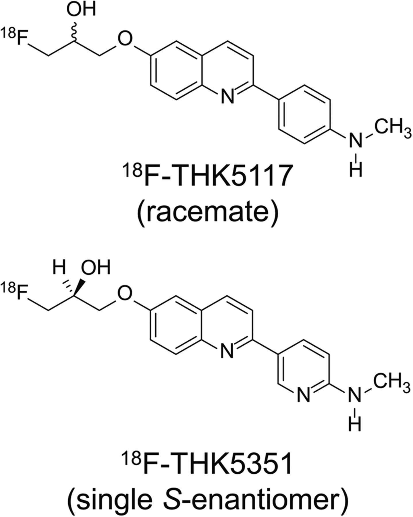
To reduce nonspecific tracer retention in white matter and increase the signal-to-background ratio of PET images, we replaced a benzene ring of 18F-THK5117 with pyridine and developed a novel tau PET tracer, 18F-THK5351. 18F-THK5351 is a single S-enantiomer, which should improve the pharmacokinetics of arylquinoline derivatives (Fig. 1). To evaluate the clinical usefulness of 18F-THK5351 as a tau PET tracer, we examined the in vitro binding properties, pharmacokinetics, and safety of 18F-THK5351 and performed a clinical study on AD patients.

18F-THK5351 and 18F-THK5117 chemical structures.
MATERIALS AND METHODS
Radiosynthesis of Quinoline Derivatives
18F-THK5351 was prepared from its tosylate precursor (S)-2-(2-methylaminopyrid-5-yl)-6-[[2-(tetrahydro-2H-pyran-2-yloxy)-3-tosyloxy]propoxy] quinoline (THK5352) according to the previously described method for synthesizing 18F-THK5105 and 18F-THK5117 (18). 18F-THK5351 was purified using semipreparative high-performance liquid chromatography (column: Inertsil ODS-4 [GL Sciences, Inc.]; mobile phase: 20 mmol/L NaH2PO4/acetonitrile [75/25 for THK5351]; flow rate: 5.0 mL/min). The radiolabeled product was dissolved in ethanol, dimethylsulfoxide, or saline with polysobate-80 (<0.1%) for biologic evaluation. 18F-THK5351 was obtained at a radiochemical yield of 46% ± 13% (decay-corrected), radiochemical purity of greater than 95%, and specific activity of 254 ± 47 GBq/μmol. 3H-Pittsburgh compound B (PiB) (specific activity, 2.96 GBq/μmol; radiochemical purity, 99%) was purchased from American Radiolabeled Chemicals. 3H-THK5351 (specific activity, 2.96 TBq/mmol; radiochemical purity, 98.9%) and 3H-THK5117 (specific activity, 2.78 TBq/mmol; radiochemical purity, 98.2%) were custom-labeled by Sekisui Medical Inc.
In Vitro Binding Study
Experiments were performed as per the regulations of the Ethics Committee of the Tohoku University School of Medicine. Brain samples were obtained from Tohoku University Brain Bank. The following studies were conducted as previously described: in vitro saturation binding assays (18); in vitro association and dissociation rate measurement (20,24); and in vitro binding assays, using 1 nmol/L 3H-labeled ligands (20).
Autoradiography
Experimental procedures followed the regulations of the Ethics Committee of the Tohoku University School of Medicine. Autoradiography in postmortem brain sections was conducted using 3H-THK5351, 3H-THK5117, and 3H-PiB as previously reported (20). Washing procedures were modified slightly. Briefly, after 30-min incubation at room temperature with 3 nmol/L 3H-labeled compounds, sections were washed sequentially with phosphate-buffered saline containing 1% bovine serum albumin (5 min) and phosphate-buffered saline (5 min, twice). Dried sections were exposed to an imaging plate for 3 d. High-resolution autoradiography of 3H-labeled sections was performed as before (20).
Small-Animal PET Studies
All animal experiment protocols were approved by the Laboratory Animal Care Committee of Tohoku University. In vivo PET studies were performed using male SLC:ICR mice, as previously described (24).
Biodistribution Studies
Biodistribution was investigated after intravenous injection of 18F-THK5351 or 18F-THK5117 into male ICR mice as previously described (18). On the basis of the biodistribution data from mice (percentage injected dose per gram), we estimated the radiation dose and mass dose for humans.
Animal Toxicity Studies
A 14-d toxicity study involving a single-dose THK5351 test-article administration through intravenous injection in ICR mice was performed at Mitsubishi Chemical Medience Corp., as previously described (18).
Radiosynthesis for Clinical PET Study
18F-THK5351, 18F-THK5117, and 11C-PiB were prepared at the Cyclotron and Radioisotope Center, Tohoku University. 18F-THK5351 was radiosynthesized using a semiautomated system developed in-house. No-carrier-added 18F-fluoride (18F−) produced by the HM-12 cyclotron (30 min/25 μA; Sumitomo Heavy Industries) was separated from the irradiated target water using a Sep-Pak Light Accell Plus QMA cartridge (Waters). The trapped 18F− was eluted using a Kryptofix solution (Kryptofix 222 [20 mg], K2CO3 [4 mg], MeCN [0.7 mL], H2O [0.3 mL]) into a reaction vial. The solution was evaporated to dryness through azeotropic distillation with acetonitrile. After drying, THK5352 (3 mg, 5.3 μmol) dissolved in dimethylsulfoxide (0.7 mL) was transferred into the reaction vial and stirred at 110°C for 10 min, and then aqueous HCl (2 mol/L, 0.2 mL) was added to the reaction solution and stirred at 110°C for another 3 min. The reaction was then quenched with aqueous AcOK (0.8 mol/L, 1 mL) and distilled water (7 mL), after which solid-phase extraction was performed using a Sep-Pak tC18 Plus cartridge (Waters). The trapped radioactive products were eluted using 60% EtOH, and then the eluate was mixed with H2O and subjected to semipreparative high-performance liquid chromatography separation under the same conditions as those described above. The high-performance liquid chromatography fraction of 18F-THK5351 was collected in glassware containing H2O (30 mL) and ascorbic acid (25%, 1.0 mL; NIPRO Pharma), and 18F-THK5351 was isolated from the solution through solid-phase extraction performed using a Sep-Pak tC18 Plus cartridge. The ethanol eluate from the cartridge was transferred into a flask containing polysorbate-80 (5% in ethanol, 0.8 mL) and ascorbic acid (25%, 0.2 mL), and then the solution was evaporated to dryness. The radioactive residue was dissolved in saline and sterilized through filtration using a Millex-GV Syringe Filter Unit (Millipore). 18F-THK5117 was synthesized as previously described (20). 11C-PiB was synthesized using a loop-method with 11C-methyl triflate, as previously reported (25). The radiochemical purity of the injectable solutions of 18F-THK5351, 18F-THK5117, and 11C-PiB was greater than 95%, and their specific activities were 254 ± 47, 357 ± 270, and 240 ± 48 GBq/μmol, respectively.
Clinical PET Study Participants
Three AD patients and 3 healthy elderly study participants underwent 18F-THK5351 PET scans. Participant demographic data are shown in Supplemental Table 1 (supplemental materials are available at http://jnm.snmjournals.org). Two AD patients underwent additional 18F-THK5117 PET scans within 2-wk intervals and additional 11C-PiB PET scans within 3-mo intervals. Probable AD was diagnosed on the basis of criteria from the National Institute of Neurologic and Communicative Disorders and Stroke and the Alzheimer Disease Related Disorders Association. This study was approved by the Ethics Committee of the Tohoku University Hospital. The study was fully described to the patients, and then written informed consent was obtained from the patients or their guardians.
PET and MR Image Acquisition
PET imaging was performed using an Eminence STARGATE PET scanner (Shimadzu). After intravenous injection of 18F-THK5117 (185 MBq), 18F-THK5351 (185 MBq), or 11C-PiB (296 MBq), dynamic PET images were obtained for 90 min (18F-THK5117 and 18F-THK5351) or 70 min (11C-PiB), with the patients’ eyes closed. MRI was performed on all participants. T1- and T2-weighted MR images were obtained using a SIGNA 1.5-T machine (GE Healthcare). In T1-weighted MRI, a 3-dimensional volumetric acquisition of a T1-weighted gradient echo sequence produced a gapless series of thin axial sections using a vascular time-of-flight spoiled gradient recalled echo sequence (echo time/repetition time, 2.4/50 ms; flip angle, 45°; acquisition matrix, 256 × 256; 1 excitation; field of view, 22 cm; slice thickness, 2.0 mm).
Image Analysis
SUV images of 18F-THK5117, 18F-THK5351, and 11C-PiB were obtained by normalizing tissue radioactivity concentration by injected dose and body weight. MRI T1 images were coregistered to the early PET images (0–10 min after injection) for each participant using statistical parametric-mapping software (SPM8; Wellcome Department of Imaging Neuroscience, UCL). PET images were processed using a semiautomatic region-of-interest method, as previously described (19). The regional SUV to cerebellar cortex SUV ratio (SUVR) was used as an index of tracer retention. Coregistered MR and PET images were spatially normalized to an MRI T1 template in Talairach space using SPM8. After spatial normalization, regional SUVs were sampled using PMOD software (PMOD Technologies). Regions of interest were placed on individual axial images in the cerebellar hemisphere, dorsolateral prefrontal cortex (Brodmann’s area [BA] 9), ventrolateral prefrontal cortex (BA 10, 44, 45, and 46), orbitofrontal cortex (BA 11 and 12), superior temporal cortex (BA 22), inferior temporal cortex (BA 20 and 37), parietal cortex (BA 39 and 40), occipital cortex (BA 17, 18, and 19), anterior cingulate cortex, posterior cingulate cortex, parahippocampal gyrus, and subcortical white matter.
Statistical Analysis
Pearson correlation coefficients were calculated to access the relationship between 3H-labeled tracer binding and the amounts of insoluble protein. Spearman correlation coefficients were calculated to access the relationship between tracer retentions in AD patients.
RESULTS
In Vitro Tracer Binding to Human Brain Tissues
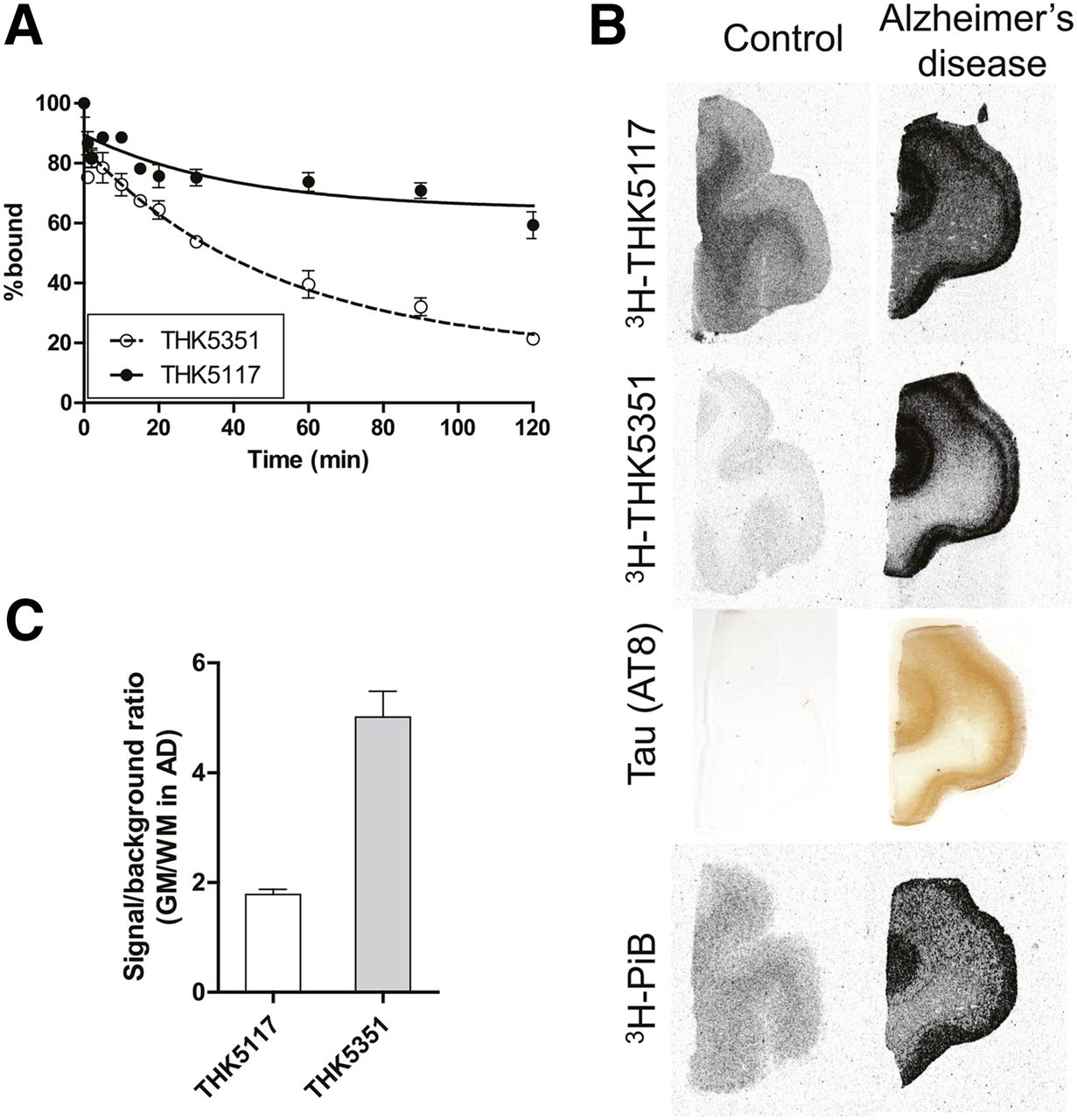
In vitro saturation binding assays were conducted to measure the binding affinity of 18F-THK5351 for postmortem tissues from AD patients. Scatchard analysis indicated 1-site binding of 18F-THK5351 for postmortem hippocampal homogenates from an AD patient (Supplemental Fig. 1A). 18F-THK5351 bound to AD hippocampal homogenates with high affinity (Kd = 2.9 nmol/L; maximum number of binding sites = 368.3 pmol/g tissue). We measured the in vitro binding of 3H-labeled THK5351 and THK5117 to postmortem tissues from 8 AD patients to compare the amount of specific binding of these tracers; the tracers were used at 1 nmol/L, the concentration typically achieved during PET scans. The specific binding of THK5351 and THK5117 was highly correlated (r = 0.98, P < 0.0001) (Supplemental Fig. 1B). The specific binding of THK5351 was also correlated with the level of insoluble tau (r = 0.71, P < 0.05) but not insoluble amyloid-β (r = −0.20, P = 0.63) or PiB (r = −0.10, P = 0.82), as observed for THK5117 (20). Furthermore, in vitro dissociation assays performed using brain white-matter homogenates revealed that THK5351 dissociated from white matter more rapidly than THK5117 did (Fig. 2A).

(A) In vitro dissociation of 18F-THK5351 and 18F-THK5117 from white-matter homogenates from normal brain. (B) Autoradiography of 3H-THK5117, 3H-THK5351, and 3H-PiB and tau (AT8) immunostaining in frontal brain sections from control participant and AD patient. (C) Signal-to-background ratios of 3H-labeled THK tracers in sections from AD patient (Braak stage V). Signal-to-background ratio was calculated as intensity of tracer binding in gray matter divided by that in subcortical white matter.
In Vitro Autoradiography in Human Brain Sections
To further evaluate binding selectivity and signal-to-background ratio, in vitro autoradiography was performed using 3H-THK5351 and 3H-THK5117 exhibiting similar specific activity; here, postmortem brain sections from a control participant and AD patients were used. Both 3H-THK5351 and 3H-THK5117 bound to the gray matter of AD brain sections in a laminar fashion, which corresponded to tau immunohistochemistry. However, in contrast to the substantial white-matter binding of 3H-THK5117, only weak 3H-THK5351 signals were detected in white matter (Fig. 2B). 3H-THK5351 showed a higher cortical–to–white-matter binding ratio than 3H-THK5117 did (Fig. 2C). 3H-THK5351 also showed little binding to control brain sections. Microautoradiography of AD brain sections provided additional evidence supporting the ability of 3H-THK5351 to selectively label neurofibrillary tangles. The 3H-THK5351 labeling patterns resembled the Gallyas–Braak staining in adjacent sections (Fig. 3). However, 3H-THK5351 did not label amyloid plaques that were labeled with 3H-PiB in an adjacent section. Furthermore, autoradiography of hemibrain sections from an AD patient demonstrated preferential 3H-THK5351 binding in the gray matter of the hippocampus, parahippocampal gyrus, fusiform gyrus, inferior and middle temporal gyri, insula, and cingulate gyrus, regions that contain a high density of tau deposits in AD (Supplemental Fig. 2). These tracer-binding patterns differed completely from the broad neocortical binding of 11C-PiB.
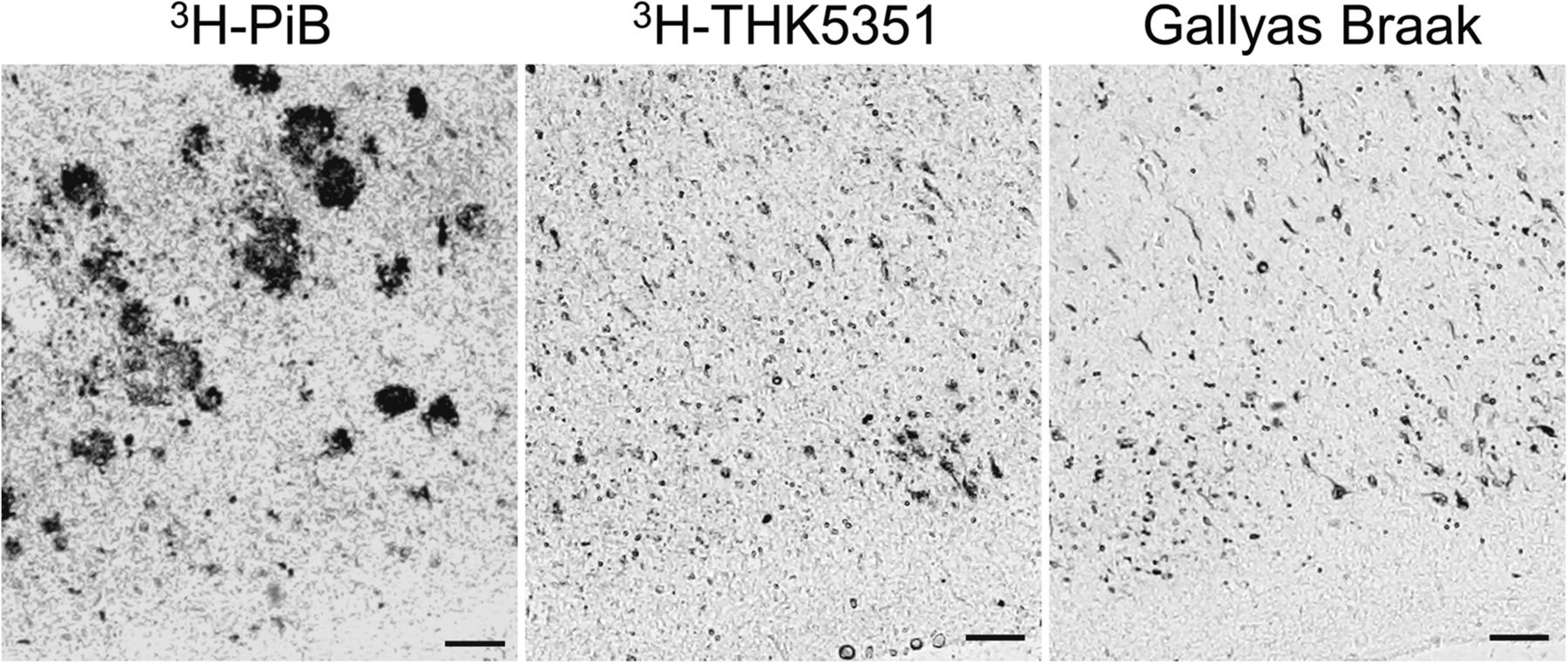
High-resolution autoradiography of 3H-PiB and 3H-THK5351 and Gallyas–Braak silver staining in entorhinal cortex of AD patient.
Pharmacokinetics in Mice
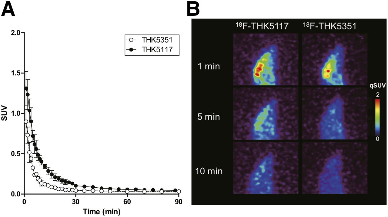
Brain pharmacokinetics of 18F-THK5351 in normal mice were investigated using a small-animal PET scanner. Although the peak brain uptake of 18F-THK5351 was slightly lower than that of 18F-THK5117, 18F-THK5351 entered the brain immediately after intravenous injection and showed faster washout from the brain than 18F-THK5117 did (Fig. 4; Supplemental Table 2). No marked radiotracer retention in bone was observed after 18F-THK5351 was injected into mice.

(A) Brain time–activity curves after intravenous administration of 18F-THK5351 and 18F-THK5117 in normal mice (n = 4). (B) Representative PET images of 18F-THK5117 and 18F-THK5351 at 1, 5, and 10 min after injection in normal mice.
Animal Acute-Toxicity Studies
At 0.1 and 1 mg/kg dosages under our study conditions, no animals died and no treatment-related changes in any animal were noted in clinical observations, body weight measurement, and pathologic examination.
Dose Estimates for Humans
18F-THK5351 radiation exposure was estimated using the biodistribution data from mice (Supplemental Table 2). The resultant whole-body effective dose equivalents were 14.4 μSv/MBq (men) and 18.4 μSv/MBq (women) (Supplemental Table 3). The organ doses for 18F-THK5351 were comparable to those associated with other common radiotracers.
Clinical PET Study
The SUV time–activity curves from 18F-THK5351 and 18F-THK5117 PET in 2 patients are shown in Figure 5. The pharmacokinetic data agreed with the small-animal PET study in normal mice. The peak uptake of 18F-THK5351 was again slightly lower than that of 18F-THK5117, but 18F-THK5351 was cleared more rapidly than 18F-THK5117 from the cerebellar cortex. In the AD patients, 18F-THK5351 binding in the inferior temporal cortex exceeded white-matter binding at all the time points after injection.

18F-THK5351 (A and B) and 18F-THK5117 (C and D) SUV time–activity curves in cerebellum (○), inferior temporal cortex (▪), and subcortical white matter (△) of 2 AD patients (AD1: 88-y-old man, Mini Mental State Examination [MMSE] score of 25; AD2: 58-y-old man, MMSE score of 19).
PET images are shown in Figure 6. 18F-THK5351 retention in the temporal lobe clearly distinguished AD patients from healthy elderly participants, although mild 18F-THK5351 retention was observed in the medial temporal cortex of elderly healthy control subjects (Fig. 6A). 18F-THK5351 showed higher contrast and lower subcortical white-matter retention than 18F-THK5117 did (Fig. 6B). 18F-THK5351 and 18F-THK5117 retention was particularly prominent in the mesial temporal lobe and the lateral temporal cortex, which differed considerably from 11C-PiB retention in the same AD patient (Table 1): 11C-PiB exhibited extremely high retention throughout broad neocortical areas except for the mesial temporal lobe. The regional SUVRs of 18F-THK5351 were higher than those of 18F-THK5117 (Table 1), and the regional SUVRs of 18F-THK5351 in 2 AD patients were significantly correlated with those of 18F-THK5117 (Spearman r = 0.839, P < 0.001) but not that of 11C-PiB (Fig. 7).

(A) SUVR images of 18F-THK5351 PET from 40 to 60 min after injection in 3 healthy control (HC) participants and 3 AD patients. (B) SUVR images of 18F-THK5117 and 18F-THK5351 PET from 50 to 60 min after injection and 11C-PiB PET from 40 to 70 min after injection in 2 AD patients (AD1: 88-y-old man, Mini Mental State Examination [MMSE] score of 25; AD2: 58-y-old man, MMSE score of 19).
Regional SUVRs (50–60 Minutes After Injection) of 18F-THK5117, 18F-THK5351, and 11C-PiB in 3 Healthy Controls and 3 AD Patients
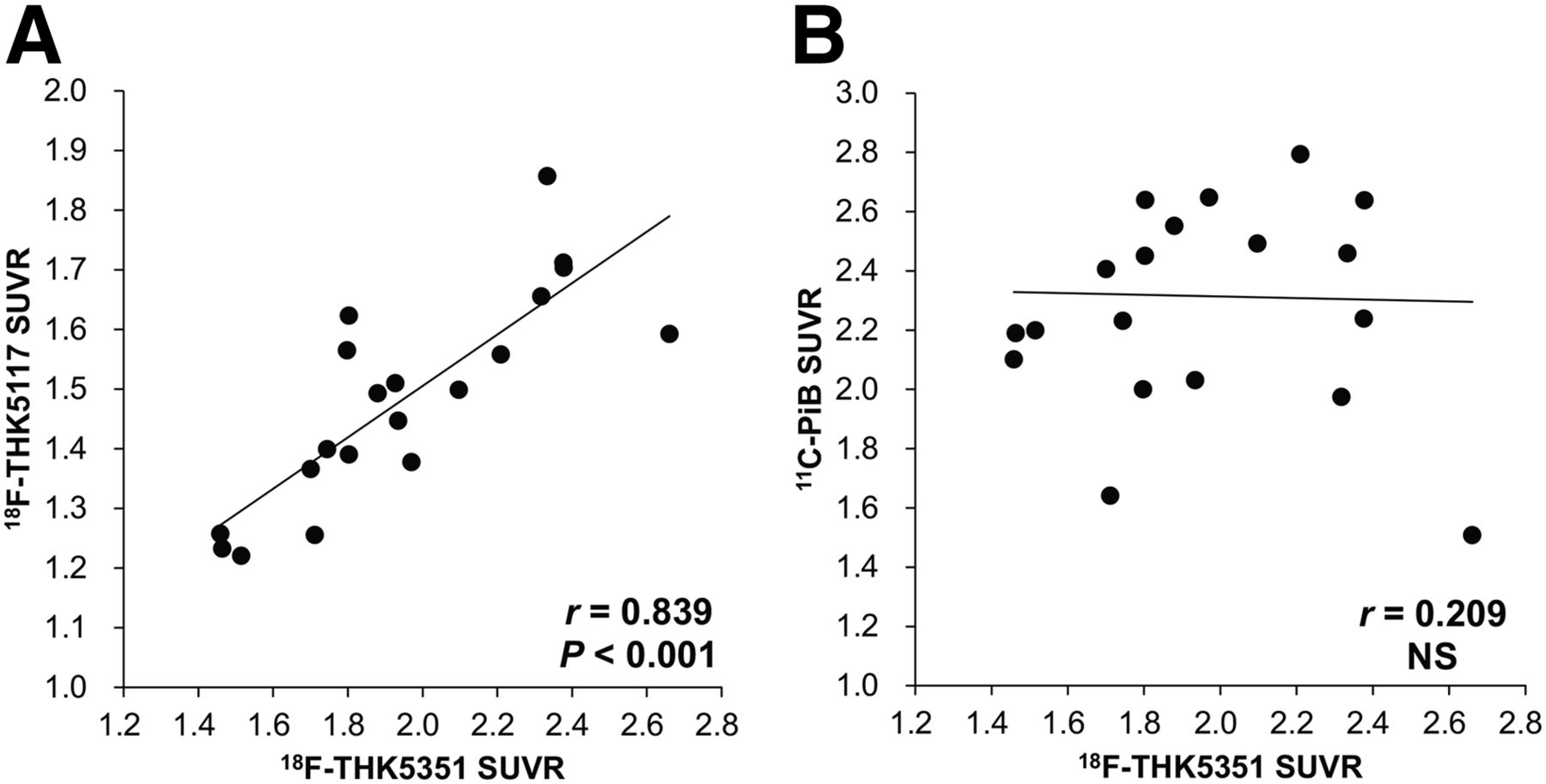
Correlation between regional SUVR of 18F-THK5351 and 18F-THK5117 (Spearman r = 0.839, P < 0.001), but not 18F-THK5351 and 11C-PiB (r = 0.209, not significant [NS]), in 2 AD patients.
DISCUSSION
18F-THK5351 is a single S-enantiomer and pyridine derivative of 18F-THK5117, and 18F-THK5351 is less lipophilic than 18F-THK5117 (Log P = 1.5 vs. 2.32). As observed with amyloid PET tracers, pyridine derivatives tend to show reduced lipophilicity, which correlates with the amount of nonspecific binding (26). Replacement of the 2-aryl group from the benzene to the pyridine ring might contribute to diminished nonspecific binding to subcortical white matter, as observed in the relationship between 11C-PiB and 11C-AZD2184 (27–29). Moreover, 18F-THK5351 is optically pure, whereas 18F-THK5105 and 18F-THK5117 are racemic mixtures. Enantiomers frequently show differences in biologic properties such as metabolism or binding affinity for targets (30). Our preclinical studies demonstrated that the pharmacokinetic profiles of the S-enantiomers of arylquinoline derivatives were more favorable than those of the R-enantiomers (20,24). Therefore, optical purification should additionally contribute toward improving THK5351 pharmacokinetics. Our preclinical data revealed that 18F-THK5351 showed high and selective binding ability for tau aggregates, low binding affinity for white matter, and rapid pharmacokinetics, which collectively resulted in the signal-to-background ratio of THK5351 being higher than that of both THK5105 and THK5117. Furthermore, autoradiography performed on human brain sections also confirmed that THK5351 did not bind to amyloid, α-synuclein, and TDP43 deposits (data not shown), suggesting that THK5351 binds to tau protein fibrils with high selectivity.
As observed in our previous 18F-THK5105 and 18F-THK5117 PET studies (19,20), 18F-THK5351 retention was prominent in the temporal lobe of AD patients. This regional distribution of 18F-THK5351 in AD patients agreed with the postmortem observation that the temporal lobe was more susceptible to tau deposition than other cortical areas (31,32). Although the cortical distributions of 18F-THK5351 and 18F-THK5117 in the same patients were almost identical, 18F-THK5351 displayed comparatively higher contrast and lower subcortical white-matter retention; this is strikingly similar to our preclinical findings (Figs. 2–4). When compared with reported tau radiotracers such as 11C-PBB3 and 18F-T807, a drawback of 18F-THK5117 is high white-matter retention, which is frequently observed with 18F-labeled amyloid radiotracers (33). The cortical–to–white matter ratio of 18F-THK5351 in AD patients was substantially higher than that of 18F-THK5117, which allows comparatively easier and more accurate visual interpretation of PET images. No remarkable retention of 18F-THK5351 was observed in the choroid plexus or venous sinus. This is one of the advantages over the other tau tracers, because off-target retention in these areas might cause a spill-in of the tracer signals into the brain. However, as observed with other radiotracers, off-target binding of 18F-THK5351 was detected in the basal ganglia; the binding target of THK5351 in this region should be clarified in future studies. Moreover, 18F-THK5351 must be directly compared with other tau radiotracers to characterize the binding property of each radiotracer and to determine how their differences affect the accuracy of visual interpretation and the sensitivity for detecting subtle changes in brain tau loads over time, as done in the case of amyloid radiotracers (34–36). Furthermore, imaging-autopsy studies are required to validate the described binding selectivity in the future.
CONCLUSION
18F-THK5351 selectively bound to pathologic tau deposits in postmortem AD brain tissues but showed weak white-matter binding. Fast pharmacokinetics, high contrast, and low white-matter retention of 18F-THK5351 allow sensitive detection of tau pathology in humans, which could facilitate early detection and longitudinal assessment of neurofibrillary pathology.
DISCLOSURE
The costs of publication of this article were defrayed in part by the payment of page charges. Therefore, and solely to indicate this fact, this article is hereby marked “advertisement” in accordance with 18 USC section 1734. This study was supported by research funds from GE Healthcare; the SEI (Sumitomo Electric Industries, Ltd.) Group CSR Foundation; the Industrial Technology Research grant program of the NEDO in Japan (09E51025a); Health and Labor Sciences Research grants from the Ministry of Health, Labor, and Welfare of Japan; grant-in-aid for Scientific Research (B) (15H04900); grant-in-aid for Scientific Research on Innovative Areas (Brain Protein Aging and Dementia Control) (26117003); grant-in-aid for Young Scientists (B) (15K19767); and grant-in-aid for JSPS Fellows and “Japan Advanced Molecular Imaging Program (J-AMP)” of the Ministry of Education, Culture, Sports, Science and Technology (MEXT), Japan. No other potential conflict of interest relevant to this article was reported.
Footnotes
Published online Nov. 5, 2015.
- © 2016 by the Society of Nuclear Medicine and Molecular Imaging, Inc.
REFERENCES
- Received for publication August 3, 2015.
- Accepted for publication October 22, 2015.


Jump to section
Related Articles
Cited By...
- Preclinical Characterization of the Tau PET Tracer [18F]SNFT-1: Comparison of Tau PET Tracers
- Preclinical Characterization of the Tau PET Tracer [18F]SNFT-1: Comparison of Tau PET Tracers
- First-in-Humans Evaluation of 18F-SMBT-1, a Novel 18F-Labeled Monoamine Oxidase-B PET Tracer for Imaging Reactive Astrogliosis
- 18F-SMBT-1: A Selective and Reversible PET Tracer for Monoamine Oxidase-B Imaging
- High-contrast in-vivo imaging of tau pathologies in Alzheimers and non-Alzheimers disease tauopathies
- Biomarkers for the diagnosis of Alzheimers disease, dementia Lewy body, frontotemporal dementia and vascular dementia
- Comparative In Vitro and In Vivo Quantifications of Pathologic Tau Deposits and Their Association with Neurodegeneration in Tauopathy Mouse Models
- Correlations of 18F-THK5351 PET with Postmortem Burden of Tau and Astrogliosis in Alzheimer Disease
- 18F-AV-1451 binds to motor-related subcortical gray and white matter in corticobasal syndrome
- Biodistribution and Radiation Dosimetry for the Tau Tracer 18F-THK-5351 in Healthy Human Subjects
- Kinetic Modeling of the Tau PET Tracer 18F-AV-1451 in Human Healthy Volunteers and Alzheimer Disease Subjects
- In Vivo Comparison of Tau Radioligands 18F-THK-5351 and 18F-THK-5317
- Modeling Strategies for Quantification of In Vivo 18F-AV-1451 Binding in Patients with Tau Pathology
- Pharmacokinetic Evaluation of the Tau PET Radiotracer 18F-T807 (18F-AV-1451) in Human Subjects
- Reference Tissue-Based Kinetic Evaluation of 18F-AV-1451 for Tau Imaging
- In vivo visualization of tau deposits in corticobasal syndrome by 18F-THK5351 PET
- Preclinical Characterization of 18F-MK-6240, a Promising PET Tracer for In Vivo Quantification of Human Neurofibrillary Tangles
- Hybrid PET/MR Imaging in Neurology: Present Applications and Prospects for the Future