


Article Figures & Data
Figures
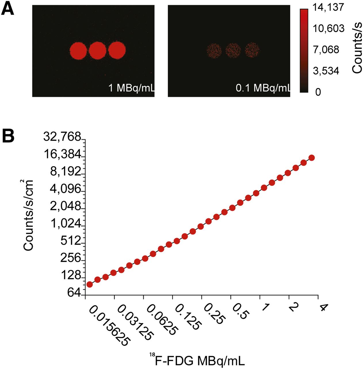
- FIGURE 1.
Sensitivity and linearity of intensified CCD to detect CL from 18F-FDG. (A) CL imaging of multiwell plate with activity at 1 and 0.1 MBq/mL. (B) Linearity analysis of background-subtracted and decay-corrected region of interest of CL photon flux from wells using intensified CCD with settings optimized for the activity concentrations.
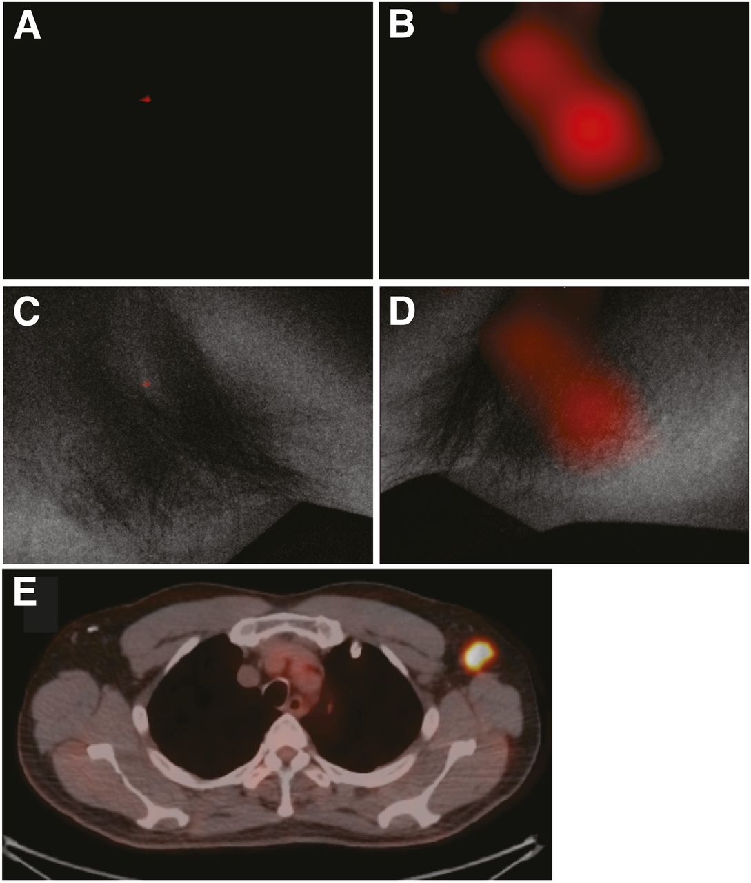
- FIGURE 2.
Representative CL and PET/CT images of 18F-FDG–positive axillary lymph node. (A and B) CL scans of right and left axillae, respectively. (C) Negative CL scan in light-protected environment of right axilla without 18F-FDG–positive lymph node, overlaid with white-light photograph. No significant CL emission from 18F-FDG decay is seen. (D) White-light photograph from left axilla, overlaid with significant CL signal (B). (E) This signal colocalized with PET/CT finding.
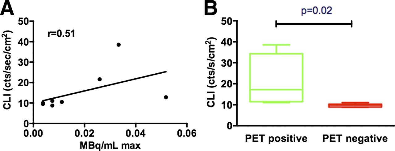
- FIGURE 3.
(A) Correlation graph between PET (MBq/mL max) and CL imaging count rate. (B) Box plot comparing Cerenkov emission from pathologic, PET-positive lymph nodes vs. contralateral PET-negative control, demonstrating significantly higher signal from PET-positive side.

Additional Files
Supplemental Data
Files in this Data Supplement:


Jump to section
Related Articles
Cited By...
- Detection of Shortwave-Infrared Cerenkov Luminescence from Medical Isotopes
- Cerenkov Luminescence Imaging in Prostate Cancer: Not the Only Light That Shines
- Quo Vadis, Molecular Imaging?
- Optical Imaging Modalities: Principles and Applications in Preclinical Research and Clinical Settings
- Trending: Radioactive and Fluorescent Bimodal/Hybrid Tracers as Multiplexing Solutions for Surgical Guidance
- Multiplexed Optical Imaging of Energy Substrates Reveals That Left Ventricular Hypertrophy Is Associated With Brown Adipose Tissue Activation
- Dynamic 18F-FDG PET Lymphography for In Vivo Identification of Lymph Node Metastases in Murine Melanoma
- Cerenkov-Activated Sticky Tag for In Vivo Fluorescence Imaging
- Cerenkov Radiation-Induced Photoimmunotherapy with 18F-FDG
- Intraoperative Assessment of Tumor Resection Margins in Breast-Conserving Surgery Using 18F-FDG Cerenkov Luminescence Imaging: A First-in-Human Feasibility Study
- In Vivo 3-Dimensional Radiopharmaceutical-Excited Fluorescence Tomography
- Optical Imaging of Ionizing Radiation from Clinical Sources
- Toward (Hybrid) Navigation of a Fluorescence Camera in an Open Surgery Setting
- {beta}-Radioluminescence Imaging: A Comparative Evaluation with Cerenkov Luminescence Imaging
- Cerenkov Luminescence Imaging for Radiation Dose Calculation of a 90Y-Labeled Gastrin-Releasing Peptide Receptor Antagonist
- Cerenkov-Specific Contrast Agents for Detection of pH In Vivo
- Cerenkov Luminescence Endoscopy: Improved Molecular Sensitivity with {beta}--Emitting Radiotracers
- In Vivo Localization of 90Y and 177Lu Radioimmunoconjugates Using Cerenkov Luminescence Imaging in a Disseminated Murine Leukemia Model
- Reply: Human Cerenkov Imaging Using 18F-FDG
- Human Cerenkov Imaging Using 18F-FDG