


Article Figures & Data
Figures
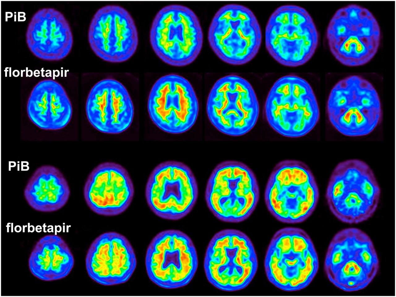
- FIGURE 1.
Axial slices of PiB and florbetapir scans are shown for 2 representative subjects, cognitively normal control with low tracer retention (top) and AD patient with high tracer retention in cortex relative to cerebellum, reflecting widespread fibrillar amyloid (bottom).
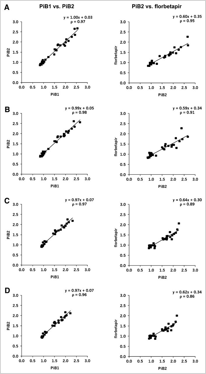
- FIGURE 2.
Cortical retention ratios are shown for consecutive PiB and florbetapir scans obtained for same participants and processed using pons (A), whole cerebellum (B), and cerebellar gray matter (C) for intensity normalization. Initial diagnosis at enrollment and any subsequent diagnostic change are represented by shape markers. Stable normal cognition or MCI diagnosis is represented with solid shapes, individuals who progressed are represented with unfilled shapes, and single individual who regressed from MCI to normal is represented with gray-filled square. Raw data were analyzed with PET-template method. Regression equations and Spearman rank correlation coefficients (ρ) are shown for each scatterplot.
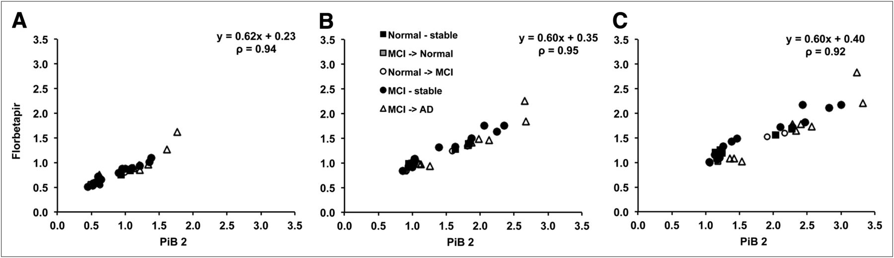
- FIGURE 3.
Cortical retention ratios for 2 consecutive PiB scans and PiB scan followed by florbetapir scan obtained for same participants are compared at different levels of preprocessing and data analysis methods. All cortical retention ratios were normalized using whole cerebellum. Top 2 rows show raw data (not at uniform voxel size or smoothing) (A) and unsmoothed data (at uniform voxel size but not uniform smoothing) (B), both processed using PET-template method. Bottom 2 rows show unsmoothed (C) and smoothed data (uniform voxel size and smoothing) (D) processed with Freesurfer method. Thus, middle 2 rows both show unsmoothed data that differs only on basis of which processing method was used (PET template [B], Freesurfer [C]). Regression equations and Spearman rank correlation coefficients (ρ) are shown for each scatterplot.
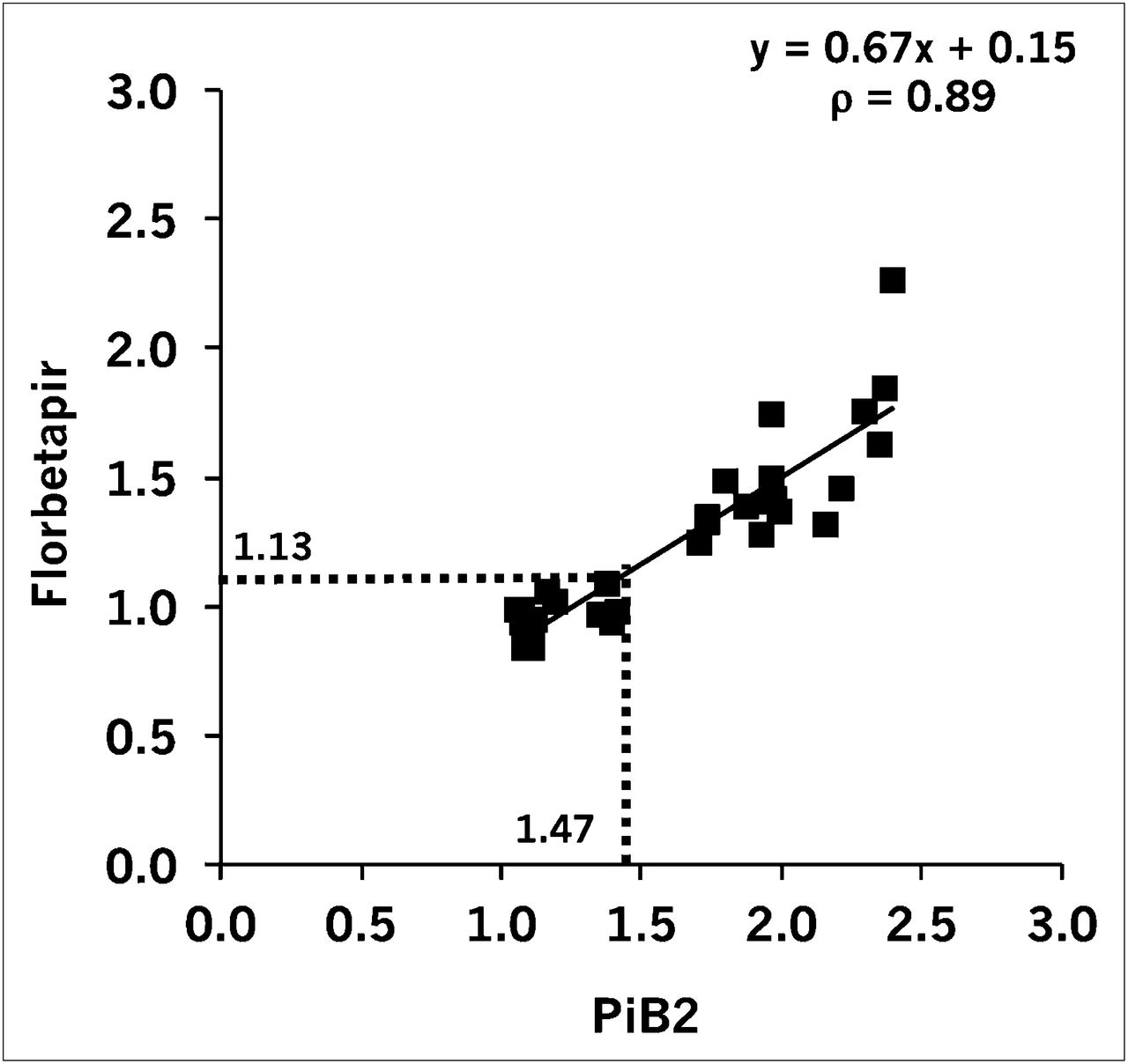
- FIGURE 4.
PiB threshold of 1.47 (14) that is based on data normalized to cerebellar gray matter can be converted to florbetapir threshold of 1.13, using raw data and whole-cerebellum normalization.


Tables
Diagnosis at enrollment Parameter Total sample (n = 32) Normal cognition (n = 8) MCI (n = 24) Duration of follow-up (y) 4.3 4.3 4.3 Mean age (±SD) at PiB2 scan (y) 75.7 ± 6.6 77.6 ± 3.9 75.1 ± 7.3 Mean age (±SD) at florbetapir scan (y) 77.3 ± 6.5 79.3 ± 3.7 76.7 ± 7.2 Mean time (±SD) between PiB1 and PiB2 scans (y) 1.1 ± 0.3 1.0 ± 0.08 1.1 ± 0.3 Mean time (±SD) between PiB2 and florbetapir scans (y) 1.5 ± 0.7 1.6 ± 0.8 1.4 ± 0.6 Sex, female (%) 31 25 33 Mean number of years (±SD) of education 16.1 ± 3.0 15.9 ± 3.0 16.1 ± 3.0 Apolipoprotein E4 carriers (%) 53 50 54 Mean MMSE score (±SD) at florbetapir scan 25.2 ± 6.0 28.3 ± 1.7 24.1 ± (.6 Mean ADAS-cog score (±SD) at florbetapir scan 13.2 ± 12.3 6.8 ± 4.7 15.4 ± 13.3 Mean PiB2 cortical retention 1.51 (95% CI, 1.31–1.71) 1.34 (95% CI, 0.99–1.68) 1.57 (95% CI, 1.31–1.82) Mean florbetapir cortical retention 1.25 (95% CI, 1.13–1.38) 1.13 (95% CI, 0.96–1.30) 1.29 (95% CI, 1.13–1.45) Diagnosis at PiB2 scan (n) Normal cognition 6 5 1 MCI 22 3 19 AD 4 0 4 Diagnosis at florbetapir scan (n) Normal cognition 6 5 1 MCI 17 3 14 AD 9 0 9 95% confidence intervals (95% CIs) are for raw data processed using PET-template method. Subject diagnostic groups are based on diagnosis at enrollment, but changes in diagnosis between enrollment, PiB2, and florbetapir scanning sessions are noted. Summary cognitive scores (mini-mental state examination [MMSE], Alzheimer’s Disease Assessment Scale–cognitive subscale [ADAS-cog]) are given for participants’ most recent imaging session.
- TABLE 2
Mean Difference and Percentage Change Between Consecutive Scans Across All Participants
PiB1 vs. PiB2 PiB2 vs. florbetapir Parameter Pons Whole cerebellum Cerebellar gray Pons Whole cerebellum Cerebellar gray Difference 0.05 (0.20) 0.10 (0.33) 0.13 (0.40) −0.12 (0.16) −0.28 (0.24) −0.37 (0.32) Percentage change 1 (6) 3 (6) 4 (7) −8 (15) −15 (11) −17 (13) Data in parentheses are SDs, reflecting increases in tracer retention between first and second PiB scans and between second PiB and florbetapir scans, separately for each reference region. Positive value represents mean increase from PiB1 to PiB2 or from PiB2 to florbetapir, whereas negative value represents decrease.


Jump to section
Related Articles
Cited By...
- Updated Appropriate Use Criteria for Amyloid and Tau PET: A Report from the Alzheimers Association and Society for Nuclear Medicine and Molecular Imaging Workgroup
- BOLD Amplitude Correlates of Preclinical Alzheimers Disease
- Evaluation of ComBat harmonization for reducing across-tracer differences in regional amyloid PET analyses
- Parallel neuroinflammatory pathways to cerebrovascular injury and amyloid-beta in Alzheimers disease
- Amyloid, Tau, and APOE in Alzheimers Disease: Impact on White Matter Tracts
- Rates of cortical thinning in Alzheimers disease signature regions associate with vascular burden but not with {beta}-amyloid status in cognitively normal adults at age 70
- Analyzing heterogeneity in Alzheimer Disease using multimodal normative modeling on imaging-based ATN biomarkers
- A Neuropathology Case Report of a Woman with Down Syndrome who Remained Cognitively Stable
- Utility of cerebrovascular imaging biomarkers to detect cerebral amyloidosis
- Exploring the genetic heterogeneity of Alzheimers disease: Evidence for genetic subtypes
- Towards cascading genetic risk in Alzheimers disease
- Clinical utility of plasma A{beta}42/40 ratio by LC-MS/MS in Alzheimers disease assessment
- Dissociable Effects of Alzheimer's Disease-Related Cognitive Dysfunction and Aging on Functional Brain Network Segregation
- Personalising Alzheimers Disease progression using brain atrophy markers
- Predicting Brain Amyloid Positivity from T1 weighted brain MRI and MRI-derived Gray Matter, White Matter and CSF maps using Transfer Learning on 3D CNNs*
- Tau-Neurodegeneration mismatch reveals vulnerability and resilience to comorbidities in Alzheimers continuum
- The RSNA QIBA Profile for Amyloid PET as an Imaging Biomarker for Cerebral Amyloid Quantification
- Directed functional brain connectivity is altered in sub-threshold amyloid-{beta} accumulators
- Plasma P-tau181 and P-tau217 in Patients With Traumatic Encephalopathy Syndrome With and Without Evidence of Alzheimer Disease Pathology
- Associations of {beta}-Amyloid and Vascular Burden With Rates of Neurodegeneration in Cognitively Normal Members of the 1946 British Birth Cohort
- Effect of reduction in brain amyloid levels on change in cognitive and functional decline in randomized clinical trials: an updated instrumental variable meta-analysis
- Medial Temporal Lobe Networks in Alzheimer's Disease: Structural and Molecular Vulnerabilities
- Validation of Plasma Amyloid-{beta} 42/40 for Detecting Alzheimer Disease Amyloid Plaques
- Extraversion is associated with lower brain amyloid deposition in cognitively normal older adults
- Diffusion MRI Metrics and their Relation to Dementia Severity: Effects of Harmonization Approaches
- Finding Treatment Effects in Alzheimer Trials in the Face of Disease Progression Heterogeneity
- Convergent abnormalities of {beta}-amyloid deposition, glucose metabolism, and fMRI activity in the dorsal precuneus in subjective cognitive decline
- Determining Amyloid-{beta} Positivity Using 18F-AZD4694 PET Imaging
- Association of vascular brain injury, neurodegeneration, amyloid, and cognitive trajectory
- Association of amyloid-{beta} CSF/PET discordance and tau load 5 years later
- Amyloid-{beta} CSF/PET discordance vs tau load 5 years later: It takes two to tangle
- Objective subtle cognitive difficulties predict future amyloid accumulation and neurodegeneration
- A Fully Automatic Technique for Precise Localization and Quantification of Amyloid-{beta} PET Scans
- The heterogeneous functional architecture of the posteromedial cortex is associated with selective functional connectivity differences in Alzheimers disease
- Alzheimer disease biomarkers may aid in the prognosis of MCI cases initially reverted to normal
- Biomarker Localization, Analysis, Visualization, Extraction, and Registration (BLAzER) Workflow for Research and Clinical Brain PET Applications
- Reproducible evaluation of classification methods in Alzheimers disease: framework and application to MRI and PET data
- Impact of Reference and Target Region Selection on Amyloid PET SUV Ratios in the Phase 1b PRIME Study of Aducanumab
- Generation of Structural MR Images from Amyloid PET: Application to MR-Less Quantification
- Spatiotemporal Distribution of {beta}-Amyloid in Alzheimer Disease Is the Result of Heterogeneous Regional Carrying Capacities
- Memory decline accompanies subthreshold amyloid accumulation
- Amyloid-PET burden and regional distribution in cerebral amyloid angiopathy: a systematic review and meta-analysis of biomarker performance
- Increased florbetapir binding in the temporal neocortex from age 20 to 60 years
- Alzheimer disease brain atrophy subtypes are associated with cognition and rate of decline
- In vivo staging of regional amyloid deposition
- APOE genotype and early {beta}-amyloid accumulation in older adults without dementia
- Relationships among Cortical Glutathione Levels, Brain Amyloidosis, and Memory in Healthy Older Adults Investigated In Vivo with 1H-MRS and Pittsburgh Compound-B PET
- Left frontal cortex connectivity underlies cognitive reserve in prodromal Alzheimer disease
- Standardized Expression of 18F-NAV4694 and 11C-PiB {beta}-Amyloid PET Results with the Centiloid Scale
- Amyloid negativity in patients with clinically diagnosed Alzheimer disease and MCI
- {beta}-Amyloid Deposition Is Associated with Decreased Right Prefrontal Activation during Task Switching among Cognitively Normal Elderly
- A Semiautomated Method for Quantification of F 18 Florbetapir PET Images
- Measurement of Longitudinal {beta}-Amyloid Change with 18F-Florbetapir PET and Standardized Uptake Value Ratios
- Brain Amyloid-{beta} Burden Is Associated with Disruption of Intrinsic Functional Connectivity within the Medial Temporal Lobe in Cognitively Normal Elderly
- Molecular Imaging of Alzheimer Disease Pathology
- Practical utility of amyloid and FDG-PET in an academic dementia center
- Head-to-Head Comparison of 11C-PiB and 18F-AZD4694 (NAV4694) for {beta}-Amyloid Imaging in Aging and Dementia
- Appropriate Use Criteria for Amyloid PET: A Report of the Amyloid Imaging Task Force, the Society of Nuclear Medicine and Molecular Imaging, and the Alzheimer's Association