
Main menu

  • Home
  • Content
    • Current
    • Ahead of print
    • Past Issues
    • JNM Supplement
    • SNMMI Annual Meeting Abstracts
    • Continuing Education
    • JNM Podcasts
  • Subscriptions
    • Subscribers
    • Institutional and Non-member
    • Rates
    • Journal Claims
    • Corporate & Special Sales
  • Authors
    • Submit to JNM
    • Information for Authors
    • Assignment of Copyright
    • AQARA requirements
  • Info
    • Reviewers
    • Permissions
    • Advertisers
  • About
    • About Us
    • Editorial Board
    • Contact Information
  • More
    • Alerts
    • Feedback
    • Help
    • SNMMI Journals
  • SNMMI
    • JNM
    • JNMT
    • SNMMI Journals
    • SNMMI

User menu

  • Subscribe
  • My alerts
  • Log in
  • Log out
  • My Cart

Search

  • Advanced search
Journal of Nuclear Medicine
  • SNMMI
    • JNM
    • JNMT
    • SNMMI Journals
    • SNMMI
  • Subscribe
  • My alerts
  • Log in
  • Log out
  • My Cart
Journal of Nuclear Medicine

Advanced Search

  • Home
  • Content
    • Current
    • Ahead of print
    • Past Issues
    • JNM Supplement
    • SNMMI Annual Meeting Abstracts
    • Continuing Education
    • JNM Podcasts
  • Subscriptions
    • Subscribers
    • Institutional and Non-member
    • Rates
    • Journal Claims
    • Corporate & Special Sales
  • Authors
    • Submit to JNM
    • Information for Authors
    • Assignment of Copyright
    • AQARA requirements
  • Info
    • Reviewers
    • Permissions
    • Advertisers
  • About
    • About Us
    • Editorial Board
    • Contact Information
  • More
    • Alerts
    • Feedback
    • Help
    • SNMMI Journals
  • View or Listen to JNM Podcast
  • Visit JNM on Facebook
  • Join JNM on LinkedIn
  • Follow JNM on Twitter
  • Subscribe to our RSS feeds
Research ArticleClinical Investigations

Biodistribution, Tumor Detection, and Radiation Dosimetry of 18F-DCFBC, a Low-Molecular-Weight Inhibitor of Prostate-Specific Membrane Antigen, in Patients with Metastatic Prostate Cancer

Steve Y. Cho, Kenneth L. Gage, Ronnie C. Mease, Srinivasan Senthamizhchelvan, Daniel P. Holt, Akimosa Jeffrey-Kwanisai, Christopher J. Endres, Robert F. Dannals, George Sgouros, Martin Lodge, Mario A. Eisenberger, Ronald Rodriguez, Michael A. Carducci, Camilo Rojas, Barbara S. Slusher, Alan P. Kozikowski and Martin G. Pomper
Journal of Nuclear Medicine December 2012, 53 (12) 1883-1891; DOI: https://doi.org/10.2967/jnumed.112.104661
Steve Y. Cho
1Russell H. Morgan Department of Radiology and Radiological Science, Johns Hopkins University, Baltimore, Maryland
2Sidney Kimmel Comprehensive Cancer Center, Johns Hopkins University, Baltimore, Maryland
  • Find this author on Google Scholar
  • Find this author on PubMed
  • Search for this author on this site
Kenneth L. Gage
1Russell H. Morgan Department of Radiology and Radiological Science, Johns Hopkins University, Baltimore, Maryland
  • Find this author on Google Scholar
  • Find this author on PubMed
  • Search for this author on this site
Ronnie C. Mease
1Russell H. Morgan Department of Radiology and Radiological Science, Johns Hopkins University, Baltimore, Maryland
2Sidney Kimmel Comprehensive Cancer Center, Johns Hopkins University, Baltimore, Maryland
  • Find this author on Google Scholar
  • Find this author on PubMed
  • Search for this author on this site
Srinivasan Senthamizhchelvan
1Russell H. Morgan Department of Radiology and Radiological Science, Johns Hopkins University, Baltimore, Maryland
  • Find this author on Google Scholar
  • Find this author on PubMed
  • Search for this author on this site
Daniel P. Holt
1Russell H. Morgan Department of Radiology and Radiological Science, Johns Hopkins University, Baltimore, Maryland
  • Find this author on Google Scholar
  • Find this author on PubMed
  • Search for this author on this site
Akimosa Jeffrey-Kwanisai
1Russell H. Morgan Department of Radiology and Radiological Science, Johns Hopkins University, Baltimore, Maryland
  • Find this author on Google Scholar
  • Find this author on PubMed
  • Search for this author on this site
Christopher J. Endres
1Russell H. Morgan Department of Radiology and Radiological Science, Johns Hopkins University, Baltimore, Maryland
  • Find this author on Google Scholar
  • Find this author on PubMed
  • Search for this author on this site
Robert F. Dannals
1Russell H. Morgan Department of Radiology and Radiological Science, Johns Hopkins University, Baltimore, Maryland
  • Find this author on Google Scholar
  • Find this author on PubMed
  • Search for this author on this site
George Sgouros
1Russell H. Morgan Department of Radiology and Radiological Science, Johns Hopkins University, Baltimore, Maryland
  • Find this author on Google Scholar
  • Find this author on PubMed
  • Search for this author on this site
Martin Lodge
1Russell H. Morgan Department of Radiology and Radiological Science, Johns Hopkins University, Baltimore, Maryland
  • Find this author on Google Scholar
  • Find this author on PubMed
  • Search for this author on this site
Mario A. Eisenberger
2Sidney Kimmel Comprehensive Cancer Center, Johns Hopkins University, Baltimore, Maryland
  • Find this author on Google Scholar
  • Find this author on PubMed
  • Search for this author on this site
Ronald Rodriguez
2Sidney Kimmel Comprehensive Cancer Center, Johns Hopkins University, Baltimore, Maryland
3Brady Urological Institute, Johns Hopkins University, Baltimore, Maryland
  • Find this author on Google Scholar
  • Find this author on PubMed
  • Search for this author on this site
Michael A. Carducci
3Brady Urological Institute, Johns Hopkins University, Baltimore, Maryland
  • Find this author on Google Scholar
  • Find this author on PubMed
  • Search for this author on this site
Camilo Rojas
4Brain Sciences Institute of Johns Hopkins University School of Medicine, Johns Hopkins University, Baltimore, Maryland; and
  • Find this author on Google Scholar
  • Find this author on PubMed
  • Search for this author on this site
Barbara S. Slusher
4Brain Sciences Institute of Johns Hopkins University School of Medicine, Johns Hopkins University, Baltimore, Maryland; and
  • Find this author on Google Scholar
  • Find this author on PubMed
  • Search for this author on this site
Alan P. Kozikowski
5Department of Medicinal Chemistry and Pharmacognosy, University of Illinois at Chicago College of Pharmacy, Chicago, Illinois
  • Find this author on Google Scholar
  • Find this author on PubMed
  • Search for this author on this site
Martin G. Pomper
1Russell H. Morgan Department of Radiology and Radiological Science, Johns Hopkins University, Baltimore, Maryland
2Sidney Kimmel Comprehensive Cancer Center, Johns Hopkins University, Baltimore, Maryland
  • Find this author on Google Scholar
  • Find this author on PubMed
  • Search for this author on this site
  • Article
  • Figures & Data
  • Supplemental
  • Info & Metrics
  • PDF
Loading

Article Figures & Data

Figures

  • Tables
  • Additional Files
  • FIGURE 1.
    • Download figure
    • Open in new tab
    • Download powerpoint
    FIGURE 1.

    Mean biodistribution curves plotted for decay-corrected percentage injected dose per gram of organ mass vs. time. Time axis represents average time over all patients for each PET scan because some patients (i.e., tall patients) required more bed positions. (A) Organs with higher uptake. (B) Organs with lower uptake. (C) Increasing urinary bladder activity. LLI = lower large intestine; SI = small intestine; ULI = upper large intestine. %ID/g = percentage injected dose per gram of organ mass.

  • FIGURE 2.
    • Download figure
    • Open in new tab
    • Download powerpoint
    FIGURE 2.

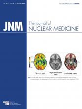
    18F-DCFBC PET anterior projection maximal-intensity-projection images at 2 h after injection in patient 1, with several bone metastases (arrow) (A), and patient 5, with LN metastases (arrow) (B), as confirmed by correlation to CT portion of PET/CT exam.

  • FIGURE 3.
    • Download figure
    • Open in new tab
    • Download powerpoint
    FIGURE 3.

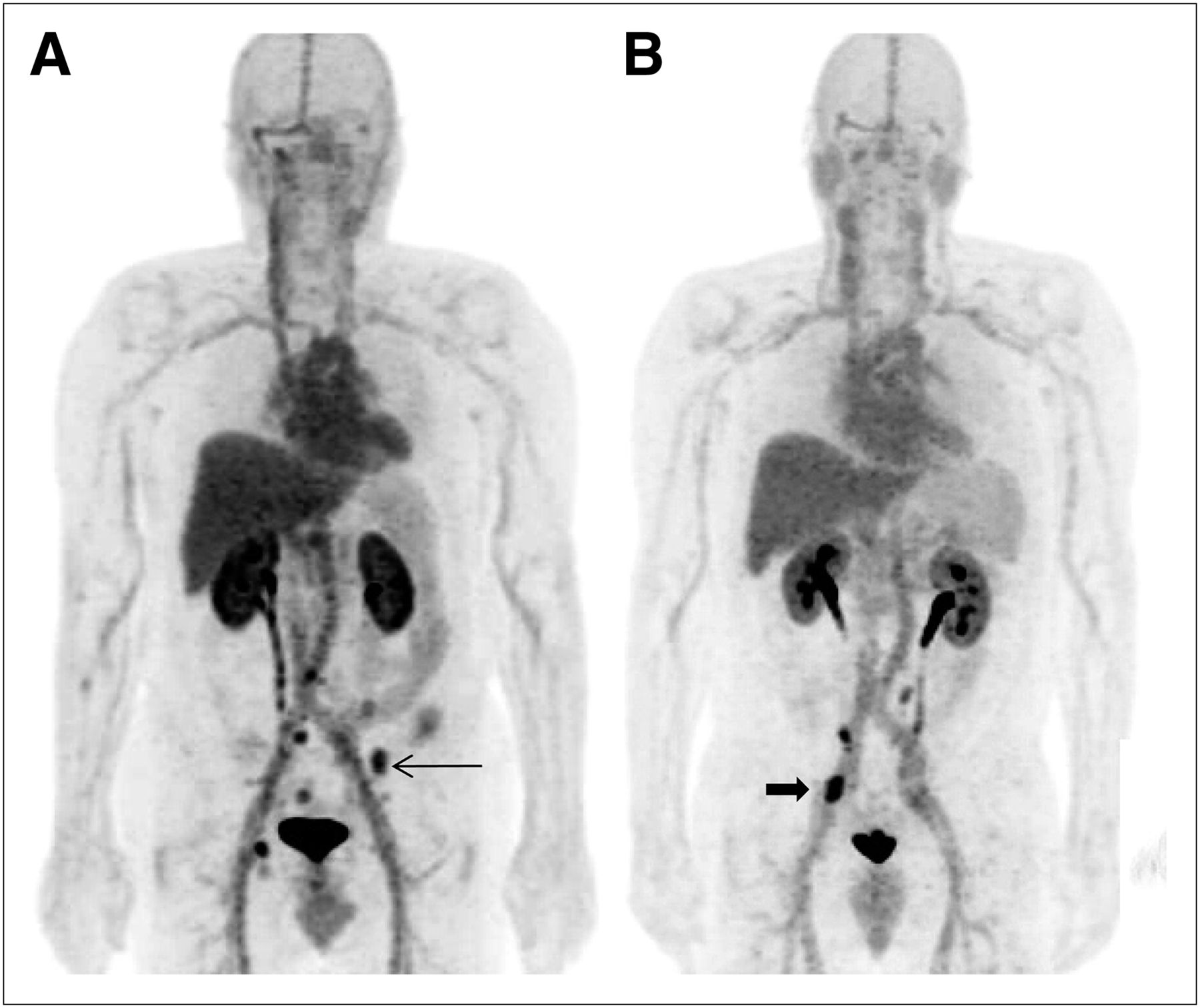
    Examples of concordant findings on 18F-DCFBC PET and CIM: (A) T12 bone metastasis (arrows) seen on bone scan (far right) and (B) retroperitoneal right external iliac LN (arrow) seen on CT (arrow).

  • FIGURE 4.
    • Download figure
    • Open in new tab
    • Download powerpoint
    FIGURE 4.

    (A and B) Focal 18F-DCFBC PET uptake at aortic bifurcation (arrow, A) with correlative small LN seen on concurrent contrast-enhanced CT (arrow, B), not considered to be nodal metastasis by CT but positive by PET. (C) Retrospective review of prior contrast-enhanced CT scan obtained 1 y previously demonstrates LN in this region (arrow).

  • FIGURE 5.
    • Download figure
    • Open in new tab
    • Download powerpoint
    FIGURE 5.

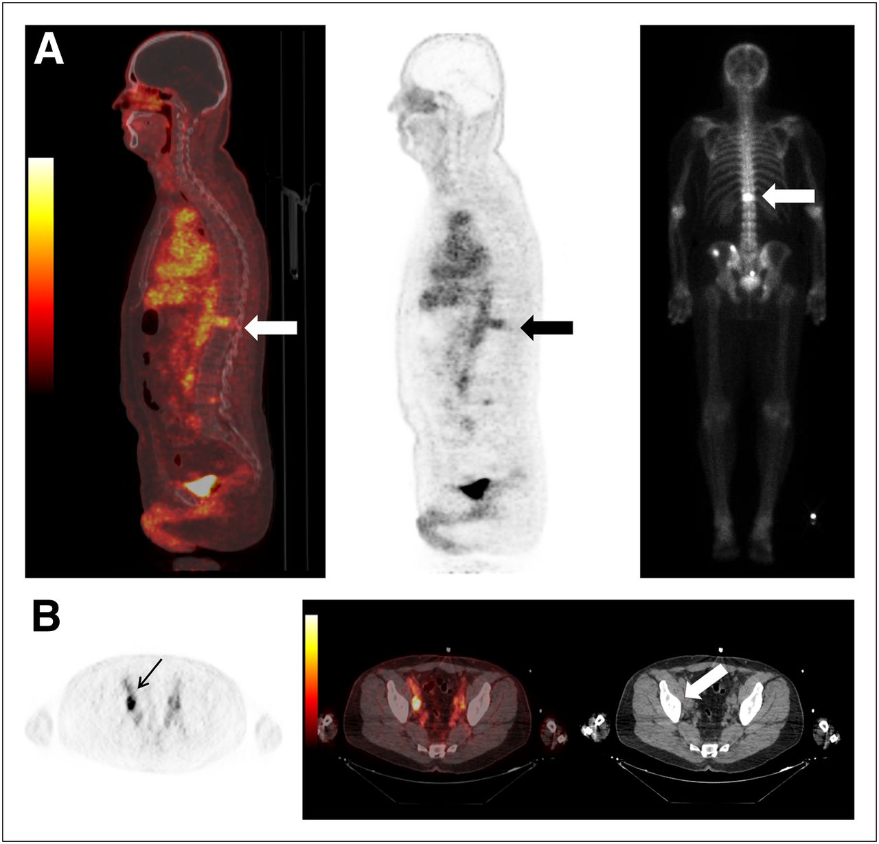
    Focal 18F-DCFBC PET uptake in L4 vertebral body on PET and fused PET/CT (thick arrows, A) with no correlative abnormality on CT (thin arrow, A) or bone scan (arrow, B).

  • FIGURE 6.
    • Download figure
    • Open in new tab
    • Download powerpoint
    FIGURE 6.

    (A) New sclerotic lesion in right posterior iliac bone seen on CT (thin white arrow) but not on 18F-DCFBC PET (black arrow) or PET/CT (thick white arrow). (B) Corresponding asymmetric uptake on bone scan (arrow).

Tables

  • Figures
  • Additional Files
    • View popup
    TABLE 1

    Average TIACs (Bq-h/Bq)

    OrganAverageSDPercentage coefficient of variation
    Adrenals9.26E−042.35E−0425.43
    Brain1.20E−021.30E−0310.78
    Gallbladder contents2.50E−036.87E−0427.47
    Lower large intestine contents1.40E−023.96E−0328.29
    Small intestine contents4.93E−025.35E−0310.85
    Stomach1.96E−023.06E−0315.55
    Upper large intestine contents1.52E−022.55E−0316.76
    Heart contents4.78E−027.26E−0315.20
    Heart wall2.10E−022.58E−0312.31
    Kidneys3.50E−025.84E−0316.68
    Liver1.59E−013.29E−0220.62
    Lungs1.09E−011.87E−0217.13
    Pancreas5.34E−038.93E−0416.73
    Spleen1.01E−028.92E−048.80
    Testes2.42E−031.00E−0341.50
    Thyroid8.00E−048.23E−0510.29
    Bladder contents1.26E−014.94E−0239.13
    Red marrow (femoral head)1.08E−011.06E−029.86
    Red marrow (spine)6.92E−027.70E−0311.12
    Total body2.37E+009.09E−023.83
    Remainder of body1.72E+001.01E−015.86
    • View popup
    TABLE 2

    Average Organ-Absorbed Dose (mGy/MBq) and Estimated Effective Dose (mSv/MBq)

    OrganAverageSDPercentage coefficient of variation
    Adrenals1.85E−022.83E−0315.32
    Brain4.21E−032.83E−046.73
    Breasts8.51E−033.22E−043.78
    Gallbladder wall1.79E−021.95E−0310.90
    Lower large intestine wall2.47E−023.69E−0314.92
    Small intestine wall2.36E−021.72E−037.31
    Stomach wall3.02E−023.24E−0310.72
    Upper large intestine wall2.34E−022.20E−039.39
    Heart wall2.92E−023.24E−0311.12
    Kidneys2.84E−023.81E−0313.45
    Liver2.46E−024.16E−0316.88
    Lungs2.45E−022.99E−0312.22
    Muscle9.69E−033.97E−044.10
    Ovaries1.32E−025.26E−043.99
    Pancreas1.92E−022.15E−0311.19
    Red marrow1.70E−029.81E−045.79
    Osteogenic cells1.82E−028.92E−044.90
    Skin7.30E−033.50E−044.79
    Spleen1.72E−021.05E−036.08
    Testes1.54E−024.19E−0327.23
    Thymus1.10E−024.53E−044.12
    Thyroid1.17E−026.87E−045.88
    Bladder wall3.24E−027.24E−0322.35
    Uterus1.34E−022.95E−042.20
    Total body1.09E−024.28E−043.91
    Effective dose1.99E−021.34E−036.73

Additional Files

  • Figures
  • Tables
  • Supplemental Data

    Files in this Data Supplement:

    • Supplemental Data
PreviousNext
Back to top

In this issue

Journal of Nuclear Medicine: 53 (12)
Journal of Nuclear Medicine
Vol. 53, Issue 12
December 1, 2012
  • Table of Contents
  • Table of Contents (PDF)
  • About the Cover
  • Index by author
Print
Download PDF
Article Alerts
Sign In to Email Alerts with your Email Address
Email Article

Thank you for your interest in spreading the word on Journal of Nuclear Medicine.

NOTE: We only request your email address so that the person you are recommending the page to knows that you wanted them to see it, and that it is not junk mail. We do not capture any email address.

Enter multiple addresses on separate lines or separate them with commas.
Biodistribution, Tumor Detection, and Radiation Dosimetry of 18F-DCFBC, a Low-Molecular-Weight Inhibitor of Prostate-Specific Membrane Antigen, in Patients with Metastatic Prostate Cancer
(Your Name) has sent you a message from Journal of Nuclear Medicine
(Your Name) thought you would like to see the Journal of Nuclear Medicine web site.
Citation Tools
Biodistribution, Tumor Detection, and Radiation Dosimetry of 18F-DCFBC, a Low-Molecular-Weight Inhibitor of Prostate-Specific Membrane Antigen, in Patients with Metastatic Prostate Cancer
Steve Y. Cho, Kenneth L. Gage, Ronnie C. Mease, Srinivasan Senthamizhchelvan, Daniel P. Holt, Akimosa Jeffrey-Kwanisai, Christopher J. Endres, Robert F. Dannals, George Sgouros, Martin Lodge, Mario A. Eisenberger, Ronald Rodriguez, Michael A. Carducci, Camilo Rojas, Barbara S. Slusher, Alan P. Kozikowski, Martin G. Pomper
Journal of Nuclear Medicine Dec 2012, 53 (12) 1883-1891; DOI: 10.2967/jnumed.112.104661

Citation Manager Formats

  • BibTeX
  • Bookends
  • EasyBib
  • EndNote (tagged)
  • EndNote 8 (xml)
  • Medlars
  • Mendeley
  • Papers
  • RefWorks Tagged
  • Ref Manager
  • RIS
  • Zotero
Share
Biodistribution, Tumor Detection, and Radiation Dosimetry of 18F-DCFBC, a Low-Molecular-Weight Inhibitor of Prostate-Specific Membrane Antigen, in Patients with Metastatic Prostate Cancer
Steve Y. Cho, Kenneth L. Gage, Ronnie C. Mease, Srinivasan Senthamizhchelvan, Daniel P. Holt, Akimosa Jeffrey-Kwanisai, Christopher J. Endres, Robert F. Dannals, George Sgouros, Martin Lodge, Mario A. Eisenberger, Ronald Rodriguez, Michael A. Carducci, Camilo Rojas, Barbara S. Slusher, Alan P. Kozikowski, Martin G. Pomper
Journal of Nuclear Medicine Dec 2012, 53 (12) 1883-1891; DOI: 10.2967/jnumed.112.104661
Twitter logo Facebook logo LinkedIn logo Mendeley logo
  • Tweet Widget
  • Facebook Like
  • Google Plus One
Bookmark this article

Jump to section

  • Article
    • Abstract
    • MATERIALS AND METHODS
    • RESULTS
    • DISCUSSION
    • CONCLUSION
    • DISCLOSURE STATEMENT
    • Acknowledgments
    • REFERENCES
  • Figures & Data
  • Supplemental
  • Info & Metrics
  • PDF

Related Articles

  • This Month in JNM
  • PubMed
  • Google Scholar

Cited By...

  • The History of Prostate-Specific Membrane Antigen as a Theranostic Target in Prostate Cancer: The Cornerstone Role of the Prostate Cancer Foundation
  • Evaluation of an Automated Module Synthesis and a Sterile Cold Kit-Based Preparation of 68Ga-PSMA-11 in Patients with Prostate Cancer
  • Quantitative and Qualitative Analyses of Biodistribution and PET Image Quality of a Novel Radiohybrid PSMA, 18F-rhPSMA-7, in Patients with Prostate Cancer
  • Prospective Comparison of PET Imaging with PSMA-Targeted 18F-DCFPyL Versus Na18F for Bone Lesion Detection in Patients with Metastatic Prostate Cancer
  • Radiation Dosimetry and Biodistribution of 18F-PSMA-11 for PET Imaging of Prostate Cancer
  • Preclinical Evaluation and Pilot Clinical Study of Al18F-PSMA-BCH for Prostate Cancer PET Imaging
  • Phase I Study of CTT1057, an 18F-Labeled Imaging Agent with Phosphoramidate Core Targeting Prostate-Specific Membrane Antigen in Prostate Cancer
  • Detection Efficacy of 18F-PSMA-1007 PET/CT in 251 Patients with Biochemical Recurrence of Prostate Cancer After Radical Prostatectomy
  • A Prospective Comparison of 18F-Sodium Fluoride PET/CT and PSMA-Targeted 18F-DCFBC PET/CT in Metastatic Prostate Cancer
  • PET Using a GRPR Antagonist 68Ga-RM26 in Healthy Volunteers and Prostate Cancer Patients
  • Biochemical Recurrence of Prostate Cancer: Initial Results with [18F]PSMA-1007 PET/CT
  • Low-Level Endogenous PSMA Expression in Nonprostatic Tumor Xenografts Is Sufficient for In Vivo Tumor Targeting and Imaging
  • Why Targeting PSMA Is a Game Changer in the Management of Prostate Cancer
  • Reply: PSMA Ligands for Imaging Prostate Cancer: Alternative Labeling by Complex Formation with Al18F2+
  • PSMA Ligands for PET Imaging of Prostate Cancer
  • Dual-Target Binding Ligands with Modulated Pharmacokinetics for Endoradiotherapy of Prostate Cancer
  • Prostate-Specific Membrane Antigen Ligands for Imaging and Therapy
  • Glu-Ureido-Based Inhibitors of Prostate-Specific Membrane Antigen: Lessons Learned During the Development of a Novel Class of Low-Molecular-Weight Theranostic Radiotracers
  • Acceleration of PSMA-Targeted Theranostics to the Clinic: Can Common Sense Prevail?
  • Semiquantitative Parameters in PSMA-Targeted PET Imaging with 18F-DCFPyL: Variability in Normal-Organ Uptake
  • Preclinical Evaluation of 18F-PSMA-1007, a New Prostate-Specific Membrane Antigen Ligand for Prostate Cancer Imaging
  • Synthesis and Biologic Evaluation of Novel 18F-Labeled Probes Targeting Prostate-Specific Membrane Antigen for PET of Prostate Cancer
  • Prostate-Specific Membrane Antigen-Targeted Radiohalogenated PET and Therapeutic Agents for Prostate Cancer
  • (2S)-2-(3-(1-Carboxy-5-(4-211At-Astatobenzamido)Pentyl)Ureido)-Pentanedioic Acid for PSMA-Targeted {alpha}-Particle Radiopharmaceutical Therapy
  • Comparison of Prostate-Specific Membrane Antigen-Based 18F-DCFBC PET/CT to Conventional Imaging Modalities for Detection of Hormone-Naive and Castration-Resistant Metastatic Prostate Cancer
  • New Strategies in Prostate Cancer: Prostate-Specific Membrane Antigen (PSMA) Ligands for Diagnosis and Therapy
  • The Theranostic PSMA Ligand PSMA-617 in the Diagnosis of Prostate Cancer by PET/CT: Biodistribution in Humans, Radiation Dosimetry, and First Evaluation of Tumor Lesions
  • 18F-Fluoride PET in the Assessment of Malignant Bone Disease
  • Auger Radiopharmaceutical Therapy Targeting Prostate-Specific Membrane Antigen
  • 68Ga- and 177Lu-Labeled PSMA I&T: Optimization of a PSMA-Targeted Theranostic Concept and First Proof-of-Concept Human Studies
  • 18F-DCFBC PET/CT for PSMA-Based Detection and Characterization of Primary Prostate Cancer
  • A Novel 111In-Labeled Anti-Prostate-Specific Membrane Antigen Nanobody for Targeted SPECT/CT Imaging of Prostate Cancer
  • Imaging Active Urokinase Plasminogen Activator in Prostate Cancer
  • Preclinical Evaluation of 86Y-Labeled Inhibitors of Prostate-Specific Membrane Antigen for Dosimetry Estimates
  • Real-time, Near-Infrared Fluorescence Imaging with an Optimized Dye/Light Source/Camera Combination for Surgical Guidance of Prostate Cancer
  • 99mTc-Labeled Small-Molecule Inhibitors of Prostate-Specific Membrane Antigen: Pharmacokinetics and Biodistribution Studies in Healthy Subjects and Patients with Metastatic Prostate Cancer
  • Development of Targeted Near-Infrared Imaging Agents for Prostate Cancer
  • AEG-1 Promoter-Mediated Imaging of Prostate Cancer
  • PET/MR Imaging: A Critical Appraisal
  • Google Scholar

More in this TOC Section

  • Feasibility of Ultra-Low-Activity 18F-FDG PET/CT Imaging Using a Long–Axial-Field-of-View PET/CT System
  • Cardiac Presynaptic Sympathetic Nervous Function Evaluated by Cardiac PET in Patients with Chronotropic Incompetence Without Heart Failure
  • Validation and Evaluation of a Vendor-Provided Head Motion Correction Algorithm on the uMI Panorama PET/CT System
Show more Clinical Investigations

Similar Articles

Keywords

  • prostate-specific membrane antigen
  • prostate cancer
  • 18F
  • urea
  • PET/CT
SNMMI

© 2025 SNMMI

Powered by HighWire