


Abstract
Prostate-specific membrane antigen (PSMA) is a membrane protein highly expressed on prostate cancer cells and a potential imaging target for diagnosis. 18F-DCFPyL has been recently developed as an effective probe with high diagnostic accuracy for prostate cancer imaging. However, its radiochemical yield is low. We developed new PSMA probes using succinimidyl 4-18F-fluorobenzoate (18F-SFB), a rapid and effective 18F-labeling agent, taking advantage of the high radiochemical yield of this compound. We evaluated the probes as PET probes for PSMA imaging. Methods: Four 18F-labeled probes, 18F-8a, 18F-8b, 18F-10a, and 18F-10b, were synthesized using 18F-SFB, and their affinity for PSMA and partition coefficients (log D) were evaluated in vitro. Biodistribution studies were performed in human prostate cancer xenograft–bearing mice. PET images were obtained using 2 compounds, 18F-8a and 18F-10a, and a toxicologic study of 18F-10a was performed. Results: Four 18F-labeled asymmetric urea compounds, conjugated with 18F-SFB, were synthesized at a radiochemical yield of 30%–50% (decay-corrected), with a radiochemical purity greater than 95%. The radiochemical yield was 10–15 times higher than that of 18F-DCFPyL, the probe currently used in clinical studies. All 4 compounds showed high affinity for PSMA. 18F-8a and 18F-10a had a particularly high binding affinity (Ki values of 3.35 and 2.23 nM, respectively). In the biodistribution study, the accumulation of 18F-8a (13.3 ± 2.2 percentage injected dose per gram [%ID/g]) and 18F-10a (14.0 ± 3.1 %ID/g) in PSMA-positive human prostate (LNCaP) tumors was higher than that of the other 2 compounds and similar to that of 18F-DCFPyL (16.0 ± 2.9 %ID/g). 18F-10a showed the lowest hepatic and intestinal accumulation among the 4 compounds and slightly slower blood clearance than others. In the PET imaging studies, 18F-8a and 18F-10a were clearly visualized in LNCaP in xenograft-bearing mice. 18F-10a showed higher LNCaP-to-liver ratios than 18F-8a. We confirmed the safety profiles of 18F-10a; the no-observed-adverse-effects level was larger than 13.2 μg/kg. Conclusion: A novel 18F-labeled asymmetric urea compound, 18F-10a, had a high radiochemical yield, high binding affinity for PSMA, and pharmacokinetic profiles suitable for a PSMA imaging probe. We believe that 18F-10a can be effectively and safely used in this type of imaging.
Prostate cancer is the second leading cause of tumor-related death in Europe and the United States (1). Targeted molecular imaging by nuclear medicine can be used for the early detection of cancers, staging of cancers, and monitoring of anticancer therapy. Although prostate-specific antigen, a blood marker of prostate cancer, is useful for early diagnosis of prostate cancer (2), it provides no information about the location of cancer. Prostate cancer imaging offers additional benefits for the detection, staging, prognosis, and posttherapy monitoring.
Prostate-specific membrane antigen (PSMA) is an attractive target for diagnosis and radioimmunotherapy of this cancer. The expression of this membrane-anchored protein is high in prostate cancer (3–5); it is upregulated in patients with hormone-refractory prostate cancer (6). Moreover, PSMA is homologous with glutamate carboxypeptidase-II (7–9). Asymmetric urea compounds (X-CO-Glu; where X is an amino acid) have been reported as glutamate carboxypeptidase-II inhibitors (10). Therefore, several asymmetric urea compounds have been reported as promising probes for imaging prostate cancer, for example, 123I-MIP-1072 (11,12), 18F-DCFBC (13,14), 68Ga-PSMA-HBED-CC (15), 68Ga-PSMA-617 (16), 18F-DCFPyL (17,18), and 99mTc-MIP-1404 (19), and some of them are currently under development.
18F-labeled compounds are particularly attractive as they produce PET images of high quality because of the low positron energy of 18F. 18F-DCFPyL is considered a promising imaging probe for prostate cancers, with high diagnostic accuracy. This compound, however, has a low radiochemical yield (2.8% ± 1.2%) (18). Recently, an 18F-labeling reagent, 18F-SFB, with high radiochemical yield, obtained using automated facile synthesis has become available (20–25). Thus, we decided to design novel PSMA-targeted probes labeled with 18F-SFB.
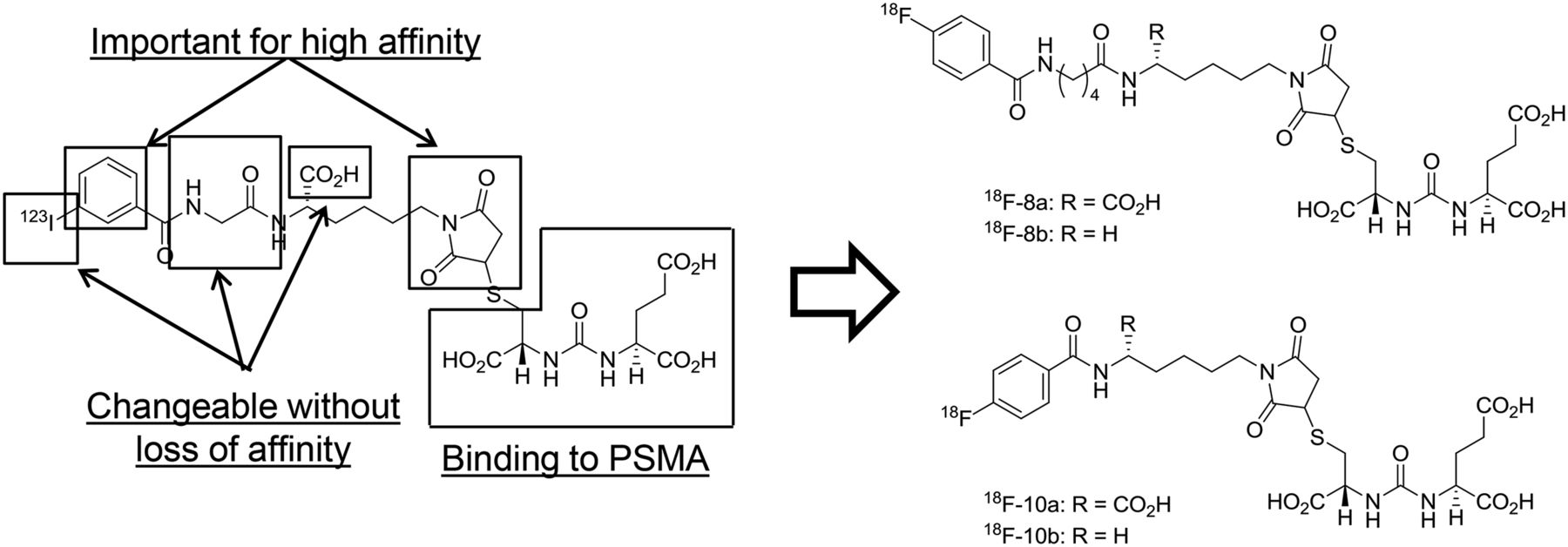
We have previously reported a radioiodinated asymmetric urea compound with high affinity for PSMA, (2S)-2-(3-((1R)-1-carboxy-2-((1-((R)-5-carboxy-5-(2-(3-123I-iodobenzamido)acetamido)pentyl)-2,5-dioxopyrrolidin-3-yl)thio)ethyl)ureido)pentanedioic acid (123I-IGLCE) (26). This compound is obtained by a nucleophilic conjugate addition reaction between maleimide (m-123I-iodohippuryl Nε-maleoyl-L-Lys) and the thiol group of (S)-2-[3-[(R)-1-carboxy-2-mercaptoethyl]ureido-pentanedioic acid (Cys-CO-Glu) (27). In our structure–activity relationship studies of 123I-IGLCE, we have found that the aromatic ring and succinimidyl moiety are associated with high affinity. The presence of iodine and length of the linker between the aromatic ring and succinimidyl moiety do not affect the affinity for PSMA. On the basis of these findings, we synthesized 18F-labeled compounds by substituting the iodobenzamido group with fluorobenzamido group of 123I-IGLCE derivatives using 18F-SFB (Fig. 1). We performed the biologic evaluations including measurement of in vitro binding affinity for PSMA, biodistribution study, and PET imaging in xenograft-bearing mice.

Design strategy for novel 18F-labeled PSMA-targeting probes.
MATERIALS AND METHODS
The supplemental materials provide additional details (available at http://jnm.snmjournals.org).
Chemistry
The methods of chemical syntheses are shown in the Supplemental Schemes 1 and 2. All final compounds were purified by reversed-phase high-performance liquid chromatography (HPLC) to a purity of greater than 95%. LC-20AD (Shimadzu Corp.) was used for HPLC with SPD-20A (Shimadzu) and NDW-351 (Hitachi Aloka Medical, Ltd.) as an ultraviolet detector (ρ; 220 and 254 nm) and radioisotope detector, respectively. The eluents were 0.1% trifluoroacetic acid in H2O and 0.1% trifluoroacetic acid in methanol.
Radiochemistry
The methods of radiosynthesis are shown in Supplemental Scheme 3. 18F-SFB was prepared according to a previously described procedure (22) with brief modifications. 18F-SFB was reacted with maleimide-based aminium precursors (3a, 3b, 13a, and 13b) at an ambient temperature for 5 min, followed by Cys-CO-Glu at an ambient temperature for 5 min, to obtain the final 18F-labeled probes. Each 18F-labeled probe was purified by reversed-phase HPLC to the radiochemical purity of greater than 95%. Each probe was prepared in approximately 100 min, and the total radiochemical yields from 18F− (decay-corrected) of 18F-8a, 18F-8b, 18F-10a, and 18F-10b were 36%, 50%, 30%, and 35%, respectively. The total time required for preparation was approximately 100 min. 18F-DCFPyL was prepared according to a previously described procedure (17) and used as a positive control probe in the biodistribution study.
Partition Coefficient
Partition coefficients (log D) of 18F-labeled probes were evaluated as previously reported (28). Each 18F-labeled compound was dissolved in 500 μL of 0.1 M phosphate buffer (pH 7.4). The test solution and 500 μL of 1-octanol were added to a test tube, which was then shaken vigorously and incubated at ambient temperature for approximately 5 min. After the mixture had been separated into 2 phases, the test tube was shaken again and centrifuged at 1,500g. A 50-μL aliquot of each layer was collected and counted using a γ-counter.
Cell Lines and Mouse Models
Two human prostate carcinoma cell lines were purchased from DS Pharma Biomedical: LNCaP (PSMA-positive) and PC-3 (PSMA-negative) (27). The cells were cultured as previously reported, in RPMI 1640 medium supplemented with 10% fetal bovine serum, glutamine, and antibiotics (penicillin/streptomycin), in a humidified CO2 incubator (37°C/5% CO2) (7).
Animal studies were conducted in accordance with institutional guidelines (Regulations for Animal Experimentation at Kyoto University), and the experimental procedures were approved by the Kyoto University Animal Care Committee. C.B-17/Icr +/+ Jcl mice and C.B.-17/Icr scid/scid Jcl mice weighing 22–24 g were purchased from CLEA Japan. Cultured cells were treated with 2.5 g/L trypsin with 1 mM ethylenediaminetetraacetic acid and resuspended in phosphate-buffered saline. Each mouse was injected with 100 μL of a 1:1 mixture of cell suspension and BD Matrigel Basement Membrane Matrix (1–5 × 10⁶ cells/mouse) in the left (PC-3) or right (LNCaP) flank. Tumor-bearing mice were used for studies when tumors reached a diameter of approximately 5–10 mm.
In Vitro Cell Binding Assay (Inhibition Assay)
The affinities of the novel compounds 8a–8 d, 10a, and 10b were determined by an in vitro binding inhibition assay as previously reported (10,26). N-[N-[(S)-1,3-dicarboxypropyl]carbamoyl]-S-3-iodo-L-Tyr (125I-DCIT) was used as the radioligand for the assay. 125I-DCIT was prepared according to a previously described procedure (29,30). LNCaP cells in 12-well plates (4 × 10⁵ cells/well) were incubated for 48 h (37°C/5% CO2). The medium was removed, and each well was washed twice with 500 μL of assay medium (RPMI 1640 supplemented with 0.5% bovine serum albumin). Next, 500 μL of 125I-DCIT (29.6 kBq/mL) and the test compound were added to each well, and the plates were incubated at 37°C for 1 h. Nonspecific binding was evaluated by adding 0.5 mM 2-(phosphonomethyl)pentanedioic acid (2-PMPA) (Tocris Bioscience). After incubation, each well was washed twice with 500 μL of fresh assay medium, and the cells were lysed with 0.2 N NaOH. Radioactivity bound to the cells was measured with a γ-counter. The half maximal inhibitory concentrations (IC50) were calculated using the GraphPad Prism 5 program (GraphPad Software Inc.). Ki values were calculated using the Cheng–Prusoff equation (31).
Biodistribution Studies
In the biodistribution studies, 100-μL aliquots of 18F-labeled probes were injected into tumor-bearing mice via the tail vein (n = 3–4 for each time point). Mice were sacrificed by decapitation at the following fixed time points: 2 min, 15 min, and 1 h. The weight and radioactivity of tissues were obtained, and the uptake of radioactivity was calculated as a percentage injected dose per gram of tissue (%ID/g). Biodistribution of 18F-DCFPyL was evaluated using the same procedure.
In Vivo Blocking Study
We selected 18F-10a as a representative probe for the in vivo blocking study because it showed the highest accumulation in the tumors. Briefly, 100 μL of 18F-10a was coinjected with 2-PMPA (50 mg/kg weight) into tumor-bearing mice via the tail vein. The mice were decapitated 1 h after injection. The weight and radioactivity of each tissue were measured, and the uptake of 18F-10a was calculated as %ID/g.
PET Imaging
The dynamic small-animal PET studies were conducted with 18F-8a and 18F-10a. All scans were obtained using a small-animal scanner (FX3300 imager; SII NanoTechnology Inc.). LNCaP and PC-3 xenograft–bearing mice were used in the PET studies after a 3-h fast (16.6–20.0 g of body weight after fasting). All mice were injected with each 18F-labeled probe (3.7–11.1 MBq in 0.1–0.15 mL of isotonic saline) via the tail vein and immediately anesthetized with isoflurane (2.5% in an air mixture). A competition experiment was performed by coinjection of 18F-10a with 50 mg/kg of 2-PMPA. The mice were placed on a heating pad to maintain their body temperature throughout the procedure. PET scans were acquired over 4.5–64.5 min, after which CT scans were acquired for anatomic reference (spatial resolution, 50 μm; 60 kV; and 310 μA). PET images were reconstructed using 3-dimensional ordered-subset expectation maximization (5 min/frame). CT images were reconstructed using a modified 3-dimensional cone-beam Feldkamp algorithm resulting in a 0.177 × 0.177 × 0.177-mm voxel size for a 512 × 512 × 512 image volume. The acquired PET and CT datasets were processed with PMOD Biomedical Image Quantification (PMOD Technologies Ltd.). Volume-of-interest analysis was based on the maximum SUVs.
Assessment of Toxicity in Mice
We performed an extended single-dose toxicity study to investigate the toxicity of 10a. Because the estimated dose of 10a for a human was 13.2 ng/kg, the toxicity was investigated at vehicle doses of 4.0 μg/kg (300-fold) and 13.2 μg/kg (1,000-fold). For each dose, 30 mice were injected, and body weight change was monitored for 1 d (n = 20) or 2 wk (n = 10). After the monitoring period, all mice were euthanized, and the hematologic, biochemical, and pathologic examinations were performed.
Statistics
GraphPad Prism 5 software was used for statistical analyses. The Bonferroni multiple-comparison test was used to assess statistical differences in the biodistribution study. A P value of less than 0.05 was considered statistically significant. The relationship between accumulation of the probes in the LNCaP tumor and log D values of the probes was evaluated as a Pearson product-moment correlation coefficient.
RESULTS
Chemistry
Four 18F-labeled asymmetric urea compounds, 18F-8a, 18F-8b, 18F-10a, and 18F-10b, were synthesized by reacting 18F-SFB with maleimide-based precursors (3a, 3b, 13a, and 13b) followed by Cys-CO-Glu, at a radiochemical yield of 30%–50% (decay-corrected) with a radiochemical purity of greater than 95% (after purification by HPLC) in approximately 100 min.
Partition Coefficient
The log D values of the 4 18F-labeled compounds are shown in Table 1. 18F-10a had the highest hydrophilicity, and 18F-8a had the second-highest hydrophilicity. Removal of a carboxylic group from 18F-8a and 18F-10a led to lower hydrophilicity as evidenced by the fact that 18F-8b and 18F-10b showed larger log D values than 18F-8a and 18F-10a.
Summary of In Vitro Studies of Each 18F-Labeled Compound
Inhibition Assay
Using the reference compounds 18F-DCFPyL and PSMA, we measured the affinity of the 4 compounds by examining the competition with 125I-DCIT for PSMA-positive LNCaP cells. Table 1 summarizes inhibition constant (Ki) values determined from the IC50 of competitive binding curves of these compounds. All 4 18F-labeled probes had a high affinity for PSMA. Among these probes, 18F-8a, 18F-8b, and 18F-10a had higher affinity than 18F-DCFPyL and 2-PMPA, a typical PSMA inhibitor. 18F-10b had slightly lower affinity than 18F-DCFPyL. The affinities of 18F-8a and 18F-10a for PSMA were approximately 4.6 and 7 times higher than the affinity of 18F-DCFPyL, respectively.
Biodistribution Study
Biodistribution data of 18F-labeled probes in mice are shown in Figure 2 and Supplemental Tables 1–3. All 18F-labeled probes showed rapid blood clearance and low accumulation in normal, nonspecific tissues (e.g., pancreas, stomach, heart, lung, muscle, and bone). High accumulation was observed in PSMA-expressing tissues (e.g., kidney and spleen). Differences between the excretion routes were observed for the different 18F-labeled probes. 18F-8a, 18F-8b, and 18F-10b were excreted from the kidney and liver, but 18F-10a was excreted almost solely from the kidney.

Biodistribution data of 18F-labeled PSMA probes: 18F-8a (A), 18F-8b (B), 18F-10a (C), 18F-10b (D), and 18F-DCFPyL (E). (F) Accumulations of 18F-labeled PSMA probes in LNCaP tumors. Mice were sacrificed at following fixed time points: 2 min, 15 min, and 1 h. Vertical axes are in %ID/g, except for stomach (shown as %ID). Columns and error bars indicate mean ± SD.
The radioactivity in the PSMA-negative PC-3 tumors decreased in a time-dependent manner. However, high levels of radioactivity accumulation and retention were observed in the PSMA-positive LNCaP tumors for all 4 compounds, 18F-8a, 18F-8b, 18F-10a, and 18F-10b. In particular, 18F-10a showed the highest accumulation in LNCaP tumors (14.0 ± 3.1 %ID/g at 60 min), almost the same as 18F-DCFPyL in LNCaP tumors (16.0 ± 2.9 %ID/g at 60 min) (Supplemental Table 3).
In Vivo Blocking Study
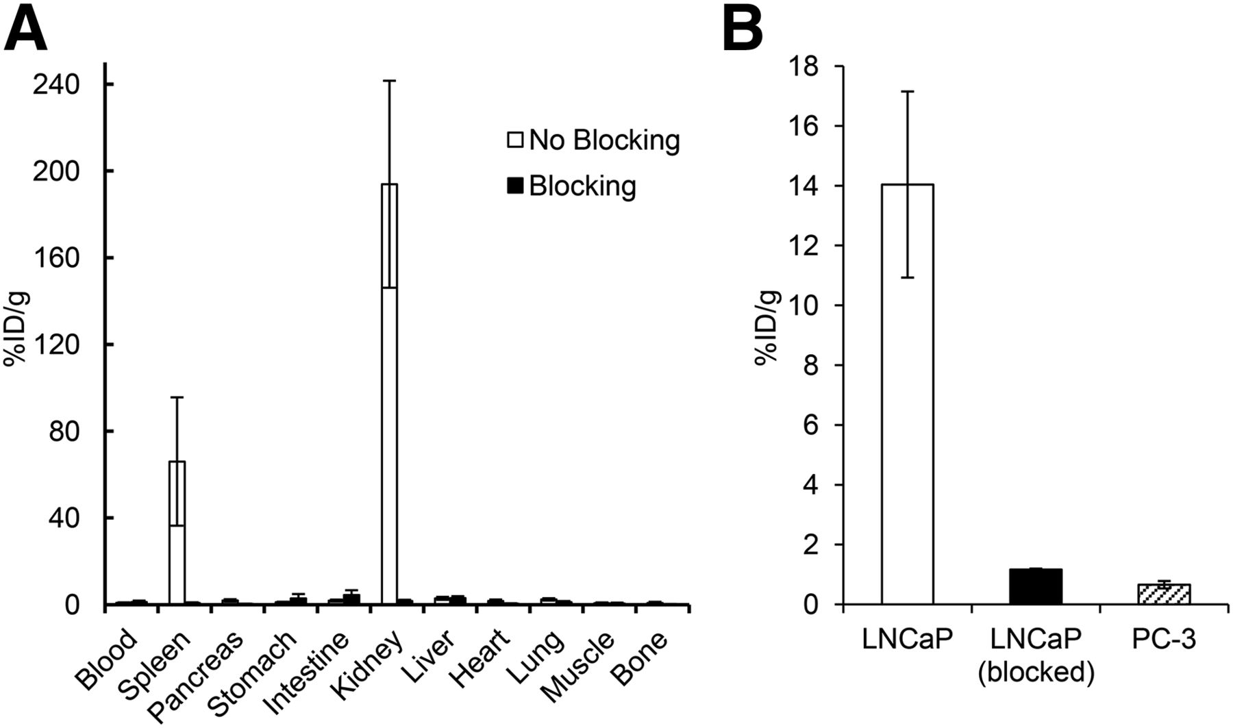
Binding of 18F-10a on PMSA was inhibited in vivo by coinjection of 2-PMPA (Fig. 3). Accumulation of 18F-10a in PSMA-specific tissues (e.g., LNCaP tumor, kidney, and spleen) was inhibited by 2-PMPA, a typical PSMA inhibitor. The LNCaP-to-blood, LNCaP-to-muscle, LNCaP-to-bone, and LNCaP-to-PC-3 ratios were decreased by 2-PMPA from 16.90, 20.09, 18.40, and 18.97 to 0.95, 2.26, 4.78, and 1.78, respectively.

In vivo blocking study with 18F-10a in normal tissues (A) and tumors (B) at 60 min. Data shown are averages of data of 4 mice, and error bars indicate SD. Data for stomach are expressed as %ID.
PET Imaging
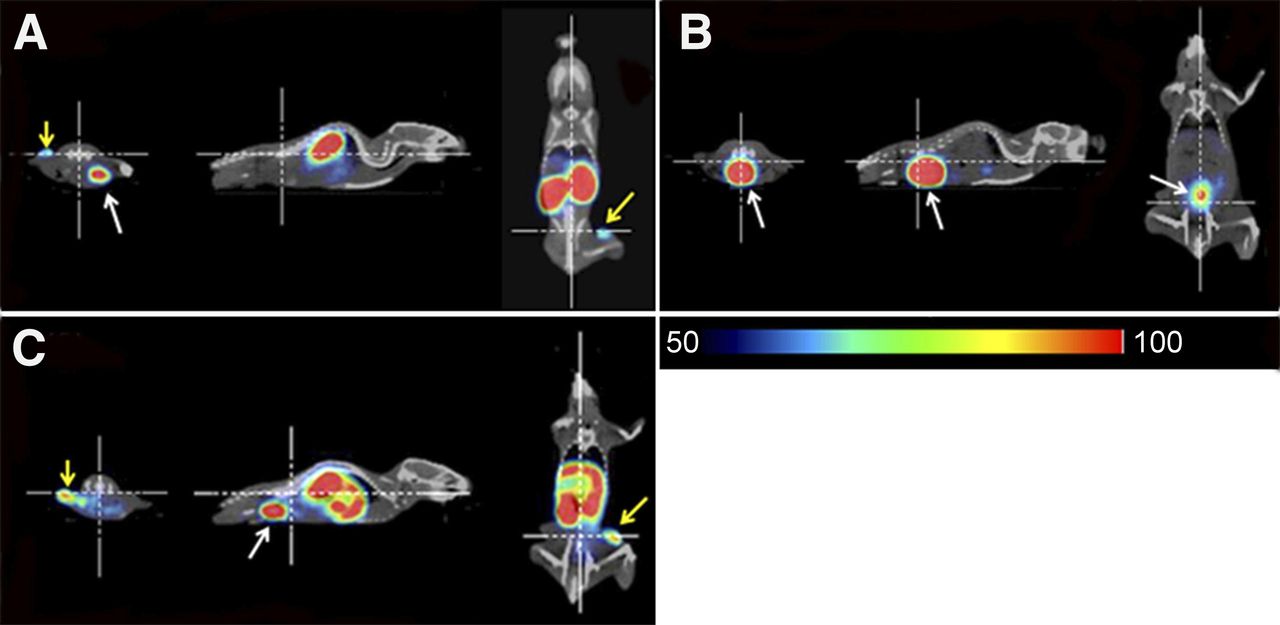
18F-8a and 18F-10a clearly visualized the LNCaP tumors and distinguished them from PC-3 tumors in mice. Both compounds showed rapid clearance from the body muscle (Fig. 4). Volume-of-interest analysis revealed the low hepatic accumulation of 18F-10a, with a higher LNCaP-to-liver ratio than 18F-8a. We performed a blocking PET study to confirm the PSMA-specific distribution of 18F-10a; coinjection with 2-PMPA completely prevented the renal uptake and accumulation in LNCaP tumors and resulted in a rapid excretion to the bladder.

Twelfth frame of PET images (59.5–64.5 min) with 18F-10a (A), with coinjection of 2-PMPA and 18F-10a (B), and with 18F-8a (C). Yellow and white arrows indicate LNCaP tumors and bladders, respectively.
Assessment of Toxicity in Mice
No lethal toxicity was observed after a single-dose injection of 10a (13.2 μg/kg). There were also no hematologic, biochemical, or pathologic effects in the animals administered with 13.2 μg/kg of 10a. Thus, the no-observed-adverse-effects level of 10a was estimated as larger than 13.2 μg/kg.
DISCUSSION
18F-DCFPyL has been developed as a promising imaging probe for prostate cancers, with high diagnostic accuracy, and its clinical studies are still being conducted. However, the radiochemical yield of 18F-DCFPyL is low (2.8% ± 1.2%) (18). To overcome this disadvantage, we designed novel PSMA-targeting probes using 18F-SFB, which has been obtained with a high radiochemical yield using the automated facile synthesis (20–25). The design of these novel PSMA imaging probes was based on the results of our previous study. The study, examining the structure–activity relationship of an asymmetric urea compound 123I-GLCE, has found that the aromatic ring and succinimidyl moiety are associated with high affinity. Moreover, the presence of iodine and length of the linker between the aromatic ring and succinimidyl moiety do not affect the affinity for PSMA. Thus, in the present study, we designed 18F-labeled compounds to be obtained by reacting 18F-SFB with maleimide-based aminium precursors followed by an asymmetric urea moiety, Cys-CO-Glu (Fig. 1).
We succeeded in synthesizing 4 18F-labeled asymmetric urea compounds, 18F-8a, 18F-8b, 18F-10a, and 18F-10b, at high radiochemical yields of 30%–50% (decay-corrected) that were more than 10–15 times higher than the yield of 18F-DCFPyL.
The affinities of the synthesized compounds were evaluated by an in vitro binding inhibition assay for PSMA-expressing cells (LNCaP cells). These 4 novel probes had a high affinity for PSMA. Among the compounds tested, 18F-8a and 18F-10a had a particularly high affinity. Their affinities were 4.5 and 7 times higher than that of 18F-DCFPyL, respectively (Table 1). This result supported the validity of our drug design.
Partition coefficients of these compounds were also evaluated. We found that our 18F-labeled probes had lower hydrophilicity than 18F-DCFPyL. 18F-10a had the highest hydrophilicity, and 18F-8a had the second highest. We believed that the lower hydrophilicity caused the hepatic accumulations of 18F-8a, 18F-8b, and 18F-10b in the biodistribution study (Fig. 2).
Furthermore, we confirmed the in vitro stability of the probes in the mouse plasma (Supplemental Figs. 1–4). In the biodistribution studies, none of the 18F-labeled probes accumulated in normal, nonspecific tissues (pancreas, stomach, heart, lung, muscle, and bone). Low accumulation in the bone indicates little defluorination of 18F-labeled probes. Such low accumulation in the bone is important for PSMA-targeting probes because prostate cancer has the propensity to metastasize to this tissue. Our results demonstrate that all the tested 18F-labeled probes have good stability and show rapid clearance from nontargeted tissues.
In the biodistribution studies, 18F-10a and 18F-DCFPyL showed similar biodistribution profiles. Namely, 18F-10a exhibited a rapid blood clearance, low accumulation in the bone, and high accumulation in the LNCaP tumor, as did the 18F-DCFPyL. Moreover, 18F-10a and 18F-DCFPyL were excreted from the kidney, whereas 18F-8a, 18F-8b, and 18F-10b accumulated in the liver (Fig. 3; Supplemental Tables 1–3). Although 18F-8a also showed high accumulation in LNCaP tumors and high LNCaP–to–nonspecific tissue ratios (i.e., high LNCaP-to-blood, LNCaP-to-muscle, and LNCaP-to-PC-3 ratios), it had a lower LNCaP-to-liver ratio than 18F-10a. Babich et al. have emphasized the importance of a high tumor-to-liver ratio for prostate cancer imaging; this cancer is likely to metastasize to the pelvic and abdominal cavities (32). Hence, among our 18F-labeled probes, 18F-10a is the most suitable for PSMA imaging of metastatic prostate cancer.
We performed an in vivo blocking study with 18F-10a to confirm PSMA-specific binding (Fig. 3). Accumulation of 18F-10a in the LNCaP tumors, kidneys, and spleen was significantly inhibited by coinjection of 2-PMPA, a PSMA inhibitor. High expression of glutamate carboxypeptidase-II has been reported in the murine kidney and spleen, and other PSMA-targeting probes accumulate in these tissues (10,13,17,33). Therefore, the accumulation of 18F-10a in the LNCaP tumors, kidneys, and spleens might be attributed to PSMA-specific binding.
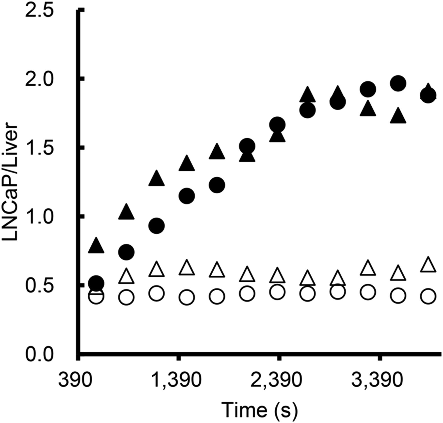
In the PET study, 18F-10a immediately accumulated in LNCaP tumors and revealed them clearly (Fig. 4). According to the results of the biodistribution study, 18F-10a was mostly excreted via the renal route, and the LNCaP-to-background ratio was better in the later part of the experiment than at the beginning (Fig. 4; Supplemental Fig. 5). Moreover, a rapid accumulation of 18F-10a in the bladder was observed in the competition experiment, indicating mostly PSMA-specific accumulation and little nonspecific accumulation of 18F-10a. Furthermore, we compared the PET images obtained using 18F-10a and 18F-8a. 18F-10a showed little accumulation in the liver; in contrast, 18F-8a accumulated in this organ (Fig. 4). These images corresponded to the results of the biodistribution study. According to volume-of-interest analysis, the LNCaP-to-liver ratio of 18F-8a was less than 1 throughout the 1-h scanning period. However, the LNCaP-to-liver ratio of 18F-10a increased with time and reached a value of more than 1.9, 1 h after injection (Fig. 5). These results suggest that 18F-10a has the potential to serve as a highly specific PSMA-targeting PET probe, achieving high accumulation levels. Because imaging mice for 60 min does not necessarily provide sufficient properties of 18F-labeled probes, we are planning further evaluation of 18F-10a.

Time-dependent alteration of quantified LNCaP-to-liver ratios of 18F-8a (empty markers) and 18F-10a (filled markers). LNCaP-to-liver ratios were evaluated using 2 mice for each probe.
CONCLUSION
We designed 18F-labeled asymmetric urea compounds as novel PSMA-targeting probes. These probes can be prepared using 18F-SFB, one of the prevalent radiolabeling reagents obtained using the widely accessible automated synthesis. The designed compounds were synthesized at a high radiochemical yield, achieving 10- to 15-fold increases in comparison with the yield of 18F-DCFPyL, a promising PSMA-imaging probe recently tested in clinical studies. Among the 4 probes synthesized, 18F-10a showed the highest binding affinity for PSMA, much higher than the affinity of 18F-DCFPyL. Furthermore, 18F-10a demonstrated high accumulation in PSMA-positive LNCaP tumors and a biodistribution pattern similar to that of 18F-DCFPyL. PET imaging using 18F-10a obtained clear images of LNCaP tumors, 60 min after injection. These results establish 18F-10a as a promising PSMA probe.
DISCLOSURE
The costs of publication of this article were defrayed in part by the payment of page charges. Therefore, and solely to indicate this fact, this article is hereby marked “advertisement” in accordance with 18 USC section 1734. This work was partly supported by a grant-in-aid for Young Scientists (B) from the Japan Society for the Promotion of Science and New Energy, Industrial Technology Development Organization (NEDO), and Jana Agency for Medical Research and Development (AMED). 18F− was supplied by the Kyoto University Hospital (Kyoto, Japan). We entrusted the toxicological study to CMIC BIORESEARCH CENTER Co., Ltd. No other potential conflict of interest relevant to this article was reported.
Footnotes
Published online Jul. 14, 2016.
- © 2016 by the Society of Nuclear Medicine and Molecular Imaging, Inc.
REFERENCES
- Received for publication March 22, 2016.
- Accepted for publication June 17, 2016.


Jump to section
Related Articles
Cited By...
- Clinical Outcomes of Metastasis-directed Therapy for Oligo-metastatic Prostate Cancer Diagnosed Using PSMA-PET/CT or Whole-body MRI
- Clinical Characterization of [18F]T-008, a Cholesterol 24-Hydroxylase PET Ligand: Dosimetry, Kinetic Modeling, Variability, and Soticlestat Occupancy
- Preclinical Evaluation and Pilot Clinical Study of Al18F-PSMA-BCH for Prostate Cancer PET Imaging
- One-Step 18F-Labeling and Preclinical Evaluation of Prostate-Specific Membrane Antigen Trifluoroborate Probes for Cancer Imaging
- Bicyclic Peptides as a New Modality for Imaging and Targeting of Proteins Overexpressed by Tumors