
Main menu

  • Home
  • Content
    • Current
    • Ahead of print
    • Past Issues
    • JNM Supplement
    • SNMMI Annual Meeting Abstracts
    • Continuing Education
    • JNM Podcasts
  • Subscriptions
    • Subscribers
    • Institutional and Non-member
    • Rates
    • Journal Claims
    • Corporate & Special Sales
  • Authors
    • Submit to JNM
    • Information for Authors
    • Assignment of Copyright
    • AQARA requirements
  • Info
    • Reviewers
    • Permissions
    • Advertisers
  • About
    • About Us
    • Editorial Board
    • Contact Information
  • More
    • Alerts
    • Feedback
    • Help
    • SNMMI Journals
  • SNMMI
    • JNM
    • JNMT
    • SNMMI Journals
    • SNMMI

User menu

  • Subscribe
  • My alerts
  • Log in
  • Log out
  • My Cart

Search

  • Advanced search
Journal of Nuclear Medicine
  • SNMMI
    • JNM
    • JNMT
    • SNMMI Journals
    • SNMMI
  • Subscribe
  • My alerts
  • Log in
  • Log out
  • My Cart
Journal of Nuclear Medicine

Advanced Search

  • Home
  • Content
    • Current
    • Ahead of print
    • Past Issues
    • JNM Supplement
    • SNMMI Annual Meeting Abstracts
    • Continuing Education
    • JNM Podcasts
  • Subscriptions
    • Subscribers
    • Institutional and Non-member
    • Rates
    • Journal Claims
    • Corporate & Special Sales
  • Authors
    • Submit to JNM
    • Information for Authors
    • Assignment of Copyright
    • AQARA requirements
  • Info
    • Reviewers
    • Permissions
    • Advertisers
  • About
    • About Us
    • Editorial Board
    • Contact Information
  • More
    • Alerts
    • Feedback
    • Help
    • SNMMI Journals
  • View or Listen to JNM Podcast
  • Visit JNM on Facebook
  • Join JNM on LinkedIn
  • Follow JNM on Twitter
  • Subscribe to our RSS feeds
OtherFOCUS ON MOLECULAR IMAGING

PET/MRI: Paving the Way for the Next Generation of Clinical Multimodality Imaging Applications

Bernd J. Pichler, Armin Kolb, Thomas Nägele and Heinz-Peter Schlemmer
Journal of Nuclear Medicine March 2010, 51 (3) 333-336; DOI: https://doi.org/10.2967/jnumed.109.061853
Bernd J. Pichler
  • Find this author on Google Scholar
  • Find this author on PubMed
  • Search for this author on this site
Armin Kolb
  • Find this author on Google Scholar
  • Find this author on PubMed
  • Search for this author on this site
Thomas Nägele
  • Find this author on Google Scholar
  • Find this author on PubMed
  • Search for this author on this site
Heinz-Peter Schlemmer
  • Find this author on Google Scholar
  • Find this author on PubMed
  • Search for this author on this site
  • Article
  • Figures & Data
  • Info & Metrics
  • PDF
Loading

Article Figures & Data

Figures

  • FIGURE 1. 
    • Download figure
    • Open in new tab
    • Download powerpoint
    FIGURE 1. 

    PET/CT and PET/MRI of 71-y-old woman with frontobasal meningioma in olfactory region. PET/CT images were acquired 20 min and PET/MR images 100 min after injection of 135 MBq of 68Ga-[1,4,7,10-tetraazacyclododecane-N,N′,N″,N″′-tetraacetic acid]-d-Phe1,Tyr3-octreotide. Tracer uptake in the tumor is seen on PET images. In addition, second smaller and previously unknown frontal meningioma was seen on PET and possibly corresponded to small mass demonstrated on T2-weighted turbo spin-echo MR images. This finding was not detected by CT.

  • FIGURE 2. 
    • Download figure
    • Open in new tab
    • Download powerpoint
    FIGURE 2. 

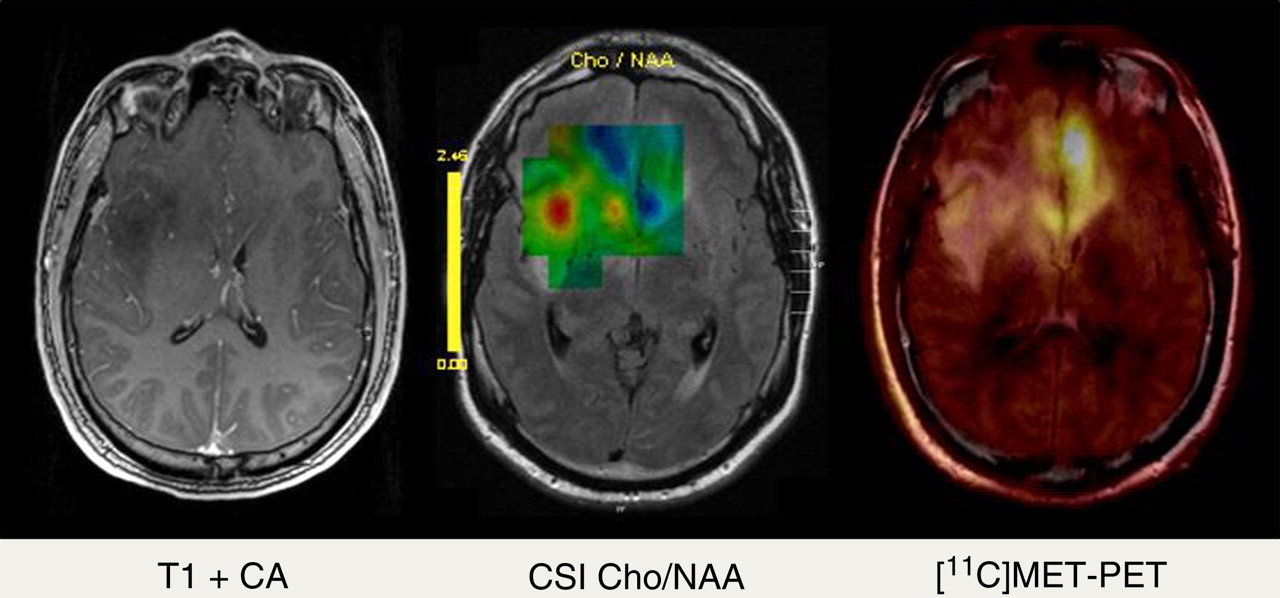
    A 36-y-old patient with brain gliomatosis was admitted for tumor biopsy. In addition to standard contrast-enhanced T1-weighted MRI (left), chemical shift imaging (CSI) (echo time = 135 ms, center) and PET/MRI with 11C-methionine (right) were performed. On standard MRI, no contrast enhancement in any part of tumor was found, indicating low-grade tumor. On CSI, mapping of choline/N-acetylaspartate (Cho/NAA) quotient showed hot spot in right insular region, whereas 11C-methionine uptake was most pronounced in basal frontal lobe on left side. Because of discrepancy between CSI and PET/MRI, biopsy was performed in both locations and revealed anaplastic glioma (World Health Organization grade III) in frontal left region and low-grade glioma (World Health Organization grade II) in right insular region. CA = contrast agent.

  • FIGURE 3. 
    • Download figure
    • Open in new tab
    • Download powerpoint
    FIGURE 3. 

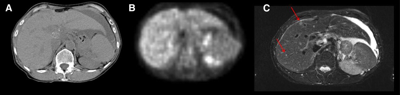
    Abdominal unenhanced 18F-FDG PET/CT and MRI of 55-y-old woman with ovarian cancer showing liver metastases detected by MRI (C) but neither by PET (B) nor native CT (A). MRI is most sensitive for detecting small liver lesions because of its superb soft-tissue contrast, whereas PET and CT are limited because of lower contrast and physiologic 18F-FDG liver uptake.

PreviousNext
Back to top

In this issue

Journal of Nuclear Medicine: 51 (3)
Journal of Nuclear Medicine
Vol. 51, Issue 3
March 2010
  • Table of Contents
  • Table of Contents (PDF)
  • About the Cover
  • Index by author
Print
Download PDF
Article Alerts
Sign In to Email Alerts with your Email Address
Email Article

Thank you for your interest in spreading the word on Journal of Nuclear Medicine.

NOTE: We only request your email address so that the person you are recommending the page to knows that you wanted them to see it, and that it is not junk mail. We do not capture any email address.

Enter multiple addresses on separate lines or separate them with commas.
PET/MRI: Paving the Way for the Next Generation of Clinical Multimodality Imaging Applications
(Your Name) has sent you a message from Journal of Nuclear Medicine
(Your Name) thought you would like to see the Journal of Nuclear Medicine web site.
Citation Tools
PET/MRI: Paving the Way for the Next Generation of Clinical Multimodality Imaging Applications
Bernd J. Pichler, Armin Kolb, Thomas Nägele, Heinz-Peter Schlemmer
Journal of Nuclear Medicine Mar 2010, 51 (3) 333-336; DOI: 10.2967/jnumed.109.061853

Citation Manager Formats

  • BibTeX
  • Bookends
  • EasyBib
  • EndNote (tagged)
  • EndNote 8 (xml)
  • Medlars
  • Mendeley
  • Papers
  • RefWorks Tagged
  • Ref Manager
  • RIS
  • Zotero
Share
PET/MRI: Paving the Way for the Next Generation of Clinical Multimodality Imaging Applications
Bernd J. Pichler, Armin Kolb, Thomas Nägele, Heinz-Peter Schlemmer
Journal of Nuclear Medicine Mar 2010, 51 (3) 333-336; DOI: 10.2967/jnumed.109.061853
Twitter logo Facebook logo LinkedIn logo Mendeley logo
  • Tweet Widget
  • Facebook Like
  • Google Plus One
Bookmark this article

Jump to section

  • Article
    • Abstract
    • PET/MRI: TECHNICAL EVOLUTION
    • CLINICAL POTENTIAL OF PET/MRI
    • WHOLE-BODY PET/MRI
    • CONCLUSION
    • Acknowledgments
    • Footnotes
    • References
  • Figures & Data
  • Info & Metrics
  • PDF

Related Articles

  • This Month in JNM
  • PubMed
  • Google Scholar

Cited By...

  • Development of a non-invasive method for testicular toxicity evaluation using a novel compact magnetic resonance imaging system
  • 18F-FDG PET/MRI for Therapy Response Assessment of Isolated Limb Perfusion in Patients with Soft-Tissue Sarcomas
  • Exact Topological Inference for Paired Brain Networks via Persistent Homology
  • Evaluation of 11C-Methionine PET and Anatomic MRI Associations in Diffuse Intrinsic Pontine Glioma
  • Generation of Structural MR Images from Amyloid PET: Application to MR-Less Quantification
  • Comparison of PET/CT with Sequential PET/MRI Using an MR-Compatible Mobile PET System
  • Regarding "Subjecting Radiologic Imaging to the Linear No-Threshold Hypothesis: A Non Sequitur of Non-Trivial Proportion"
  • Simultaneous Multiparametric PET/MRI with Silicon Photomultiplier PET and Ultra-High-Field MRI for Small-Animal Imaging
  • Impact of MR-Based Attenuation Correction on Neurologic PET Studies
  • ImmunoPET/MR imaging allows specific detection of Aspergillus fumigatus lung infection in vivo
  • Imaging-Based Treatment Adaptation in Radiation Oncology
  • Reconstruction-Incorporated Respiratory Motion Correction in Clinical Simultaneous PET/MR Imaging for Oncology Applications
  • MR-Based Attenuation Correction Using Ultrashort-Echo-Time Pulse Sequences in Dementia Patients
  • Techniques, Benefits, and Challenges of PET-MR
  • PET/MR Imaging and Optical Imaging of Metastatic Rhabdomyosarcoma in Mice
  • Imaging and Nanomedicine in Inflammatory Atherosclerosis
  • Potential Pediatric Applications of PET/MR
  • Preclinical and Translational PET/MR Imaging
  • Thoracic Staging in Lung Cancer: Prospective Comparison of 18F-FDG PET/MR Imaging and 18F-FDG PET/CT
  • Tumor-specific Localization of Self-assembled Nanoparticle PET/MR Modalities
  • MR-Based Attenuation Correction Methods for Improved PET Quantification in Lesions Within Bone and Susceptibility Artifact Regions
  • Multimodality Assessment of Brain Tumors and Tumor Recurrence
  • Complementary Roles of Whole-Body Diffusion-Weighted MRI and 18F-FDG PET: The State of the Art and Potential Applications
  • Cerenkov Luminescence Imaging of Medical Isotopes
  • Google Scholar

More in this TOC Section

  • From Genome to Phenome: Opportunities and Challenges of Molecular Imaging
  • Imaging the Activity of Efflux Transporters at the Blood–Brain Barrier in Neurologic Diseases: Radiotracer Selection Criteria
  • Molecular Imaging of Pulmonary Fibrosis
Show more Focus on Molecular Imaging

Similar Articles

SNMMI

© 2025 SNMMI

Powered by HighWire