


Article Figures & Data
Figures
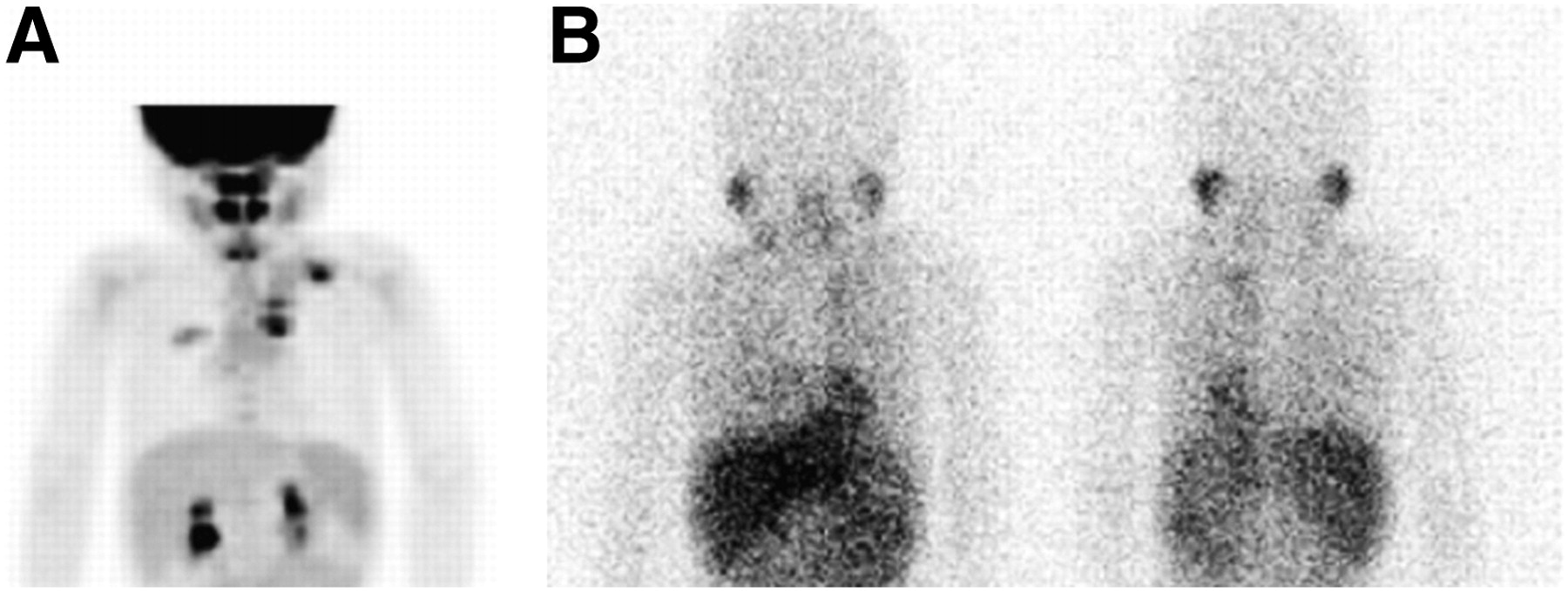
- FIGURE 1.
A 3-year-old boy with stage 2 neuroblastoma at diagnosis. (A) 18F-FDG anterior maximum-intensity-projection image demonstrates marked uptake in left thoracic paraspinal tumor and left supraclavicular metastatic lymph nodes. Uptake is also seen within dependent right upper lobe, corresponding to inflammatory airspace disease. (B) 123I-MIBG anterior and posterior planar images demonstrate minimal uptake in left thoracic paraspinal tumor.
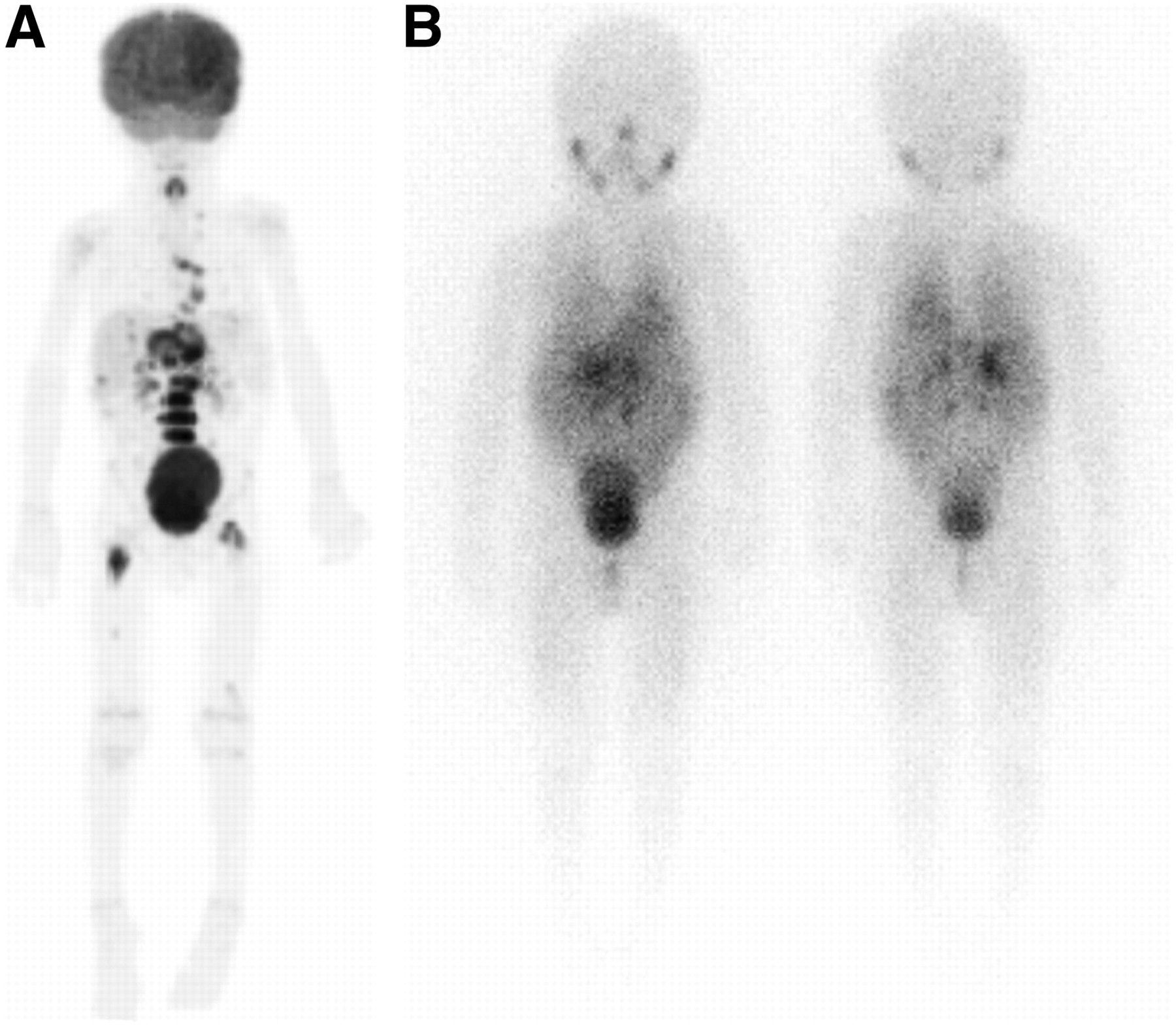
- FIGURE 2.
A 3-month-old boy with stage 3 neuroblastoma at diagnosis. (A) 18F-FDG anterior maximum-intensity-projection image demonstrates marked uptake in the abdominal mass and mild diffuse physiologic uptake in the bone marrow. (B) 123I-MIBG anterior planar image demonstrates minimal uptake in abdominal mass, slightly above background.
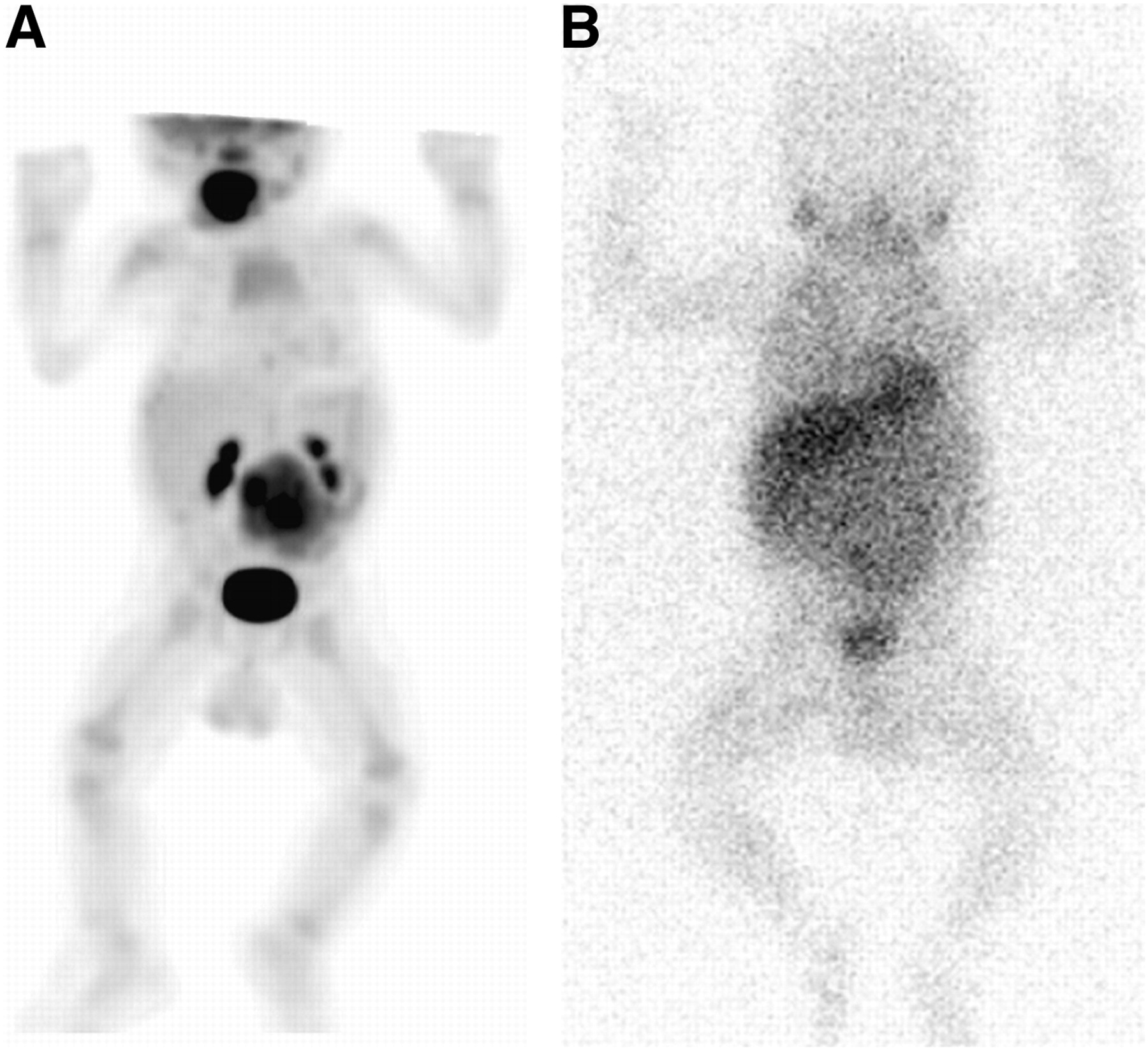
- FIGURE 3.
A 3-year-old boy with stage 4 neuroblastoma before bone marrow transplantation. (A) 18F-FDG anterior maximum-intensity-projection image demonstrates marked uptake in retroperitoneal and mediastinal disease. Uptake is also seen in bony disease, most marked in lumbar spine and proximal femurs. (B) 123I-MIBG anterior and posterior planar images demonstrate retroperitoneal uptake.
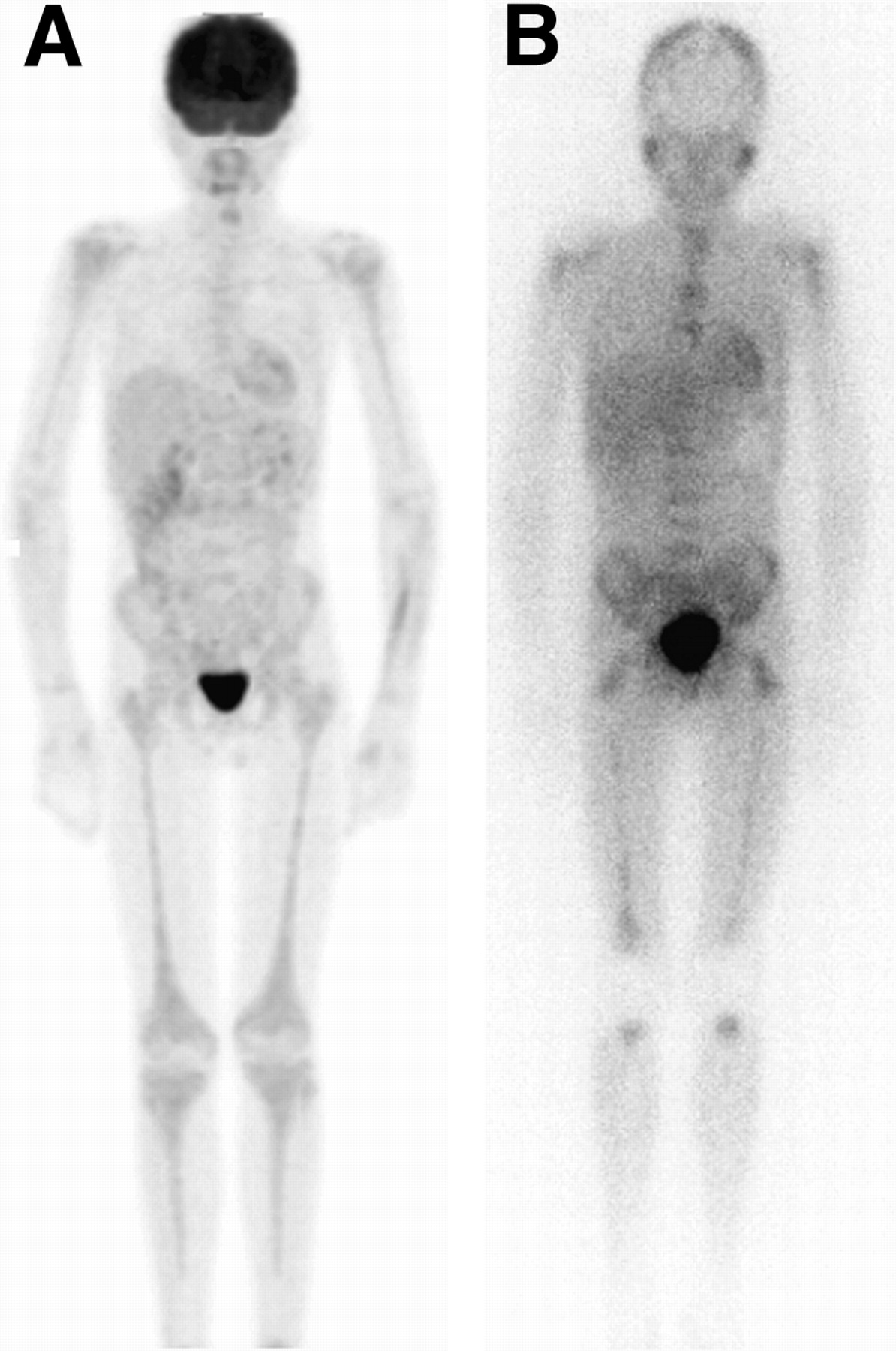
- FIGURE 4.
A 13-year-old boy with stage 4 neuroblastoma after initial chemotherapy. (A) 18F-FDG anterior maximum-intensity-projection image demonstrates mild diffuse bone marrow uptake extending throughout axial and appendicular skeleton in response to marrow expansion with some G-CSF stimulation. (B) 123I-MIBG anterior planar image demonstrates uptake in diffuse bone or marrow metastases. Note absence of 123I-MIBG uptake in mid- or distal humerus and mid- or distal tibia. G-CSF = granulocyte colony-stimulating factor.
- FIGURE 5.
A 6-year-old girl with stage 4 neuroblastoma. (A) 18F-FDG anterior maximum-intensity-projection image obtained at diagnosis demonstrates uptake in primary left retroperitoneal tumor (arrows) and multiple discrete bone or marrow metastases. 123I-MIBG imaging at diagnosis (not shown) demonstrated uptake in primary tumor and multiple bone or marrow metastases. (B) 18F-FDG anterior maximum-intensity-projection image obtained after chemotherapy and G-CSF therapy demonstrates intense diffuse bone marrow uptake, obscuring or mimicking metastatic disease. 123I-MIBG images at follow-up (not shown) demonstrated resolution of uptake in primary tumor and bone or marrow metastases. Bone marrow sampling at time of follow-up scan showed no evidence of metastatic tumor. G-CSF = granulocyte colony-stimulating factor.


Tables
Result Stage 123I-MIBG > 18F-FDG 18F-FDG > 123I-MIBG Equivalent Negative Stage 1 and 2 neuroblastoma (13 scans/10 patients*) Diagnosis — 4 scans/4 pts 1 scan/1 pt — Follow-up — 5 scans/3 pts — 3 scans/3 pts Total — 9 scans/6 pts 1 scan/1 pt 3 scans/3 pts Stage 3 neuroblastoma (15 scans/10 patients*) Diagnosis — 1 scan/1 pt 2 scans/2 pts — Follow-up 5 scans/4 pts 3 scans/3 pts — 4 scans/3 pts Total 5 scans/4 pts 4 scans/4 pts 2 scans/2 pts 4 scans/3 pts Stage 4 neuroblastoma (85 scans/40 patients*) Diagnosis 8 scans/8 pts 3 scans/3 pts 5 scans/5 pts — Follow-up ≤ 12 months 19 scans/12 pts 5 scans/3 pts 5 scans/5 pts 6 scans/5 pts Follow-up > 12 months 17 scans/10 pts 3 scans/3 pts 3 scans/3 pts 11 scans/10 pts Total 44 scans/24 pts 11 scans/8 pts 13 scans/11 pts 17 scans/14 pts ↵* Patients with more than 1 scan during study period may be listed in different categories (i.e., patients scanned at both diagnosis and follow-up or patients with multiple follow-up scans with different results).
123I-MIBG > 18F-FDG = numbers of scans and patients for which 123I-MIBG detected more lesions; pts = patients; 18F-FDG > 123I-MIBG = numbers of scans and patients for which 18F-FDG detected more lesions; Equivalent = numbers of scans and patients for which 123I-MIBG and 18F-FDG detected similar or complementary numbers and distributions of lesions; Negative = numbers of scans and patients for which 123I-MIBG and 18F-FDG study results were normal.
Result Stage 123I-MIBG > 18F-FDG 18F-FDG > 123I-MIBG Equivalent Negative Stage 1 neuroblastoma (5 scans/3 patients*) Diagnosis — 2 scans/2 pts — — Follow-up — 3 scans/1 pt — — Total — 5 scans/3 pts — — Stage 4 neuroblastoma (8 scans/5 patients*) Diagnosis 1 scan/1 pt — — — Follow-up ≤ 12 months 1 scan/1 pt — — — Follow-up > 12 months 2 scans/2 pts 1 scan/1 pt 1 scan/1 pt 2 scans/2 pts Total 4 scans/4 pts 1 scan/1 pt 1 scan/1 pt 2 scans/2 pts ↵* Patients with more than 1 scan during study period may be listed in different categories (i.e., patients scanned at both diagnosis and follow-up or patients with multiple follow-up scans with different results).
123I-MIBG > 18F-FDG = numbers of scans and patients for which 123I-MIBG detected more lesions; pts = patients; 18F-FDG > 123I-MIBG = numbers of scans and patients for which 18F-FDG detected more lesions; Equivalent = numbers of scans and patients for which 123I-MIBG and 18F-FDG detected similar or complementary numbers and distributions of lesions; Negative = numbers of scans and patients for which 123I-MIBG and 18F-FDG study results were normal.


Jump to section
Related Articles
Cited By...
- SNMMI Procedure Standard/EANM Practice Guideline on Pediatric 18F-FDG PET/CT for Oncology 1.0
- Is True Whole-Body 18F-FDG PET/CT Required in Pediatric Lymphoma? An IAEA Multicenter Prospective Study
- Radiolabeled (4-Fluoro-3-Iodobenzyl)Guanidine Improves Imaging and Targeted Radionuclide Therapy of Norepinephrine Transporter-Expressing Tumors
- Value of 18F-FDG PET and PET/CT for Evaluation of Pediatric Malignancies
- Potential Pediatric Applications of PET/MR
- Imaging the Norepinephrine Transporter in Neuroblastoma: A Comparison of [18F]-MFBG and 123I-MIBG
- Characterization of Neuroblastic Tumors Using 18F-FDOPA PET
- 64Cu-p-NH2-Bn-DOTA-hu14.18K322A, a PET Radiotracer Targeting Neuroblastoma and Melanoma
- Nuclear Medicine in the First Year of Life
- 18F-FDG PET/CT and 123I-Metaiodobenzylguanidine Imaging in High-Risk Neuroblastoma: Diagnostic Comparison and Survival Analysis
- Clinical presentations and imaging findings of neuroblastoma beyond abdominal mass and a review of imaging algorithm
- PET Imaging of Norepinephrine Transporter-Expressing Tumors Using 76Br-meta-Bromobenzylguanidine
- 123I-MIBG Versus 18F-FDG: Which Is Better, or Which Can Be Eliminated?
- Reply: 123I-MIBG Scintigraphy and 18F-FDG PET in Neuroblastoma
- 123I-MIBG Scintigraphy and 18F-FDG PET in Neuroblastoma