
Main menu

  • Home
  • Content
    • Current
    • Ahead of print
    • Past Issues
    • JNM Supplement
    • SNMMI Annual Meeting Abstracts
    • Continuing Education
    • JNM Podcasts
  • Subscriptions
    • Subscribers
    • Institutional and Non-member
    • Rates
    • Journal Claims
    • Corporate & Special Sales
  • Authors
    • Submit to JNM
    • Information for Authors
    • Assignment of Copyright
    • AQARA requirements
  • Info
    • Reviewers
    • Permissions
    • Advertisers
  • About
    • About Us
    • Editorial Board
    • Contact Information
  • More
    • Alerts
    • Feedback
    • Help
    • SNMMI Journals
  • SNMMI
    • JNM
    • JNMT
    • SNMMI Journals
    • SNMMI

User menu

  • Subscribe
  • My alerts
  • Log in
  • Log out
  • My Cart

Search

  • Advanced search
Journal of Nuclear Medicine
  • SNMMI
    • JNM
    • JNMT
    • SNMMI Journals
    • SNMMI
  • Subscribe
  • My alerts
  • Log in
  • Log out
  • My Cart
Journal of Nuclear Medicine

Advanced Search

  • Home
  • Content
    • Current
    • Ahead of print
    • Past Issues
    • JNM Supplement
    • SNMMI Annual Meeting Abstracts
    • Continuing Education
    • JNM Podcasts
  • Subscriptions
    • Subscribers
    • Institutional and Non-member
    • Rates
    • Journal Claims
    • Corporate & Special Sales
  • Authors
    • Submit to JNM
    • Information for Authors
    • Assignment of Copyright
    • AQARA requirements
  • Info
    • Reviewers
    • Permissions
    • Advertisers
  • About
    • About Us
    • Editorial Board
    • Contact Information
  • More
    • Alerts
    • Feedback
    • Help
    • SNMMI Journals
  • View or Listen to JNM Podcast
  • Visit JNM on Facebook
  • Join JNM on LinkedIn
  • Follow JNM on Twitter
  • Subscribe to our RSS feeds
Research ArticleOncology

Is True Whole-Body 18F-FDG PET/CT Required in Pediatric Lymphoma? An IAEA Multicenter Prospective Study

Juliano J. Cerci, Elba C. Etchebehere, Helen Nadel, Anita Brink, Chandrasekhar S. Bal, Venkatesh Rangarajan, Thomas Pfluger, Olga Kagna, Omar Alonso, Fatima K. Begum, Kahkashan Bashir Mir, Vincent P. Magboo, Leon J. Menezes, Diana Paez and Thomas N. Pascual
Journal of Nuclear Medicine August 2019, 60 (8) 1087-1093; DOI: https://doi.org/10.2967/jnumed.118.222299
Juliano J. Cerci
1QUANTA–Diagnóstico e Terapia, Curitiba, Brazil
  • Find this author on Google Scholar
  • Find this author on PubMed
  • Search for this author on this site
Elba C. Etchebehere
2Medicina Nuclear de Campinas, Campinas, Brazil
  • Find this author on Google Scholar
  • Find this author on PubMed
  • Search for this author on this site
Helen Nadel
3University of British Columbia, Vancouver, Canada
  • Find this author on Google Scholar
  • Find this author on PubMed
  • Search for this author on this site
Anita Brink
4University of Cape Town, Cape Town, South Africa
  • Find this author on Google Scholar
  • Find this author on PubMed
  • Search for this author on this site
Chandrasekhar S. Bal
5All India Institute of Medical Sciences, New Delhi, India
  • Find this author on Google Scholar
  • Find this author on PubMed
  • Search for this author on this site
Venkatesh Rangarajan
6Tata Memorial Centre, Mumbai, India
  • Find this author on Google Scholar
  • Find this author on PubMed
  • Search for this author on this site
Thomas Pfluger
7Ludwig-Maximillian University of Munich, Munich, Germany
  • Find this author on Google Scholar
  • Find this author on PubMed
  • Search for this author on this site
Olga Kagna
8Rambam Health Care Campus, Rambam, Israel
  • Find this author on Google Scholar
  • Find this author on PubMed
  • Search for this author on this site
Omar Alonso
9Centro Uruguayo de Imagenología Molecular, Montevideo, Uruguay
  • Find this author on Google Scholar
  • Find this author on PubMed
  • Search for this author on this site
Fatima K. Begum
10National Institute of Nuclear Medicine and Allied Sciences, Bangladesh, Bangladesh
  • Find this author on Google Scholar
  • Find this author on PubMed
  • Search for this author on this site
Kahkashan Bashir Mir
11Nuclear Medicine, Oncology and Radiotherapy Institute, Islamabade, Pakistan
  • Find this author on Google Scholar
  • Find this author on PubMed
  • Search for this author on this site
Vincent P. Magboo
12University of the Philippines Manila, Manila, Phillipines
  • Find this author on Google Scholar
  • Find this author on PubMed
  • Search for this author on this site
Leon J. Menezes
13Institute of Nuclear Medicine, London, United Kingdom; and
  • Find this author on Google Scholar
  • Find this author on PubMed
  • Search for this author on this site
Diana Paez
14International Atomic Energy Agency, Vienna, Austria
  • Find this author on Google Scholar
  • Find this author on PubMed
  • Search for this author on this site
Thomas N. Pascual
14International Atomic Energy Agency, Vienna, Austria
  • Find this author on Google Scholar
  • Find this author on PubMed
  • Search for this author on this site
  • Article
  • Figures & Data
  • Info & Metrics
  • PDF
Loading

Abstract

Guidelines recommend true whole-body 18F-FDG PET/CT scans from vertex to toes in pediatric lymphoma patients, although this suggestion has not been validated in large clinical trials. The objective of the study was to evaluate the incidence and clinical impact of lesions outside the “eyes to thighs” regular field of view (R-FOV) in 18F-FDG PET/CT staging (sPET) and interim (iPET) scans in pediatric lymphoma patients. Methods: True whole-body sPET and iPET scans were prospectively obtained in pediatric lymphoma patients (11 worldwide centers). Expert panel central review of sPET and iPET scans were evaluated for lymphoma lesions outside the R-FOV and clinical relevance of these findings. Results: A total of 610 scans were obtained in 305 patients. The sPET scans did not show lesions outside the R-FOV in 91.8% of the patients, whereas in 8.2% patients the sPET scans demonstrated lesions also outside the R-FOV (soft tissue, bone, bone marrow, and skin); however, the presence of these lesions did not change the clinical stage of any patient and did not affect treatment decision. Among the 305 iPET scans, there were no new positive 18F-FDG–avid lesions outside the R-FOV, when compared with their paired sPET scans. A single lesion outside the R-FOV on iPET occurred in 1 patient (0.3%), with the primary lesion diagnosed in the femur on sPET that persisted on iPET. Conclusion: The identification of additional lesions outside the R-FOV (eyes to thighs) using 18F-FDG PET/CT has no impact in the definition of the clinical stage of disease and minimal impact in the treatment definition of patients with pediatric lymphoma. As so, R-FOV for both sPET and iPET scans could be performed.

  • 18F-FDG PET/CT
  • pediatric
  • lymphoma
  • Hodgkin’s lymphoma
  • Non-Hodgkin’s lymphoma

In the last decades, 18F-FDG PET/CT has become the established modality for pediatric staging of all solid tumors (1), Hodgkin lymphoma (HL), non-Hodgkin lymphoma (NHL) (2–4), sarcomas (5,6), and neuroblastomas (7). The literature on pediatric oncologic 18F-FDG PET/CT advocates performance of true whole-body field-of-view (TWB-FOV) examinations, with imaging from vertex to toes (8,9), as tumors in children might have systemic involvement that can include the distal extremities (10–12). In adults, the reduced field-of-view (R-FOV) of 18F-FDG PET/CT (so-called eyes-to-thighs) is limited to the neck, chest, abdomen, pelvis, and upper thighs, which provide significant advantages. Exceptions are made for some sarcomas, melanomas, or cases of primary or suspected extremity involvement (13) in which TWB-FOV PET/CT images are also obtained. Early articles describing the use of 18F-FDG PET/CT in pediatrics were more likely to assume that R-FOVs done in adults may be enough in some cases of pediatric lymphoma (3,9,14).

The R-FOV images provide significant advantages for patients: they decrease radiation exposure and scanning time, potentially improving image quality and reducing anesthesia time.

HL has a relatively predictable progression of metastases, with rare extremity involvement in the absence of disseminated disease or bone marrow involvement within the chest, abdomen, and pelvis (15–19). Indications for 18F-FDG PET/CT have also been established in certain types of NHL (20–23).

There are certain “trues” in science, based mainly in common sense, without solid empiric evidence as TWB for pediatric patients. The purpose of this multicenter international investigation, coordinated by the International Atomic Energy Agency (IAEA), was to evaluate whether there is a clinically significant incidence of lesions outside the R-FOV in PET/CT staging (sPET) and interim (iPET) scans in pediatric lymphoma patients.

MATERIALS AND METHODS

This IAEA project is registered as the Coordinated Research Project E12017. The protocol was developed jointly at 2 investigator’s meetings in 2012 and 2013. The data collection and partial analysis investigator’s meeting was performed in 2015, and the final investigator meeting occurred in 2017.

Research Regulation and Data Protection

Each center obtained research ethics approval for the study protocol and patient information from the appropriate Ethics Review Board. Fully informed consent was an inclusion criterion for recruitment. Signed parental consent was kept by the local investigators. To ensure confidentiality while sharing data internationally, cases and forms were anonymous.

Eligibility Criteria and Treatment Protocol

Pediatric patients (age < 18 y) with newly diagnosed HL or NHL were recruited.

Exclusion criteria were pregnancy, breast-feeding, prior cancer, prior radiation therapy or chemotherapy, concurrent HIV infection or history of tuberculosis, and non-18F-FDG–avid disease on staging PET/CT scans (sPET). Diagnosis was based on biopsy with immunohistochemistry according to the World Health Organization classification criteria (24). Furthermore, because guidelines and 18F-FDG dosing are comparatively broad worldwide, rest period protocols could vary between 50 and 90 min. All imaging data acquired outside the preestablished criteria were excluded.

PET/CT Scheduling, Acquisition, and Reporting

All patients underwent TWB-FOV PET/CT studies, from the top of the skull to the toes. All PET/CT studies were performed for sPET and iPET. Scans were obtained according to the procedure guidelines of the Society of Nuclear Medicine and Molecular Imaging or European Association of Nuclear Medicine (EANM), which are standard for all nuclear medicine practice (13,25).

Clinical data were collected, including for initial staging using the Ann Arbor classification and for NHL using the St. Jude classification. All patients were staged by the local pediatric oncologists.

The iPET scan was recommended after 2 cycles of chemotherapy, at a maximum interval from the preceding treatment (1–5 d before the next chemotherapy cycle). In recognition of technical and scheduling constraints, iPET after 3 cycles was permitted and in no circumstances after 4 cycles.

An expert panel composed of 11 certified nuclear medicine physicians working together on a common platform at the final collaborator’s meeting reviewed all sPET and iPET scans. Any discrepancy among scans after all reviews were undertaken were resolved by consensus.

The R-FOV and the TWB-FOV were scored separately. Reviewers analyzed the presence of lesions outside the eye to thighs in both scans. Discrepancies were resolved by consensus.

All 18F-FDG uptake above the background and outside the areas of normal biodistribution were considered abnormal. Abnormalities including focal lesions within the bone/bone marrow, focal soft-tissue lesions, or diffuse bone marrow involvement were classified as positive for disease. We also recorded whether these abnormalities changed the stage or disease management in any individual patient.

The above classification was applied for both sPET and iPET scans.

RESULTS

A total of 305 patients underwent paired sPET and iPET examinations, totalling 610 PET/CT scans (Table 1).

View this table:
  • View inline
  • View popup
TABLE 1

Patient Demographics

sPET

All 305 patients underwent sPET examinations. TWB-FOV PET images revealed disease limited to the R-FOV in most of the patients (n = 280; 91.8%), with no lymphoma lesion outside the R-FOV (Fig. 1).

FIGURE 1.
  • Download figure
  • Open in new tab
  • Download powerpoint
FIGURE 1.

A 12-y-old male patient was diagnosed with clinical stage 2 NHL and underwent sPET and iPET PET/CT studies. sPET (left image) shows no lymphoma lesions outside the R-FOV. iPET (right image), obtained after 2 cycles of chemotherapy, shows a complete metabolic response (Lugano Classification score 1). He was disease free at 12 mo of follow-up.

TWB-FOV sPET images of the remaining 25 (8.2%) patients revealed disease outside the R-FOV. These lesions outside the R-FOV were noted in the soft tissue (1 patient with NHL), bone (2 patients with NHL), bone marrow (23 patients, 9 with HL and 14 NHL), and skin (1 patient with NHL) (Table 2). All patients with lesions outside R-FOV PET presented lesions in the extremities; only 2 patients also presented lesions in the vertex.

View this table:
  • View inline
  • View popup
TABLE 2

Patients with Lesion Outside R-FOV PET/CT Clinical Characteristics

In the 25 patients, age varied from 0.6 to 17 y (median age, 9.31 y), 9 patients with HL (5 nodular sclerosis, 2 lymphocyte-rich classical, and 2 mixed cells) and 16 patients with NHL (9 Burkit, 3 diffuse large B cell lymphoma [DLBCL], 2 T cell, and 2 anaplastic lymphoma).

Twenty-four of these patients (24/25) presented also with lesions in the R-FOV and were already classified as having advanced-stage disease based on the R-FOV sPET images. All patients presented with bone marrow disease (confirmed by bone marrow biopsy in 14 patients, with negative bone marrow biopsy in 11 patients), with more than 1 extranodal site in 15 patients with commitment in lung (6), pleura (3), liver (3), gastrointestinal (3), soft tissue (2), kidney (2), pancreas (1), and skin (1). Therefore, the additional findings of disease outside the R-FOV were not impactful and did not alter the clinical stage or treatment.

Only 1 remaining (1/25) NHL patient (DLBCL) with immunodeficiency presented with a single bone lesion in the right distal femur, exclusively outside the R-FOV. This patient also presented with focal 18F-FDG uptake in the lung, which was related to Pneumocystis carinii pneumonia.

iPET

The same 305 patients who underwent sPET examinations also underwent paired iPET scans. Among the 280 patients in whom sPET revealed disease limited to the R-FOV, no additional disease was noted outside the R-FOV. Therefore, the iPET scans of roughly 92% of patients were also negative for disease outside the R-FOV (Fig. 2).

FIGURE 2.
  • Download figure
  • Open in new tab
  • Download powerpoint
FIGURE 2.

A 15-y-old male patient was diagnosed with clinical stage 4 HL. In addition to the bone marrow lesions identified within the R-FOV, sPET (left image) shows lesions outside the R-FOV, including bone marrow lesions in both humeri, femur and tibiae. iPET (right image), obtained after 2 cycles of chemotherapy, shows a complete metabolic response (Lugano Classification score 1). The patient is still disease-free at 33 mo of follow-up.

Among the 25 patients in whom the TWB-FOV sPET images revealed disease outside the R-FOV, the paired TWB-FOV iPET images did not identify additional disease outside the R-FOV.

However, the 1 patient (1/25) in whom the sPET images had identified a lymphoma in the right distal femur, the iPET revealed a partial response to therapy with no impact on subsequent patient management as the initial proposed treatment was not modified (Fig. 3). Therefore, in only 1 (0.3%) of the 305 iPET scans was the TWB-FOV important for subsequent patient management (Fig. 4).

FIGURE 3.
  • Download figure
  • Open in new tab
  • Download powerpoint
FIGURE 3.

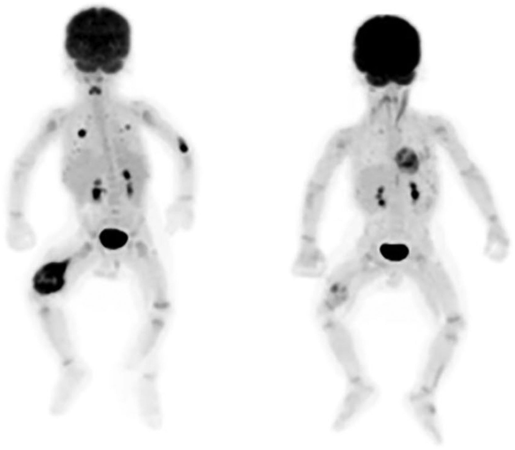
A 7-mo-old male patient was diagnosed with clinical stage 4 NHL. sPET (left image) shows 18F-FDG–avid lesions in the lungs and the right distal femur. Further investigation revealed an infectious process due to Pneumocystis carinii in lungs (18F-FDG uptake in left elbow is site of injection). iPET (right image), obtained after 2 cycles of chemotherapy, shows partial response in lymphoma in right distal femur (Lugano Classification score 4). At the end of treatment, there was a complete response and patient was still alive after 38 mo of follow-up.

FIGURE 4.
  • Download figure
  • Open in new tab
  • Download powerpoint
FIGURE 4.

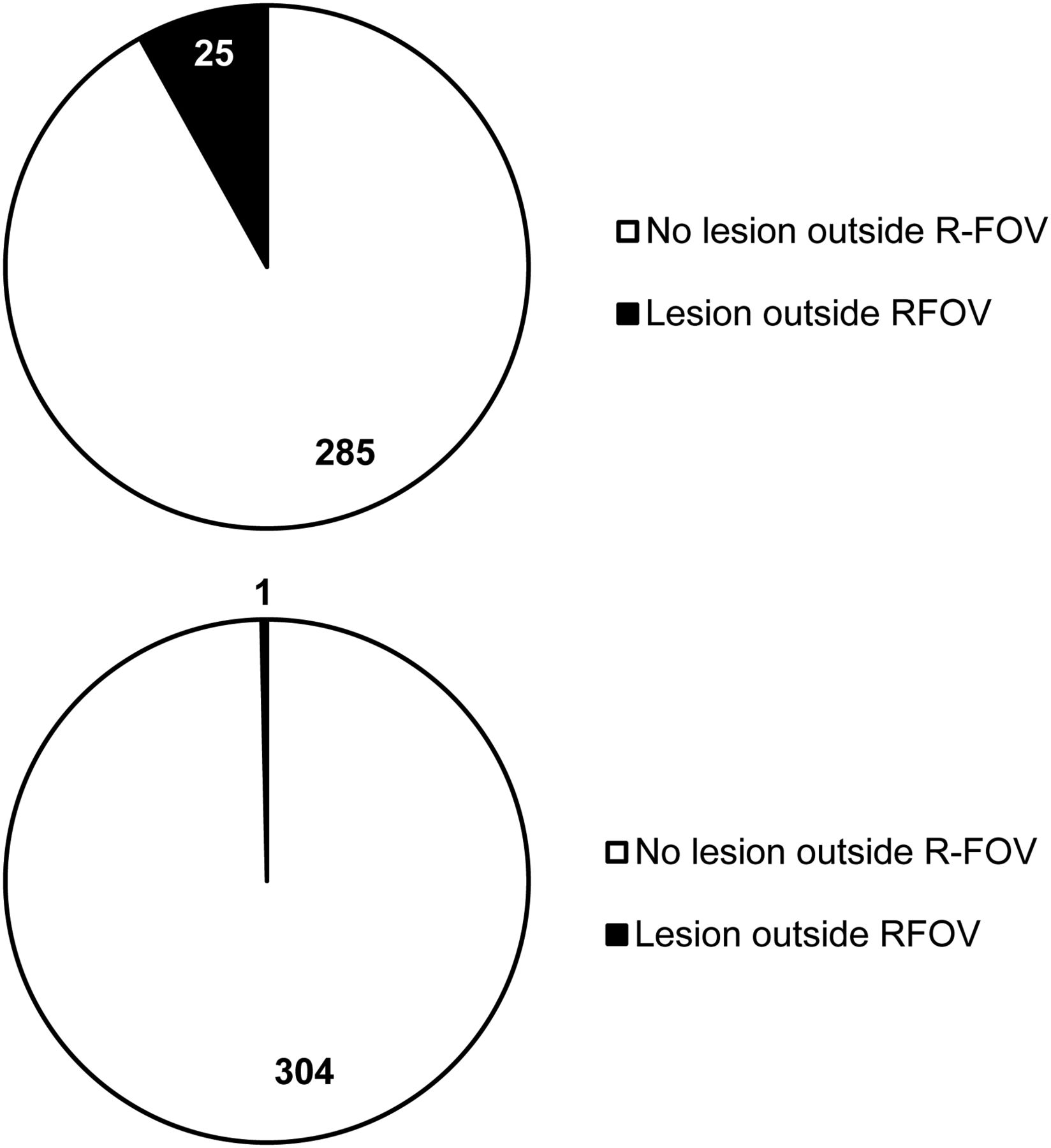
Results of the presence of lesions inside and outside the R-FOV on sPET (A) and iPET (B).

DISCUSSION

In this multicenter international study coordinated by the IAEA, for 610 scans obtained in 305 patients (305 sPET and 305 iPET scans), the sPET scans did not show lesions outside the R-FOV in 92% of the patients. In 8% of the patients, the sPET scans showed lesions outside the R-FOV. These lesions outside the R-FOV were in the soft tissue, bone (2 patients with NHL), bone marrow (23 patients, 9 with HL and 14 NHL), and the skin (1 patient with NHL). There were no changes in the clinical stage of any patient or treatment decision. Although current guidelines for pediatric oncologic 18F-FDG PET/CT empirically suggest use of TWB examinations (8,9), these guidelines and other articles (3,14,26) acknowledge that R-FOV PET/CT is likely to be sufficient for some cases of pediatric lymphoma. Our evidence reinforces that R-FOV PET/CT is sufficient in pediatric lymphoma both at staging and interim assessments.

An R-FOV PET/CT has the advantages of decreased radiation exposure due to a reduction in the area of the body that is irradiated outside of R-FOV. Ionizing radiation has been implicated in conferring a latent increased risk of malignancy (27,28). This is especially important in pediatric patients who, given their age, have longer to manifest these adverse effects (29,30). In pediatric oncology, recent reports have shown a significant cumulative radiation dose in lymphoma patients (31,32). PET/CT applies the “as low as reasonably achievable” principle (ALARA) (33), to both the 18F-FDG dose and the acquisition parameters for the CT scan (34–39). A possibility for decreasing dose is eliminating the CT dose to the extremities and to the brain if an R-FOV PET/CT scan is performed rather than a TWB PET/CT scan. Using published pediatric PET/CT acquisition protocols (40), the R-FOV acquisition imparts a whole-body effective dose of, at most, 5.5 mSv, whereas TWB acquisition imparts 5.7 mSv, resulting in a 5% reduction in whole-body effective dose. In terms of organ-specific doses, compared with the TWB examination, the R-FOV acquisition reduces the CT dose to the brain by 70%, to the skin by 15%, and to the remaining organs (primarily oral mucosa) by 7%.

Another advantage of R-FOV over TWB PET/CT examinations is a shorter scanning time, possibly reducing the risk of movement during the scan (41). Even if the pediatric population represents a reasonably broad spectrum of ages and corresponding sizes, reducing the PET/CT FOV could potentially reduce to half of the scanning time. A shorter imaging time may also increase accessibility of PET/CT examinations in centers at which high demand on facility resources is present. This is a significant consideration in developing countries where there are limited pediatric PET/CT scanners available.

Decreasing imaging time could also result in a similarly decreased length of anesthesia time for those patients requiring sedation. The need for shorter sedation could allow use of a more desirable anesthetic, which would be determined according to pediatric sedation guidelines (42). Depth of sedation may also potentially be reduced, which could result in similar advantages. As we now consider a shift to the use of PET/MRI in children, we will further reduce radiation dose to the child with the CT but there will be a concomitant potential increase in sedation time for the MRI portion of the studies. A Food and Drug Administration drug safety communication stated that there is the probability of increasing neurodevelopmental risks in children younger than 3 y and in women in the third trimester of pregnancy with prolonged and repeated procedural sedation of greater than 3 h (43).

Although our findings challenge conventional clinical practice, for many years consolidated without empiric basis, a similar finding in another study, with a smaller series of cases, supports a reduced FOV practice. Sammer et al. (44) is the only previous paper to have evaluated R-FOV versus TWB-FOV differences in PET/CT studies in 170 lymphoma patients. No disease outside the R-FOV sPET was found on iPET scans in the 150 patients, while 12% of patients had disease outside the R-FOV in staging. In only 1 patient with lymphoblastic NHL was the imaging stage altered with the additional TWB-FOV. They concluded that it was appropriate to perform R-FOV iPET when sPET did not show disease beyond eyes to thighs. Likewise, in our study with a substantially larger cohort, only 8.3% of patients had disease outside the R-FOV in sPET, and also in only 1 case on both sPET and iPET (0.3%) was a clinically relevant bone lesion outside of R-FOV found. It is important to note that this lesion was the primary lesion biopsied in this immunodeficiency related lymphoma patient (DLBCL). The location of his disease would make this case an exception and so this isolated case would have a priori called for a TWB PET scan. This case, although unique, would not be missed because common sense and clinical experience still rule in the clinical decision-making process. Similar to practice in adults, exceptions should probably include cases of primary or suspected extremity involvement (13) for which TWB-FOV PET/CT images are also obtained. Therefore, our results support the use of R-FOV PET/CT not only for iPET but also for sPET.

This paper does not suggest that R-FOV PET/CT should be used in other pediatric tumors. Just as there are certain tumors in the adult population that have more propensities to involve the extremities, tumors such as sarcomas, neuroblastomas, and leukemias are well known to have systemic involvement and TWB PET/CT imaging is still the recommendation in children who may present with these and other tumors, even though there are limited data to support this approach (8,9).

A limitation of our study is our relatively small number of patients with the different subtypes of NHL, even though we obtained 219 PET/CT scans in HL patients and in 86 NHL patients. Whereas HL behaves in a relatively predictable way with well-defined staging methodology, NHL encompasses a relatively heterogeneous group of histologies, many with different morphologies, presentations, and natural histories (45). Consequently, further studies dedicated to NHL or specific types of NHL may be helpful to more precisely define the role of R-FOV PET/CT in these patients. Also, in our study we did not perform dosimetry to determine the percentage reduction in radiation exposure or to determine the exact reduction in scanning time.

Major strengths of this study were that this is the biggest series of cases of pediatric lymphoma patients as far as we know in an international multicenter trial spanning differing socioeconomic layers as defined by the World Bank to include low-middle, upper-middle, and high-income countries.

CONCLUSION

Our results demonstrate that the identification of additional lesions outside the R-FOV (eyes to thighs) 18F-FDG PET/CT has no impact in the definition of the clinical stage of disease and minimal impact in the conduct definition of patients with pediatric lymphoma. As so, R-FOV for both staging and interim 18F-FDG PET/CT scans could be performed. This consideration may be quite impactful in developing countries where there are limited pediatric PET/CT scanners available.

DISCLOSURE

No potential conflict of interest relevant to this article was reported.

Footnotes

  • Published online Jan. 25, 2019.

  • © 2019 by the Society of Nuclear Medicine and Molecular Imaging.

REFERENCES

  1. 1.↵
    1. Murphy JJ,
    2. Tawfeeq M,
    3. Chang B,
    4. Nadel H
    . Early experience with PET/CT scan in the evaluation of pediatric abdominal neoplasms. J Pediatr Surg. 2008;43:2186–2192.
    OpenUrlCrossRefPubMed
  2. 2.↵
    1. Hudson MM,
    2. Krasin MJ,
    3. Kaste SC
    . PET imaging in pediatric Hodgkin’s lymphoma. Pediatr Radiol. 2004;34:190–198.
    OpenUrlCrossRefPubMed
  3. 3.↵
    1. Kleis M,
    2. Daldrup-Link H,
    3. Matthay K,
    4. et al
    . Diagnostic value of PET/CT for the staging and restaging of pediatric tumors. Eur J Nucl Med Mol Imaging. 2009;36:23–36.
    OpenUrlCrossRefPubMed
  4. 4.↵
    1. Riad R,
    2. Omar W,
    3. Kotb M,
    4. et al
    . Role of PET/CT in malignant pediatric lymphoma. Eur J Nucl Med Mol Imaging. 2010;37:319–329.
    OpenUrlCrossRefPubMed
  5. 5.↵
    1. Franzius C,
    2. Juergens KU,
    3. Vormoor J
    . PET/CT with diagnostic CT in the evaluation of childhood sarcoma. AJR. 2006;186:581.
    OpenUrlCrossRefPubMed
  6. 6.↵
    1. McCarville B
    . The role of positron emission tomography in pediatric musculoskeletal oncology. Skeletal Radiol. 2006;35:553–554.
    OpenUrlCrossRefPubMed
  7. 7.↵
    1. Sharp SE,
    2. Shulkin BL,
    3. Gelfand MJ,
    4. Salisbury S,
    5. Furman WL
    . 123I-MIBG scintigraphy and 18F-FDG PET in neuroblastoma. J Nucl Med. 2009;50:1237–1243.
    OpenUrlAbstract/FREE Full Text
  8. 8.↵
    1. Nadel HR,
    2. Shulkin B
    . Pediatric positron emission tomography-computed tomography protocol considerations. Semin Ultrasound CT MR. 2008;29:271–276.
    OpenUrlPubMed
  9. 9.↵
    1. Stauss J,
    2. Franzius C,
    3. Pfluger T,
    4. et al
    . Guidelines for 18F-FDG PET and PET-CT imaging in paediatric oncology. Eur J Nucl Med Mol Imaging. 2008;35:1581–1588.
    OpenUrlCrossRefPubMed
  10. 10.↵
    1. von Mehren M,
    2. Randall RL,
    3. Benjamin RS,
    4. et al
    . Soft tissue sarcoma, version 2.2016, NCCN Clinical Practice Guidelines in Oncology. J Natl Compr Canc Netw. 2016;14:758–786.
    OpenUrlAbstract/FREE Full Text
  11. 11.
    1. Schulte JH,
    2. Eggert A
    . Neuroblastoma. Crit Rev Oncog. 2015;20:245–270.
    OpenUrl
  12. 12.↵
    1. Mauz- Körholz C,
    2. Metzger ML,
    3. Kelly KM,
    4. et al
    . Pediatric Hodgkin lymphoma. J Clin Oncol. 2015;33:2975–2985.
    OpenUrlAbstract/FREE Full Text
  13. 13.↵
    1. Delbeke D,
    2. Coleman RE,
    3. Guiberteau MJ,
    4. et al
    . Procedure guideline for tumor imaging with 18F-FDG PET/CT 1.0. J Nucl Med. 2006;47:885–895.
    OpenUrlFREE Full Text
  14. 14.↵
    1. Shammas A,
    2. Lim R,
    3. Charron M
    . Pediatric FDG PET/CT: physiologic uptake, normal variants, and benign conditions. Radiographics. 2009;29:1467–1486.
    OpenUrlCrossRefPubMed
  15. 15.↵
    1. Plaza JA,
    2. Perez-Montiel D,
    3. Mayerson J,
    4. Morrison C,
    5. Suster S
    . Metastases to soft tissue: a review of 118 cases over a 30-year period. Cancer. 2008;112:193–203.
    OpenUrlPubMed
  16. 16.
    1. Rosenberg SA,
    2. Kaplan HS
    . Evidence for an orderly progression in the spread of Hodgkin’s disease. Cancer Res. 1966;26:1225–1231.
    OpenUrlAbstract/FREE Full Text
  17. 17.
    1. Elstrom R,
    2. Guan L,
    3. Baker G,
    4. et al
    . Utility of FDG-PET scanning in lymphoma by WHO classification. Blood. 2003;101:3875–3876.
    OpenUrlAbstract/FREE Full Text
  18. 18.
    1. Juweid ME,
    2. Stroobants S,
    3. Hoekstra OS,
    4. et al
    . Use of positron emission tomography for response assessment of lymphoma: consensus of the Imaging Subcommittee of International Harmonization Project in Lymphoma. J Clin Oncol. 2007;25:571–578.
    OpenUrlAbstract/FREE Full Text
  19. 19.↵
    1. Tsukamoto N,
    2. Kojima M,
    3. Hasegawa M,
    4. et al
    . The usefulness of 18F-fluorodeoxyglucose positron emission tomography (18F-FDG-PET) and a comparison of 18F-FDG-pet with 67gallium scintigraphy in the evaluation of lymphoma: relation to histologic subtypes based on the World Health Organization classification. Cancer. 2007;110:652–659.
    OpenUrlCrossRefPubMed
  20. 20.↵
    1. Jerusalem G,
    2. Beguin Y,
    3. Najjar F,
    4. et al
    . Positron emission tomography (PET) with 18F-fluorodeoxyglucose (18F-FDG) for the staging of low-grade non-Hodgkin’s lymphoma (NHL). Ann Oncol. 2001;12:825–830.
    OpenUrlCrossRefPubMed
  21. 21.
    1. Karam M,
    2. Novak L,
    3. Cyriac J,
    4. Ali A,
    5. Nazeer T,
    6. Nugent F
    . Role of fluorine-18 fluoro-deoxyglucose positron emission tomography scan in the evaluation and follow-up of patients with low-grade lymphomas. Cancer. 2006;107:175–183.
    OpenUrlCrossRefPubMed
  22. 22.
    1. Teräs M,
    2. Tolvanen T,
    3. Johansson JJ,
    4. Williams JJ,
    5. Knuuti J
    . Performance of the new generation of whole-body PET/CT scanners: Discovery STE and Discovery VCT. Eur J Nucl Med Mol Imaging. 2007;34:1683–1692.
    OpenUrlCrossRefPubMed
  23. 23.↵
    1. Seam P,
    2. Juweid ME,
    3. Cheson BD
    . The role of FDG-PET scans in patients with lymphoma. Blood. 2007;110:3507–3516.
    OpenUrlAbstract/FREE Full Text
  24. 24.↵
    1. Swerdlow SH,
    2. Campo E,
    3. Pileri SA,
    4. Harris NL,
    5. Stein H,
    6. Siebert R,
    7. et al
    . The 2016 revision of the World Health Organization (WHO) classification of lymphoid neoplasms. 2016: Blood-2016-01-643569.
  25. 25.↵
    1. Bolard G,
    2. Prior JO,
    3. Modolo L,
    4. et al
    . Performance comparison of two commercial BGO-based PET/CT scanners using NEMA NU 2-2001. Med Phys. 2007;34:2708–2717.
    OpenUrlCrossRefPubMed
  26. 26.↵
    1. Younes A,
    2. Hilden P,
    3. Coiffier B,
    4. et al
    . International Working Group consensus response evaluation criteria in lymphoma (RECIL 2017). Ann Oncol. 2017;28:1436–1447.
    OpenUrl
  27. 27.↵
    1. Brenner DJ,
    2. Hall EJ
    . Computed tomography–an increasing source of radiation exposure. N Engl J Med. 2007;357:2277–2284.
    OpenUrlCrossRefPubMed
  28. 28.↵
    1. Hall EJ,
    2. Brenner DJ
    . Cancer risks from diagnostic radiology. Br J Radiol. 2008;81:362–378.
    OpenUrlAbstract/FREE Full Text
  29. 29.↵
    1. Brenner DJ
    . Estimating cancer risks from pediatric CT: going from the qualitative to the quantitative. Pediatr Radiol. 2002;32:228-1.
    OpenUrlCrossRefPubMed
  30. 30.↵
    1. Chodick G,
    2. Ronckers CM,
    3. Shalev V,
    4. Ron E
    . Excess lifetime cancer mortality risk attributable to radiation exposure from computed tomography examinations in children. Isr Med Assoc J. 2007;9:584–587.
    OpenUrlPubMed
  31. 31.↵
    1. Chawla SC,
    2. Federman N,
    3. Zhang D,
    4. et al
    . Estimated cumulative radiation dose from PET/CT in children with malignancies: a 5-year retrospective review. Pediatr Radiol. 2010;40:681–686.
    OpenUrlCrossRefPubMed
  32. 32.↵
    1. Gelfand MJ,
    2. Sharp SE,
    3. Treves ST,
    4. Fahey FH,
    5. Parisi MT,
    6. Alessio AM
    . Estimated cumulative radiation dose from PET/CT in children with malignancies. Pediatr Radiol. 2010;40:1712–1713.
    OpenUrlCrossRefPubMed
  33. 33.↵
    1. Voss SD,
    2. Reaman GH,
    3. Kaste SC,
    4. Slovis TL
    . The ALARA concept in pediatric oncology. Pediatr Radiol. 2009;39:1142–1146.
    OpenUrlCrossRefPubMed
  34. 34.↵
    1. Fahey FH,
    2. Ziniel SI,
    3. Manion D,
    4. Baker A,
    5. Treves ST
    . Administered activities in pediatric nuclear medicine and the impact of the 2010 North American consensus guidelines on general hospitals in the United States. J Nucl Med. 2016;57:1478–1485.
    OpenUrlAbstract/FREE Full Text
  35. 35.
    1. Gelfand MJ,
    2. Parisi MT,
    3. Treves ST,
    4. Pediatric Nuclear Medicine Dose Reduction Workgroup
    . Pediatric radiopharmaceutical administered doses: 2010 North American consensus guidelines. J Nucl Med. 2011;52:318–322.
    OpenUrlFREE Full Text
  36. 36.
    1. Lassmann M,
    2. Treves ST,
    3. Group ESPDHW
    . Paediatric radiopharmaceutical administration: harmonization of the 2007 EANM paediatric dosage card (version 1.5.2008) and the 2010 North American consensus guidelines. Eur J Nucl Med Mol Imaging. 2014;41:1036–1041.
    OpenUrlCrossRefPubMed
  37. 37.
    1. Treves ST,
    2. Gelfand MJ,
    3. Fahey FH,
    4. Parisi MT
    . 2016 Update of the North American consensus guidelines for pediatric administered radiopharmaceutical activities. J Nucl Med. 2016;57:15N–18N.
    OpenUrlFREE Full Text
  38. 38.
    1. Warbey VS,
    2. Schleyer PJ,
    3. Barrington SF,
    4. O’Doherty MJ
    . The new EANM paediatric dosage card: does it conform to ALARA for PET/CT? Eur J Nucl Med Mol Imaging. 2007;34:1881–1882.
    OpenUrlCrossRefPubMed
  39. 39.↵
    1. Shrimpton PC,
    2. Jones DG
    . Normalised organ doses for x ray computed tomography calculated using Monte Carlo techniques and a mathematical anthropomorphic phantom. Radiat Prot Dosimetry. 1993;49:241–243.
    OpenUrl
  40. 40.↵
    1. Alessio AM,
    2. Kinahan PE,
    3. Manchanda V,
    4. Ghioni V,
    5. Aldape L,
    6. Parisi MT
    . Weight-based, low-dose pediatric whole-body PET/CT protocols. J Nucl Med. 2009;50:1570–1577.
    OpenUrlAbstract/FREE Full Text
  41. 41.↵
    1. Brix G,
    2. Lechel U,
    3. Glatting G,
    4. et al
    . Radiation exposure of patients undergoing whole-body dual-modality 18F-FDG PET/CT examinations. J Nucl Med. 2005;46:608–613.
    OpenUrlAbstract/FREE Full Text
  42. 42.↵
    American Academy of Pediatrics, American Academy of Pediatric Dentistry, Coté CJ, Wilson S, Work Group on Sedation. Guidelines for monitoring and management of pediatric patients during and after sedation for diagnostic and therapeutic procedures: an update. Paediatr Anaesth. 2008;18:9–10.
  43. 43.↵
    1. Gleich SJ,
    2. Flick R,
    3. Hu D,
    4. et al
    . Neurodevelopment of children exposed to anesthesia: design of the Mayo Anesthesia Safety in Kids (MASK) study. Contemp Clin Trials. 2015;41:45–54.
    OpenUrl
  44. 44.↵
    1. Sammer MB,
    2. Shulkin BL,
    3. Alessio A,
    4. Parisi MT
    . Role of limited whole-body PET/CT in pediatric lymphoma. AJR. 2011;196:1047–1055.
    OpenUrlCrossRefPubMed
  45. 45.↵
    1. Zucca E,
    2. Roggero E,
    3. Bertoni F,
    4. Cavalli F
    . Primary extranodal non-Hodgkin’s lymphomas: part 1—gastrointestinal, cutaneous and genitourinary lymphomas. Ann Oncol. 1997;8:727–737.
    OpenUrlCrossRefPubMed
  • Received for publication October 23, 2018.
  • Accepted for publication December 19, 2018.
PreviousNext
Back to top

In this issue

Journal of Nuclear Medicine: 60 (8)
Journal of Nuclear Medicine
Vol. 60, Issue 8
August 1, 2019
  • Table of Contents
  • Table of Contents (PDF)
  • About the Cover
  • Index by author
Print
Download PDF
Article Alerts
Sign In to Email Alerts with your Email Address
Email Article

Thank you for your interest in spreading the word on Journal of Nuclear Medicine.

NOTE: We only request your email address so that the person you are recommending the page to knows that you wanted them to see it, and that it is not junk mail. We do not capture any email address.

Enter multiple addresses on separate lines or separate them with commas.
Is True Whole-Body 18F-FDG PET/CT Required in Pediatric Lymphoma? An IAEA Multicenter Prospective Study
(Your Name) has sent you a message from Journal of Nuclear Medicine
(Your Name) thought you would like to see the Journal of Nuclear Medicine web site.
Citation Tools
Is True Whole-Body 18F-FDG PET/CT Required in Pediatric Lymphoma? An IAEA Multicenter Prospective Study
Juliano J. Cerci, Elba C. Etchebehere, Helen Nadel, Anita Brink, Chandrasekhar S. Bal, Venkatesh Rangarajan, Thomas Pfluger, Olga Kagna, Omar Alonso, Fatima K. Begum, Kahkashan Bashir Mir, Vincent P. Magboo, Leon J. Menezes, Diana Paez, Thomas N. Pascual
Journal of Nuclear Medicine Aug 2019, 60 (8) 1087-1093; DOI: 10.2967/jnumed.118.222299

Citation Manager Formats

  • BibTeX
  • Bookends
  • EasyBib
  • EndNote (tagged)
  • EndNote 8 (xml)
  • Medlars
  • Mendeley
  • Papers
  • RefWorks Tagged
  • Ref Manager
  • RIS
  • Zotero
Share
Is True Whole-Body 18F-FDG PET/CT Required in Pediatric Lymphoma? An IAEA Multicenter Prospective Study
Juliano J. Cerci, Elba C. Etchebehere, Helen Nadel, Anita Brink, Chandrasekhar S. Bal, Venkatesh Rangarajan, Thomas Pfluger, Olga Kagna, Omar Alonso, Fatima K. Begum, Kahkashan Bashir Mir, Vincent P. Magboo, Leon J. Menezes, Diana Paez, Thomas N. Pascual
Journal of Nuclear Medicine Aug 2019, 60 (8) 1087-1093; DOI: 10.2967/jnumed.118.222299
Twitter logo Facebook logo LinkedIn logo Mendeley logo
  • Tweet Widget
  • Facebook Like
  • Google Plus One
Bookmark this article

Jump to section

  • Article
    • Abstract
    • MATERIALS AND METHODS
    • RESULTS
    • DISCUSSION
    • CONCLUSION
    • DISCLOSURE
    • Footnotes
    • REFERENCES
  • Figures & Data
  • Info & Metrics
  • PDF

Related Articles

  • This Month in JNM
  • PubMed
  • Google Scholar

Cited By...

  • No citing articles found.
  • Google Scholar

More in this TOC Section

Oncology

  • Role of PET/CT in the management of multiple myeloma
  • FDG-PET/CT and NaF-PET/CT in the diagnosis and assessment of radiation therapy-induced vascular complications in patients with head and neck cancer
  • 18F-FDG PET/CT manifestations of three Cases of Female Desmoplastic Small Round Cell Tumor
Show more Oncology

Clinical

  • Role of PET/CT in the management of multiple myeloma
  • FDG-PET/CT and NaF-PET/CT in the diagnosis and assessment of radiation therapy-induced vascular complications in patients with head and neck cancer
  • 18F-FDG PET/CT manifestations of three Cases of Female Desmoplastic Small Round Cell Tumor
Show more Clinical

Similar Articles

Keywords

  • 18F-FDG PET/CT
  • pediatric
  • lymphoma
  • Hodgkin’s lymphoma
  • Non-Hodgkin’s lymphoma
SNMMI

© 2025 SNMMI

Powered by HighWire