


Article Figures & Data
Figures
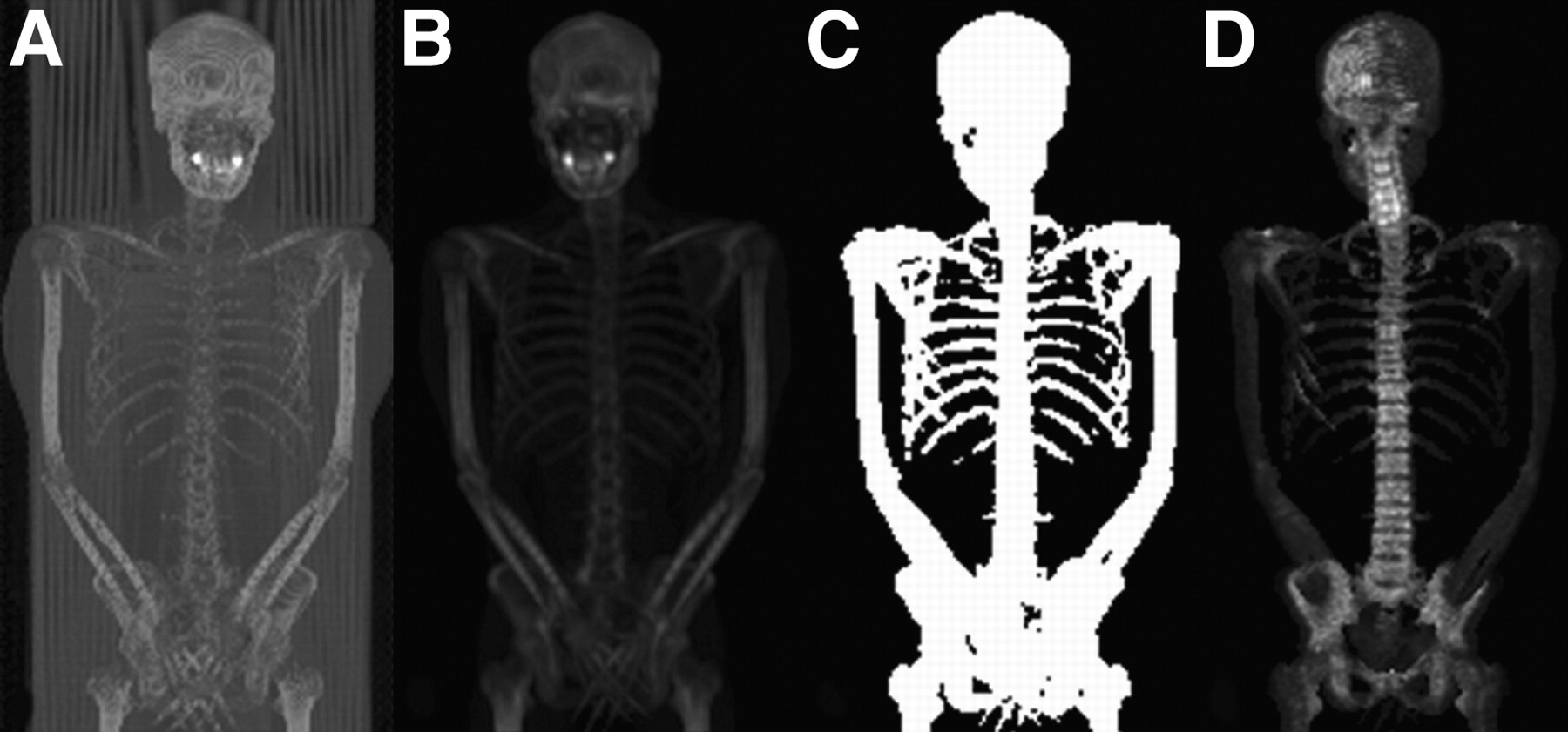
- FIGURE 1.
(A) The first step is to reformat CT image to dimensions of PET image. (B) Bed is eliminated by indicating most dependent part of body as image limit. (C) CT image is interactively thresholded to eliminate all soft tissue but keep bone densities. (D) PET image multiplied by mask leaves bone image with combined 18F/18F-FDG uptake.
- FIGURE 2.
A 44-y-old man with soft-tissue sarcoma. (A) MIP image of 18F-FDG PET shows normal radiotracer uptake. (B) MIP image of 18F PET shows intense radiotracer uptake in skull lesion (arrow) and in T10 vertebra and right pubis (arrowheads). (C) Skull lesion is missed on MIP image of combined 18F/18F-FDG PET, but MIP image of combined 18F/18F-FDG PET shows skeletal lesions in T10 vertebra and right pubis noted on 18F PET (arrowheads). Skull lesion (arrow) is seen on transaxial CT (D) and 18F PET (E) but not on combined 18F/18F-FDG PET (F). Lesion in T10 vertebra (arrowhead) is seen on transaxial CT (G), 18F PET (H), and combined 18F/18F-FDG PET (I).
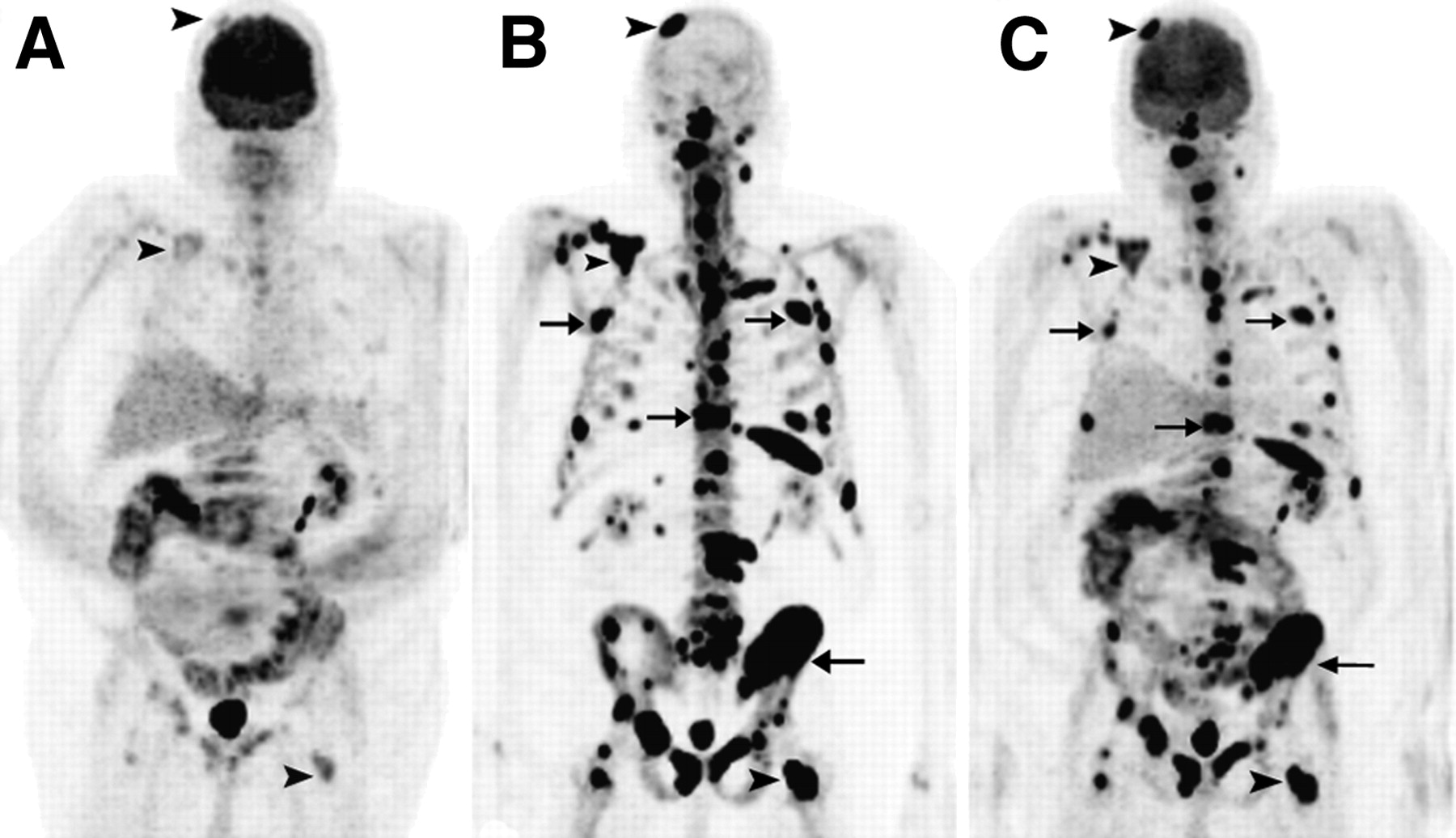
- FIGURE 3.
A 68-y-old man with colon cancer. (A) MIP image of 18F-FDG PET shows faint radiotracer uptake in several skeletal lesions (arrowheads). (B) MIP image of 18F PET shows intense radiotracer uptake in multiple bone lesions, including better visualization of lesions seen on 18F-FDG PET (arrowheads) and more extensive skeletal metastases (arrows). (C) MIP image of combined 18F/18F-FDG PET shows skeletal lesions noted on 18F PET (arrowheads).
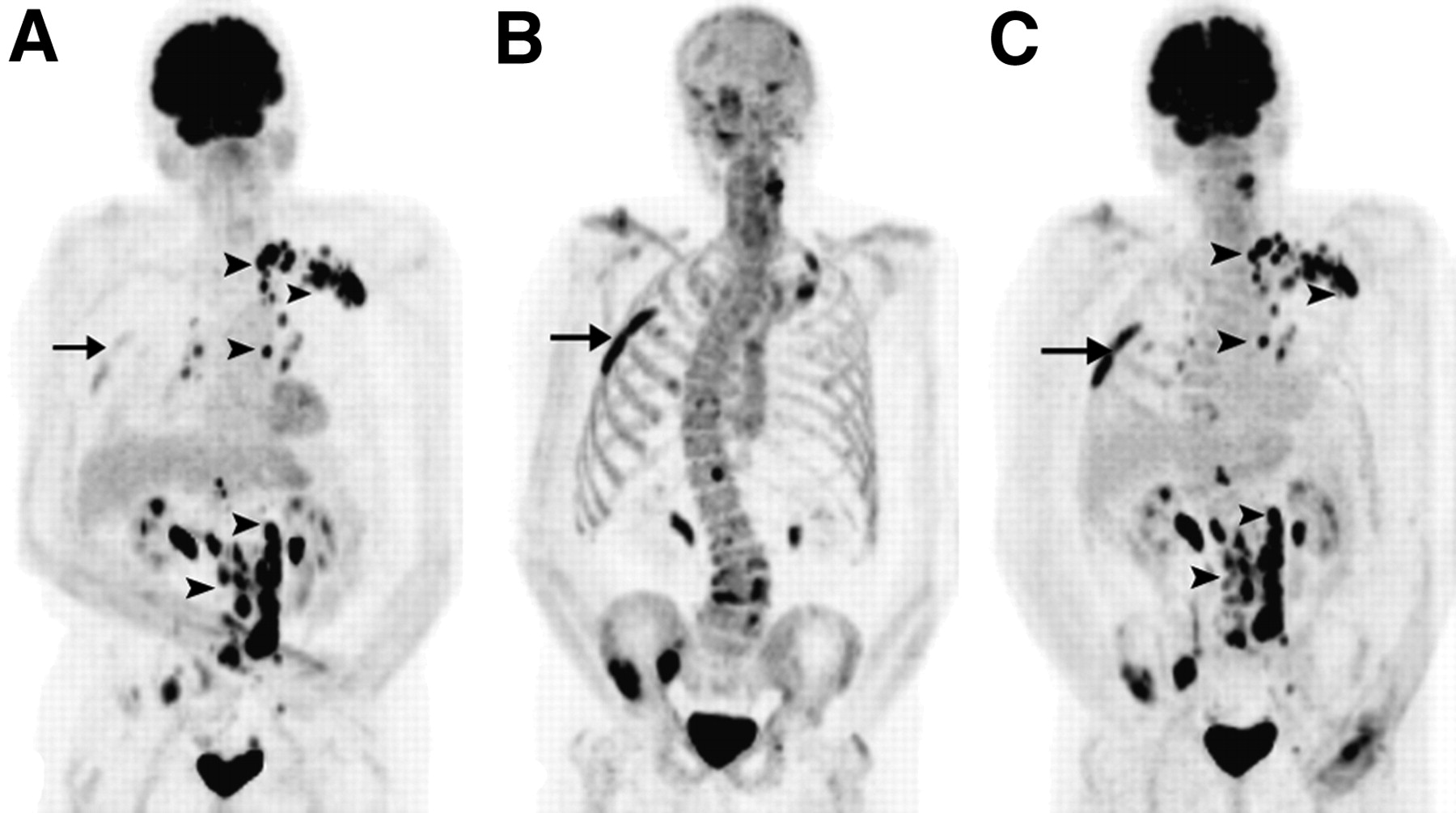
- FIGURE 4.
A 75-y-old man with prostate cancer. (A) MIP image of 18F-FDG PET shows lymph node metastases (arrowheads) and faint uptake in osseous lesions, such as a right rib (arrow). (B) MIP image of 18F PET shows intense radiotracer uptake in multiple bone lesions, including right rib lesion (arrow) seen on 18F-FDG PET. (C) MIP image of combined 18F/18F-FDG PET shows both lesions noted on 18F-FDG PET (arrowheads) and skeletal lesions noted on 18F PET (reference right rib lesion marked with arrow).


Tables
Age (y) Cancer 18F-FDG findings 18F findings Cocktail findings Cocktail vs. 18F Cocktail vs. 18F-FDG 75 Prostate Ribs, pelvis, femur, LNs Skull, ribs, T/L, pelvis, femur Skull, ribs, T/L, pelvis, femur, LNs Equal Equal 59 Lung LUL nodule, LN Negative LUL nodule, LN Equal Equal 65 Prostate Pelvic LNs Negative Pelvic LNs Equal Equal 68 Colon Liver, scapula, C/T/L, pelvis Skull, scapula, ribs, C/T/L, pelvis, femurs Liver, skull, scapula, ribs, C/T/L, pelvis, femurs Equal Equal 31 Sarcoma Right thigh, B/L lung nodules Negative Right thigh, B/L lung nodules Equal Equal 44 Sarcoma Soft-tissue mass Skull, T10, pubis Soft-tissue mass, T10, pubis Lesion missed in the skull on cocktail Equal 41 Sarcoma Right femur Right femur Right femur Equal Equal 70 Prostate Negative Scapula, ribs Scapula, ribs Equal Equal 55 Breast Negative Negative Negative Equal Equal 55 Breast Liver Negative Liver Equal Equal 19 Sarcoma Rib, soft-tissue mass Rib Rib Equal Equal 63 Sarcoma Negative Negative Negative Equal Equal 30 Sarcoma Left gluteus Negative Left gluteus Equal Equal 30 Paraganglioma Soft-tissue mass, skull, scapula, ribs, C/T/L, humerus, pelvis, femurs Skull, scapula, ribs, C/T/L, humerus, pelvis, femurs Soft-tissue mass, skull, scapula, ribs, C/T/L, humerus, pelvis, femurs Equal Equal LNs = lymph nodes; T/L = thoracic and lumbar spine; LUL = left upper lung; C/T/L = cervical, thoracic, and lumbar spine; B/L = bilateral.
Supplemental Data
Files in this Data Supplement:


Jump to section
Related Articles
Cited By...
- 18F-Sodium Fluoride PET: History, Technical Feasibility, Mechanism of Action, Normal Biodistribution, and Diagnostic Performance in Bone Metastasis Detection Compared with Other Imaging Modalities
- Nanobody-Facilitated Multiparametric PET/MRI Phenotyping of Atherosclerosis
- The Role of 18F-Sodium Fluoride PET/CT Bone Scans in the Diagnosis of Metastatic Bone Disease from Breast and Prostate Cancer
- Bone-Targeted Imaging and Radionuclide Therapy in Prostate Cancer
- Prospective Comparison of 99mTc-MDP Scintigraphy, Combined 18F-NaF and 18F-FDG PET/CT, and Whole-Body MRI in Patients with Breast and Prostate Cancer
- Semiquantitative Analysis of the Biodistribution of the Combined 18F-NaF and 18F-FDG Administration for PET/CT Imaging
- An Approach to Breast Cancer Diagnosis via PET Imaging of Microcalcifications Using 18F-NaF
- Combined 18F-Fluoride and 18F-FDG PET/CT Scanning for Evaluation of Malignancy: Results of an International Multicenter Trial
- Combined 18F-Fluoride and 18F-FDG PET/CT Scanning for Evaluation of Malignancy: Results of an International Multicenter Trial
- Proof-of-Concept Study of Monitoring Cancer Drug Therapy with Cerenkov Luminescence Imaging
- Correlation of Inflammation Assessed by 18F-FDG PET, Active Mineral Deposition Assessed by 18F-Fluoride PET, and Vascular Calcification in Atherosclerotic Plaque: A Dual-Tracer PET/CT Study
- SNM Practice Guideline for Sodium 18F-Fluoride PET/CT Bone Scans 1.0
- Combined 18F-FDG and Fluoride Approach in PET/CT Imaging: Is There a Clinical Future?
- Combined 18F-FDG and Fluoride Approach in PET/CT Imaging: Is There a Clinical Future?
- Reply: Combined 18F-FDG and Fluoride Approach in PET/CT Imaging: Is There a Clinical Future?