Main menu

  • Home
  • Content
    • Current
    • Ahead of print
    • Past Issues
    • JNM Supplement
    • SNMMI Annual Meeting Abstracts
  • Subscriptions
    • Subscribers
    • Institutional and Non-member
    • Rates
    • Corporate & Special Sales
    • Journal Claims
  • Authors
    • Submit to JNM
    • Information for Authors
    • Assignment of Copyright
    • AQARA requirements
  • Info
    • Permissions
    • Advertisers
    • Continuing Education
  • About
    • About Us
    • Editorial Board
    • Contact Information
  • More
    • Alerts
    • Feedback
    • Help
    • SNMMI Journals
  • SNMMI
    • JNM
    • JNMT
    • SNMMI Journals
    • SNMMI

User menu

  • Subscribe
  • My alerts
  • Log in
  • My Cart

Search

  • Advanced search
Journal of Nuclear Medicine
  • SNMMI
    • JNM
    • JNMT
    • SNMMI Journals
    • SNMMI
  • Subscribe
  • My alerts
  • Log in
  • My Cart
Journal of Nuclear Medicine

Advanced Search

  • Home
  • Content
    • Current
    • Ahead of print
    • Past Issues
    • JNM Supplement
    • SNMMI Annual Meeting Abstracts
  • Subscriptions
    • Subscribers
    • Institutional and Non-member
    • Rates
    • Corporate & Special Sales
    • Journal Claims
  • Authors
    • Submit to JNM
    • Information for Authors
    • Assignment of Copyright
    • AQARA requirements
  • Info
    • Permissions
    • Advertisers
    • Continuing Education
  • About
    • About Us
    • Editorial Board
    • Contact Information
  • More
    • Alerts
    • Feedback
    • Help
    • SNMMI Journals
  • Follow JNM on Twitter
  • Visit JNM on Facebook
  • Join JNM on LinkedIn
  • Subscribe to our RSS feeds
OtherThis Month in JNM

This Month in JNM

Journal of Nuclear Medicine April 2009, 50 (4) 11A-12A;
  • Article
  • Info & Metrics
  • PDF
Loading

Connecting the quantum dots: Bentolila and colleagues provide an overview of the growing contributions of quantum dots to in vivo molecular imaging in small-animal models and describe the possibilities for clinical applications and novel hybrid approaches.
Page 493

Figure

Toward routine vulnerable plaque imaging: Fox and Strauss offer a perspective on the current status of metabolic imaging of vulnerable plaque in coronary arteries and preview an article on this topic in this issue of JNM.
Page 497

Figure

Testing a PET/CT cocktail: Iagaru and colleagues report on the combined administration of 18F and 18F-FDG in a single PET/CT study for cancer detection in a range of malignancies.
Page 501

Figure

PET/CT evaluation of ascites: Zhang and colleagues compare 18F-FDG PET/CT with CT alone in determining the unknown primary cause of ascites and in detecting abdominal cavity metastases.
Page 506

Figure

18F-FDOPA PET for pheochromocytomas: Imani and colleagues investigate the sensitivity and specificity of PET with18F-FDOPA as an independent marker for diagnosis and localization of benign and malignant pheochromocytomas.
Page 513

Figure

Segmented attenuation maps for PET/MRI: Martinez-Möller and colleagues propose and evaluate a potential solution to the challenge of attenuation correction of whole-body PET data in combined PET/MRI.
Page 520

Figure

PET in diffuse large B-cell lymphoma: Itti and colleagues determine whether semiquantification of standardized uptake values improves the prognostic value of 18F-FDG PET after 4 cycles of chemotherapy for diffuse large B-cell lymphoma.
Page 527

Figure

Annexin imaging of joint prostheses: Lorberboym and colleagues explore the use of 99mTc-recombinant human annexin V imaging for differential diagnosis of aseptic loosening and low-grade infection in hip and knee prostheses.
Page 534

Figure

PET/CT in chronic lung disease: Groves and colleagues evaluate integrated 18F-FDG PET/CT in idiopathic pulmonary fibrosis and other forms of diffuse parenchymal lung disease and assess patterns of tracer metabolism in these patients.
Page 538

Figure

SPECT in stable ischemic heart disease: Gimelli and colleagues compare the capabilities of gated stress/rest myocardial perfusion SPECT with those of a complete diagnostic work-up and other indicators in predicting cardiac events in patients with stable ischemic heart disease.
Page 546

Half-time SPECT MPI with AC: Ali and colleagues describe a new algorithm and postprocessing technique facilitating half-time gated myocardial SPECT perfusion imaging with attenuation correction.
Page 554

PET/CT imaging of coronary plaque: Wykrzykowska and colleagues report on 18F-FDG PET/CT imaging of inflamed and vulnerable plaque in coronary arteries using a low-carbohydrate, high-fat preparation in patients with suppression of myocardial uptake.
Page 563

Figure

Radiotracer breast cancer imaging: Lee and colleagues provide the first in a 2-part educational overview of current and future radiotracer imaging methods for breast cancer in the context of management strategies and nonnuclear imaging approaches.
Page 569

Figure

Antisense-mediated Auger cytotoxicity: Liu and colleagues evaluate a novel 3-component streptavidin-delivery nanoparticle capable of tumor targeting, transmembrane transport, 111In nuclear migration, and specific radiation-induced cytotoxicity.
Page 582

Figure

Side-chain coordination in HYNIC peptides: King and colleagues determine the effects of assisted coordination by amino acids, such as histidine and glutamic acid, on the function of 99mTc-labeled gastrin peptide–HYNIC conjugates in targeting cholecystokinin-R in small-animal models.
Page 591

U-SPECT-II rodent imaging system: van der Have and colleagues describe a new SPECT device that enables both molecular imaging of murine organs down to resolutions of less than 0.5 mm and high-resolution total-body imaging.
Page 599

Figure

PET imaging with anti-PSMA antibody: Elsässer-Beile and colleagues elucidate the in vivo behavior and tumor uptake of a radiolabeled anti–prostate-specific membrane antigen monoclonal antibody and review its potential as a PET tracer.
Page 606

MMP imaging in atherosclerotic mice: Ohshima and colleagues detail a strategy for noninvasive assessment of the extent of matrix metalloproteinase expression in various transgenic mouse models of atherosclerosis.
Page 612

Figure

Fibronectin stimulates 18F-FDG uptake: Paik and colleagues investigate the mechanisms by which matrix fibronectin influences endothelial cell glucose uptake and describe the signaling pathways that mediate this effect.
Page 618

LLP2A tumor targeting: DeNardo and colleagues review the design, synthesis, radiolabeling, and in vivo pharmacokinetics of derivatives of a high-affinity, high-specificity peptidomimetic ligand that binds the activated α4β1 integrin found on a variety of malignant lymphoid cell lines.
Page 625

Figure

Solid-state single-photon imaging: Gambhir and colleagues report on the development and validation, including patient scanning, of a new solid-state single-photon γ-camera and compare it with a conventional SPECT Anger camera.
Page 635

Figure

Ibritumomab tiuxetan dose estimate: Fisher and colleagues evaluate organ radiation absorbed doses from intravenously administered 111In- and 90Y-ibritumomab tiuxetan, as published in MIRD Dose Estimate Report No. 20.
Page 644

Figure

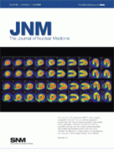
ON THE COVER

This rest/stress 99mTc-tetrofosmin SPECT study compares 2 acquisition methods. At top is a full-time acquisition reconstructed with ordered-subset expectation maximization without attenuation correction; at bottom is a half-time acquisition reconstructed with ordered-subset expectation maximization and resolution recovery with attenuation correction. No difference in image quality or clinical diagnosis was observed in 96% of cases between the 2 methods.

See Page 557.

Figure
PreviousNext
Back to top

In this issue

Journal of Nuclear Medicine: 50 (4)
Journal of Nuclear Medicine
Vol. 50, Issue 4
April 2009
  • Table of Contents
  • Table of Contents (PDF)
  • About the Cover
  • Index by author
Print
Download PDF
Article Alerts
Sign In to Email Alerts with your Email Address
Email Article

Thank you for your interest in spreading the word on Journal of Nuclear Medicine.

NOTE: We only request your email address so that the person you are recommending the page to knows that you wanted them to see it, and that it is not junk mail. We do not capture any email address.

Enter multiple addresses on separate lines or separate them with commas.
This Month in JNM
(Your Name) has sent you a message from Journal of Nuclear Medicine
(Your Name) thought you would like to see the Journal of Nuclear Medicine web site.
Citation Tools
This Month in JNM
Journal of Nuclear Medicine Apr 2009, 50 (4) 11A-12A;

Citation Manager Formats

  • BibTeX
  • Bookends
  • EasyBib
  • EndNote (tagged)
  • EndNote 8 (xml)
  • Medlars
  • Mendeley
  • Papers
  • RefWorks Tagged
  • Ref Manager
  • RIS
  • Zotero
Share
This Month in JNM
Journal of Nuclear Medicine Apr 2009, 50 (4) 11A-12A;
Twitter logo Facebook logo LinkedIn logo Mendeley logo
  • Tweet Widget
  • Facebook Like
  • Google Plus One
Bookmark this article

Jump to section

  • Article
    • ON THE COVER
  • Info & Metrics
  • PDF

Related Articles

  • Quantum Dots for In Vivo Small-Animal Imaging
  • One Step Closer to Imaging Vulnerable Plaque in the Coronary Arteries
  • Novel Strategy for a Cocktail 18F-Fluoride and 18F-FDG PET/CT Scan for Evaluation of Malignancy: Results of the Pilot-Phase Study
  • The Role of 18F-FDG PET/CT in the Evaluation of Ascites of Undetermined Origin
  • 18F-FDOPA PET and PET/CT Accurately Localize Pheochromocytomas
  • Tissue Classification as a Potential Approach for Attenuation Correction in Whole-Body PET/MRI: Evaluation with PET/CT Data
  • Prognostic Value of Interim 18F-FDG PET in Patients with Diffuse Large B-Cell Lymphoma: SUV-Based Assessment at 4 Cycles of Chemotherapy
  • The Use of 99mTc-Recombinant Human Annexin V Imaging for Differential Diagnosis of Aseptic Loosening and Low-Grade Infection in Hip and Knee Prostheses
  • Idiopathic Pulmonary Fibrosis and Diffuse Parenchymal Lung Disease: Implications from Initial Experience with 18F-FDG PET/CT
  • Stress/Rest Myocardial Perfusion Abnormalities by Gated SPECT: Still the Best Predictor of Cardiac Events in Stable Ischemic Heart Disease
  • Half-Time SPECT Myocardial Perfusion Imaging with Attenuation Correction
  • Imaging of Inflamed and Vulnerable Plaque in Coronary Arteries with 18F-FDG PET/CT in Patients with Suppression of Myocardial Uptake Using a Low-Carbohydrate, High-Fat Preparation
  • The Role of Radiotracer Imaging in the Diagnosis and Management of Patients with Breast Cancer: Part 1—Overview, Detection, and Staging
  • Auger Radiation–Induced, Antisense-Mediated Cytotoxicity of Tumor Cells Using a 3-Component Streptavidin-Delivery Nanoparticle with 111In
  • 99mTc-HYNIC-Gastrin Peptides: Assisted Coordination of 99mTc by Amino Acid Side Chains Results in Improved Performance Both In Vitro and In Vivo
  • U-SPECT-II: An Ultra-High-Resolution Device for Molecular Small-Animal Imaging
  • PET Imaging of Prostate Cancer Xenografts with a Highly Specific Antibody against the Prostate-Specific Membrane Antigen
  • Molecular Imaging of Matrix Metalloproteinase Expression in Atherosclerotic Plaques of Mice Deficient in Apolipoprotein E or Low-Density-Lipoprotein Receptor
  • Fibronectin Stimulates Endothelial Cell 18F-FDG Uptake Through Focal Adhesion Kinase–Mediated Phosphatidylinositol 3-Kinase/Akt Signaling
  • 111In-LLP2A-DOTA Polyethylene Glycol–Targeting α4β1 Integrin: Comparative Pharmacokinetics for Imaging and Therapy of Lymphoid Malignancies
  • A Novel High-Sensitivity Rapid-Acquisition Single-Photon Cardiac Imaging Camera
  • MIRD Dose Estimate Report No. 20: Radiation Absorbed-Dose Estimates for 111In- and 90Y-Ibritumomab Tiuxetan
  • Google Scholar

Cited By...

  • No citing articles found.
  • Google Scholar

More in this TOC Section

  • This Month in JNM
  • This Month in JNM
  • This Month in JNM
Show more This Month in JNM

Similar Articles

SNMMI

© 2023 Journal of Nuclear Medicine

Powered by HighWire