


Article Figures & Data
Figures
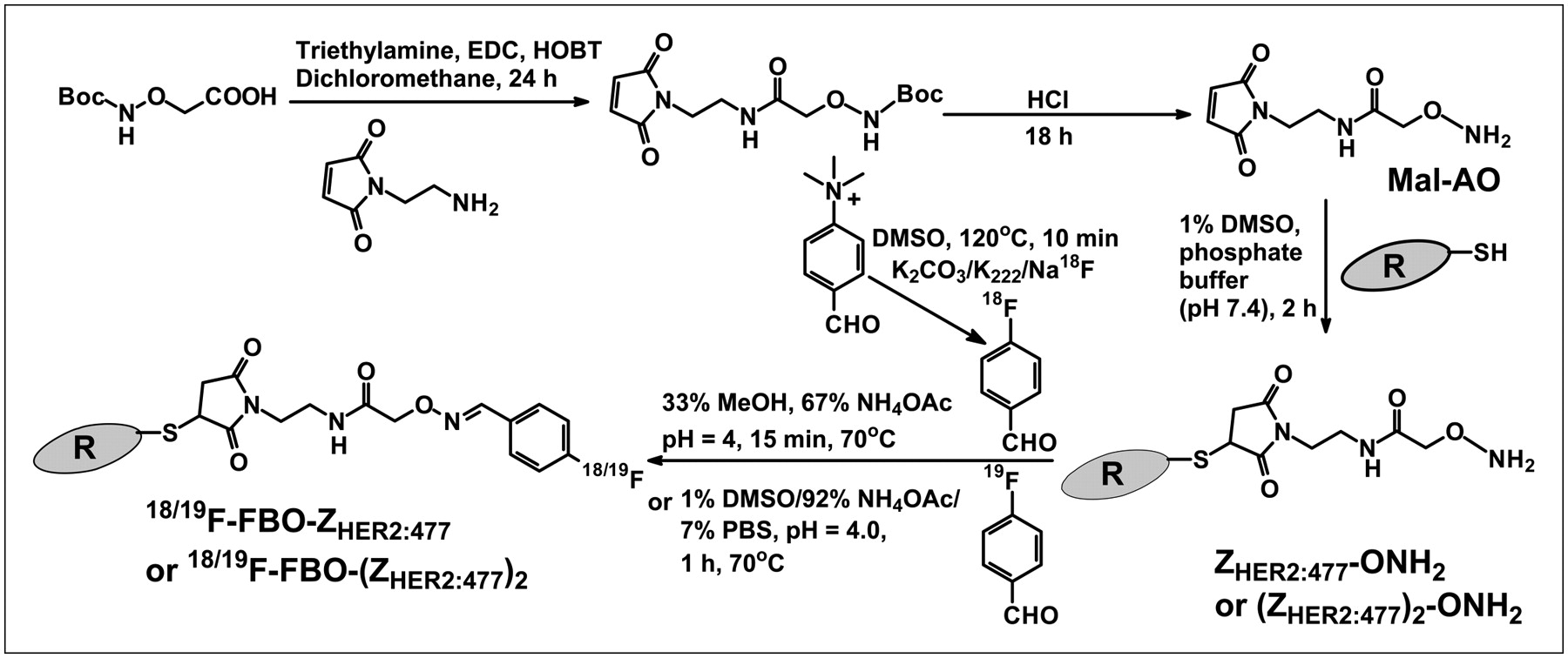
- FIGURE 1.
Synthetic schemes for bifunctional linker Mal-AO, ZHER2:477-ONH2, (ZHER2:477)2-ONH2, 18F-FBO-ZHER2:477, and 18F-FBO-(ZHER2:477)2. Boc = tert-butoxycarbonyl; DMSO = dimethyl sulfoxide; EDC = 1-ethyl-3-(3-dimethylaminopropyl)carbodiimide; HOBT = N-hydroxybenzotriazole; MeOH = methanol; NH4OAc = ammonium acetate; PBS = phosphate-buffered saline.
- FIGURE 2.
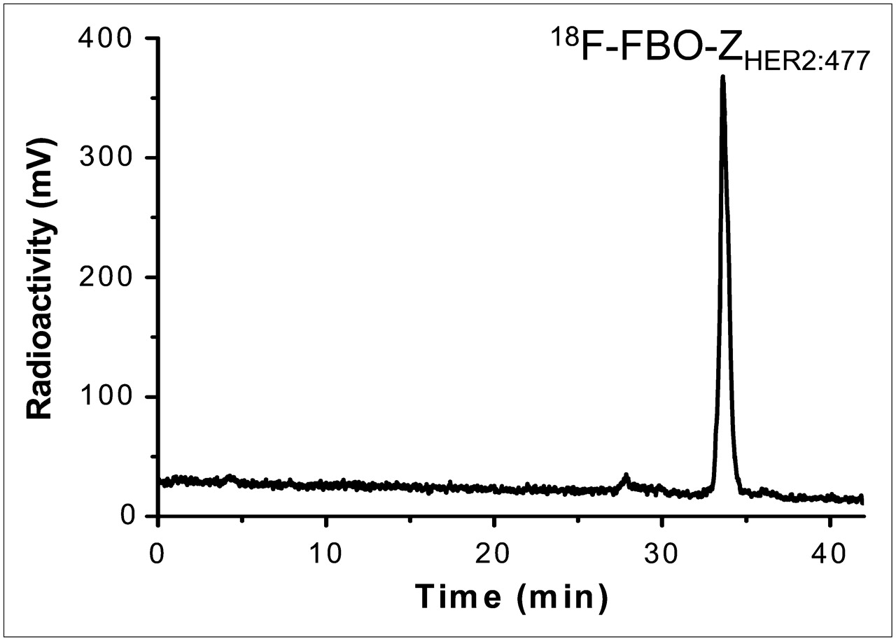
HPLC radiochromatogram of purified 18F-FBO-ZHER2:477.
- FIGURE 3.
Biosensor binding studies of ZHER2:477 (A) and FBO-(ZHER2:477)2 (B). Sensorgrams were obtained after injection of various concentrations of purified Affibody molecules onto sensor chip flow cell surface containing amine-coupled Fc–HER2 chimeric protein. Resp. Diff. = response difference.
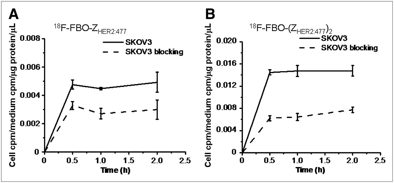
- FIGURE 4.
Uptake of 18F-FBO-ZHER2:477 (A) and 18F-FBO-(ZHER2:477)2 (B) in SKOV3 tumors over time at 37°C in presence or absence of nonradioactive Affibody molecules. All results, expressed as percentage of activity, are mean of triplicate measurements ± SD.
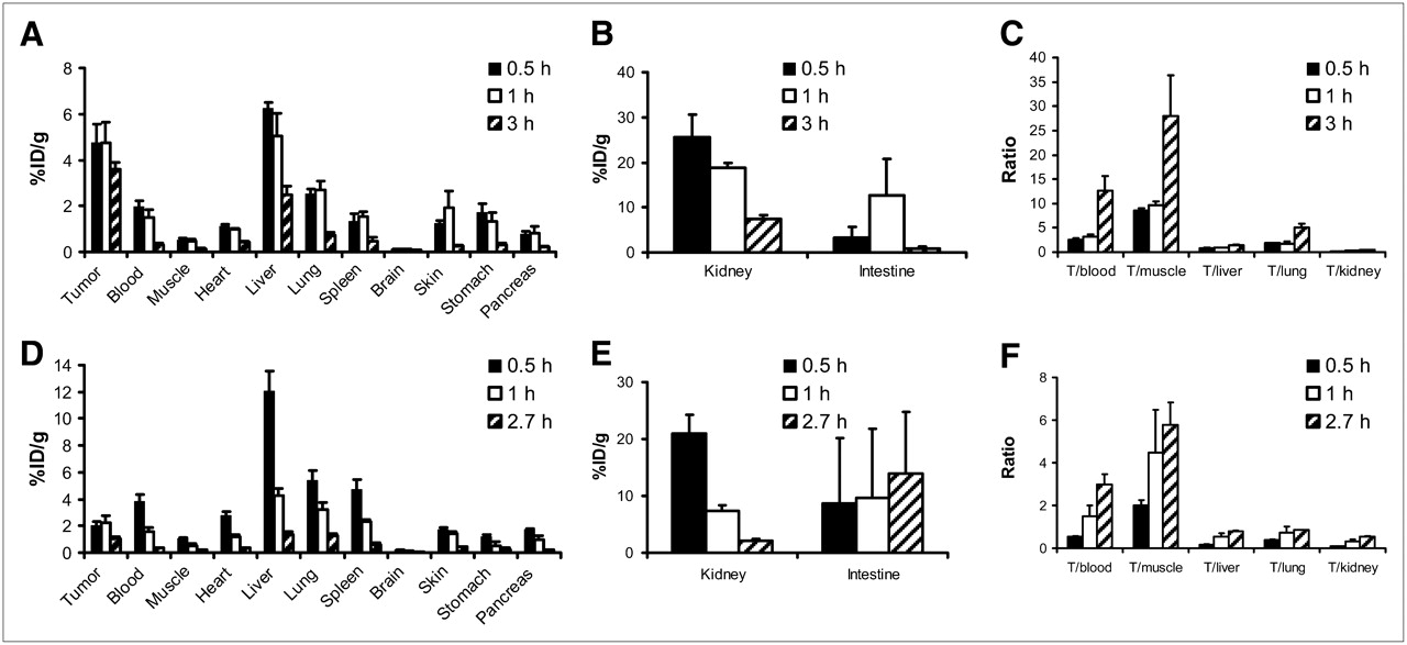
- FIGURE 5.
Biodistribution results (A, B, D, and E) and tumor-to-normal tissue ratios (C and F) for 18F-FBO-ZHER2:477 (A, B, and C) and 18F-FBO-(ZHER2:477)2 (D, E, and F) in nude mice bearing subcutaneously xenotransplanted SKOV3 tumors (human ovarian cancer) (T). Data are expressed as %ID/g at various times after intravenous injection of 18F-FBO-ZHER2:477 (0.37–1.11 MBq [10–30 μCi]) or 18F-FBO-(ZHER2:477)2 (0.74–1.67 MBq [20–45 μCi]) (n = 3 for each group). 18F-FBO-ZHER2:477 showed significantly higher uptake in SKOV3 tumors than 18F-FBO-(ZHER2:477)2 (P < 0.05).
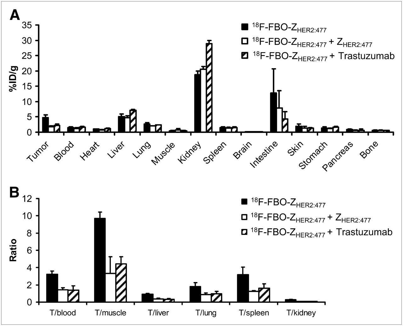
- FIGURE 6.
In vivo HER2 tumor–targeting specificity of 18F-FBO-ZHER2:477. One-hour biodistribution results (A) and tumor-to-normal tissue ratios (B) are shown for 18F-FBO-ZHER2:477 with or without pretreatment with either 300 μg of ZHER2:477 or 500 μg of trastuzumab. 18F-FBO-ZHER2:477 SKOV3 tumor (T) uptake was significantly inhibited by ZHER2:477 or trastuzumab (P < 0.05) (n = 3).
- FIGURE 7.
Decay-corrected coronal (top) and transaxial (bottom) small-animal PET images of nude mice bearing SKOV3 tumors on right shoulder at 0.5, 1, 2, and 4 h after tail vein injection of 18F-FBO-ZHER2:477 (A) and 18F-FBO-(ZHER2:477)2 (B). Arrows indicate locations of tumors. (C) Tumor and muscle time–activity curves derived from multiple-time-point small-animal PET images of mice bearing SKOV3 tumors. Data are shown as mean ± SD %ID/g (n = 3).


Additional Files
Supplemental Data
Files in this Data Supplement:


Jump to section
Related Articles
Cited By...
- First-in-Human Molecular Imaging of HER2 Expression in Breast Cancer Metastases Using the 111In-ABY-025 Affibody Molecule
- Three Methods for 18F Labeling of the HER2-Binding Affibody Molecule ZHER2:2891 Including Preclinical Assessment
- Interrogating Tumor Metabolism and Tumor Microenvironments Using Molecular Positron Emission Tomography Imaging. Theranostic Approaches to Improve Therapeutics
- 18F-Fluorobenzoate-Labeled Cystine Knot Peptides for PET Imaging of Integrin {alpha}v{beta}6
- Optical Imaging with Her2-Targeted Affibody Molecules Can Monitor Hsp90 Treatment Response in a Breast Cancer Xenograft Mouse Model
- Proof-of-Concept Study of Monitoring Cancer Drug Therapy with Cerenkov Luminescence Imaging
- Micro-CT enables microlocalisation and quantification of Her2-targeted gold nanoparticles within tumour regions
- Molecular Design and Optimization of 99mTc-Labeled Recombinant Affibody Molecules Improves Their Biodistribution and Imaging Properties
- Towards detecting the HER-2 receptor and metabolic changes induced by HER-2-targeted therapies using medical imaging
- A 2-Helix Small Protein Labeled with 68Ga for PET Imaging of HER2 Expression
- Changes in HER2 Expression in Breast Cancer Xenografts After Therapy Can Be Quantified Using PET and 18F-Labeled Affibody Molecules
- On the Selection of a Tracer for PET Imaging of HER2-Expressing Tumors: Direct Comparison of a 124I-Labeled Affibody Molecule and Trastuzumab in a Murine Xenograft Model
- Noninvasive prediction of tumor responses to gemcitabine using positron emission tomography