


Abstract
Human epidermal growth factor receptor (HER2)–targeted Affibody molecules radiolabeled with 18F allow the noninvasive assessment of HER2 status in vivo through PET imaging. Such agents have the potential to improve patient management by selecting individuals for HER2-targeted therapies and allowing therapy monitoring. The aim of this study was to assess different 18F radiolabeling strategies of the HER2-specific Affibody molecule ZHER2:2891, preclinically determine the biologic efficacy of the different radiolabel molecules, and select a preferred radiolabeling strategy to progress for automated manufacture. Methods: Cysteine was added to the C terminus of the Affibody molecule for the coupling of maleimide linkers, and 3 radiolabeling strategies were assessed: silicon-fluoride acceptor approach (18F-SiFA), 18F-AlF-NOTA, and 4-18F-fluorobenzaldehyde (18F-FBA). The biodistributions of the radiolabeled Affibody molecules were then determined in naïve CD-1 nude mice, and tumor targeting was assessed in CD-1 nude mice bearing high-HER2-expressing NCI-N87 tumors and low-HER2-expressing A431 tumors. The 111In-ABY-025 compound, which has demonstrable clinical utility, served as a reference tracer. Results: The non–decay-corrected radiochemical yields based on starting 18F-fluoride using the 18F-FBA, 18F-SiFA, and 18F-AlF-NOTA methods were 13% ± 3% (n = 5), 38% ± 2% (n = 3), and 11% ± 4% (n = 6), respectively. In naïve mice, both the 18F-AlF-NOTA-ZHER2:2891 and the 111In-ABY-025 compounds showed a significant kidney retention (70.3 ± 1.3 and 73.8 ± 3.0 percentage injected dose [%ID], respectively, at 90 min after injection), which was not observed for 18F-FBA-ZHER2:2891 or 18F-SiFA-ZHER2:2891 (4.8 ± 0.6 and 10.1 ± 0.7 %ID, respectively, at 90 min). The 18F-SiFA-ZHER2:2891 conjugate was compromised by increasing bone retention over time (5.3 ± 1.0 %ID/g at 90 min after injection), indicating defluorination. All the radiolabeled Affibody molecules assessed showed significantly higher retention in NCI-N87 tumors than A431 tumors at all time points (P < 0.05), and PET/CT imaging of 18F-FBA-ZHER2:2891 in a dual NCI-N87/A431 xenograft model demonstrated high tumor-to-background contrast for NCI-N87 tumors. Conclusion: The HER2 Affibody molecule ZHER2:2891 has been site-selectively radiolabeled by three 18F conjugation methods. Preliminary biologic data have identified 18F-FBA-ZHER2:2891 (also known as GE226) as a favored candidate for further development and radiochemistry automation.
Approximately 20%–25% of all patients with breast cancer test positive for the human epidermal growth factor receptor (HER2, also known as c-ErbB2, Neu, CD340, or p185), a transmembrane protein that belongs to the human epidermal growth factor tyrosine kinase receptor family (EGFR or ErbB) (1). HER2 expression is either absent or present at low levels in normal adult tissue (2). The occurrence of HER2 overexpression seems to be linked to tumor malignancy in breast cancer (1). HER2-overexpressing tumors can be successfully treated using the monoclonal antibody trastuzumab (Herceptin; Hoffmann-La Roche AG), and several other treatment options are available or are in development (3). The assessment of HER2 expression in biopsies from primary lesions of breast cancer patients is a standard procedure to select patients for HER2-targeted therapies. However, in recurrent disease in which HER2 status can change, determination of HER2 expression is not a standard procedure, which can complicate patient management after changes in HER2 status over time. Here, a HER2-specific probe for PET would offer a quantitative, noninvasive, and sensitive diagnostic tool to characterize HER2 status in the entire tumor burden (including metastases) and determine changes in HER2 expression that are not captured by established biopsy assessment.
Affibody (Affibody AB) molecules are peptides of approximately 6.5 kDa that originate from the 3-helical B domain scaffold of the IgG-binding region of staphylococcal protein A (4,5). High-affinity binders can be easily tailored to several specific targets by altering only a few amino acids (5). The relatively small size of an Affibody molecule improves the pharmacokinetic properties as radiotracers, compared with labeled Fabs, diabodies, and others.
We anticipated the HER2-binding Affibody molecule (MMA-DOTA-Cys61)-ZHER2:2891-Cys (ABY-025) would be an attractive radiolabeled vector for 18F. This second-generation Affibody molecule is an improved variant of ABY-002 (DOTA-ZHER2:342-pep2), with better thermostability, excellent hydrophilicity, and an affinity (KD) of 76 pM (6). Despite significant reengineering of the nonbinding surface, the preclinical in vivo pharmacokinetics of 111In-DOTA-ZHER2:2891 (111In-ABY-025) are in remarkably good agreement with the parent tracers (7). Published clinical studies with 111In-ABY-025 in a breast cancer cohort demonstrated that this agent can accurately determine HER2 status in patients (8). Compared with SPECT, for oncology PET offers improved sensitivity, resolution, and quantification that add to the utility of a HER2-targeted diagnostic imaging agent. An accurate assessment of the HER2 status could be used to guide the selection of patients most likely to benefit from HER2-targeted therapies. Even though 68Ga-labeled Affibody molecules have shown good results in preclinical animal studies and a first-in-human investigation (9,10), an 18F-labeled Affibody molecule for clinical use would be preferred because 18F is a radionuclide with established and approved good manufacturing practice production for clinical use. Furthermore, the longer half-life of 18F than 68Ga will allow image acquisition over a longer time window. In addition, both the higher abundance and the lower energy of 18F positrons can be seen as beneficial for image resolution (11). Radiohalogen-labeled tracers do not generally accumulate in the kidneys, which may improve detection of lesions in close proximity to the kidneys.
18F has been previously coupled to Affibody molecules. Kiesewetter et al. successfully used N-[2-(4-18F-fluorobenzamido)ethyl]maleimide (18F-FBEM) to label the HER2-binding ZHER2:342 (12–16). Miao et al. applied the same reagent to the related EGFR binder ZEGFR:1907 (17). The established labeling reagent 4-18F-fluorobenzaldehyde (18F-FBA) can be conveniently obtained in a single labeling step (18). Cheng et al. conjugated 18F-FBA with the aminooxy-modified HER2 Affibody molecule ZHER2:477 (19). We decided to test this radiochemistry for ZHER2:2891 in combination with the recently discovered aniline catalysis (20–22). With regard to the automation potential of the radiolabeling protocol, we also focused our attention on 2 simple 1-step methods. The silicon-fluoride acceptor approach (18F-SiFA) by Schirrmacher et al. with an isotopic 19F/18F exchange reaction by aqueous 18F-fluoride appeared to be promising (23,24). As a third method, we studied the 18F-AlF-NOTA concept developed by McBride et al. (25–29). More recently, this protocol has been exploited by Heskamp et al. to obtain the 18F-labeled HER2 Affibody molecule 18F-AlF-NOTA-ZHER2:2395 (30).
We report here the results from our development work to radiolabel the HER2 Affibody molecule ZHER2:2891 using 18F-FBA/aminooxy, 18F-SiFA, and 18F-AlF-NOTA strategies and the biologic evaluation of the new tracers regarding targeting HER2-positive mouse xenografts.
MATERIALS AND METHODS
Radiochemistry
No-carrier-added aqueous 18F-fluoride was produced from the 18O(p,n)18F reaction (PETtrace cyclotron; GE Healthcare) by the irradiation of an isotopically enriched 18O-H2O target using a 16.4-MeV proton beam. Carrier-free 111In-indium(III) chloride was either purchased from Perkin Elmer U.K. as an aqueous HCl (0.05 M) solution or obtained from GE Healthcare (Indiclor) as an aqueous 0.04 M HCl solution. (MMA-DOTA-Cys61)-ZHER2:2891-Cys (ABY-025) was obtained from Affibody AB as a solution in sodium acetate (0.2 M, pH 5.3).
Analytic high-performance liquid chromatography (HPLC) was done using a Gilson 322 pump with a UV/ViS 156 detector. The instrument was equipped with a γ-detector (Bioscan Flow-count, PMT probe, Thermo MS3083; LabLogic Systems Limited). Radioactivity measurements were done using a Capintec CRC-15R ion chamber. Radiochemical yields (RCYs) are reported as non–decay-corrected. Analytic radio HPLC used a Luna C18(2) column (Phenomenex; 50 × 4.6 mm, 3 μm). A gradient system (solvent A: ammonium acetate, 0.1 M; solvent B: acetonitrile; 18F-5 and 18F-11: gradient 5%–95% B in 15 min, 18F-12 and 18F-13: gradient, 20%–50% B in 15 min) with a flow rate of 1 mL/min was used. Analytic radio HPLC for 111In-ZHER2:2891-Cys-DOTA (111In-ABY-025) used a C4 30 nm 150 × 4.6 mm column (Ace) with a gradient system (solvent A: 0.1% trifluoroacetic acid [TFA]/H2O, solvent B: 0.1% TFA/acetonitrile; gradient, 0%–20% B in 4 min, followed by 20%–60% in 12 min) and a flow rate of 1 mL/min.
The ultraviolet absorption was monitored at 280 nm. Quality control was done using the above analytic HPLC system and by RP-18 reversed-phase thin-layer chromatography (silica gel 60 RP-18 on alumina sheets Merck KGaA) eluting water/70% acetonitrile (labeled peptide remained at origin).
111In-ZHER2:2891-Cys-DOTA (111In-ABY-025)
A solution containing ZHER2:2891-Cys-DOTA (48 μL, 104 μg, 14 μmol) in a silanized P6 vial was mixed with aqueous ammonium acetate (100 μL, 0.25 M) and 111In-InCl3 (120 μL, 115 MBq). The vial was heated at 60°C for 30 min. The product was formulated by adding saline (3.5 mL). The radiochemical purity of 111In-ABY-025 was greater than 99% (HPLC).
18F-ZHER2:2891-Cys-Aminooxy-FBA (18F-11)
18F-FBA was obtained in high radioactive concentration from a FASTlab module (GE Healthcare) (supplemental data; supplemental materials are available at http://jnm.snmjournals.org) (31). A solution of 18F-FBA in ethanol (92 μL, 740 MBq) was added to a solution of 3 (0.4 mg, 55 nmol) and aniline hydrochloride (3.2 mg, 25 μmol) in water (138 μL). The mixture was heated at 70°C for 20 min. After being cooled to room temperature, the product 18F-11 was isolated by transferring the reaction mixture onto a conditioned NAP5 (GE Healthcare U.K.) cartridge, followed by elution with saline (1.0 mL, 0.1% NaCl, 0.1% sodium ascorbate) of which a first fraction (0.25 mL) was discarded.
18F-ZHER2:2891-Cys-SiFA (18F-5)
A solution of Affibody molecule precursor 19F-5 (830 μg, 111 nmol) in acetonitrile (40 μL) was mixed with sodium acetate buffer (100 μL, pH 4.0, 0.5 M) in a P6 vial, followed by 18F-fluoride in water (100 μL, 504 MBq). The mixture was incubated for 15 min at 95°C. After being cooled to room temperature, the reaction mixture was transferred to a NAP5 cartridge, followed by saline (260 μL). The product 18F-5 was eluted with saline (750 μL) into a P6 vial.
18F-ZHER2:2891-Cys-NOTA-(COOH)2-AlF (18F-12)
A solution of the Affibody molecule precursor 7 (746 μg, 100 nmol) in sodium acetate buffer (50 μL, pH 4.0, 0.5 M) was mixed with a solution of AlCl3 (3 μL, 3.33 μg, 25 nmol in sodium acetate buffer, pH 4.0, 0.5 M) in a polypropylene centrifuge vial (1.5 mL). This mixture was added to a small volume of 18F-fluoride (50 μL, 410 MBq) in a capped P6 vial and heated for 15 min at 100°C. After being diluted with saline (100 μL), the reaction solution was transferred to a NAP5 size-exclusion cartridge. The product 18F-12 was eluted into a P6 vial using saline (750 μL).
18F-ZHER2:2891-Cys-NOTA-(COOH)3-AlF (18F-13)
A QMA SepPak light cartridge (Waters Ltd.) was activated using sodium acetate solution (10 mL, 0.5 M) and water (10 mL). The cartridge was loaded with an 18F-fluoride solution (209 MBq), followed by drying using a stream of nitrogen (30 s, 100 mL/min). The activity was eluted using saline and collected in 3 fractions of 0.2 mL. The first fraction containing most activity (59%) was used for the subsequent labeling chemistry.
A solution of NOTA(COOH)3-ZHER2:2891 Affibody molecule 9 (380 μg, 50 nmol) in sodium acetate buffer (20 μL, pH 4.0, 0.5 M) was added to a solution of AlCl3 (1.5 μL, 1.67 μg, 12.5 nmol in sodium acetate buffer, pH 4.0, 0.5 M) and 18F-fluoride (10 MBq, 25 μL) as eluted from the QMA cartridge. This mixture was added to a small volume of (50 μL) in a capped P6 vial. The vial was heated for 15 min at 100°C. After being diluted with saline (100 μL), the reaction solution was transferred to a NAP5 size-exclusion cartridge. After a first fraction (0.3 mL) was discarded, 5 fractions (0.2 mL) were collected. The combined fractions 2 and 3 contained the labeled product.
Biacore Assay
The purified Affibody molecules 5, 7, 9, and 11 were lyophilized in portions (0.1 mg) and dissolved in HBS-EP buffer (HEPES-buffered saline [contains 4-(2-hydroxyethyl)-1-piperazineethanesulfonic acid, sodium chloride, ethylenediaminetetraacetic acid, and surfactant P20]) (1 mL, 10 mM) as stock solution and diluted further in the range of 0.1–25 nM.
Human-recombinant HER2 was supplied by R&D Systems: the recombinant ErbB2/Fc chimera (cat. no. 1129-ER, 50 μg) was dissolved in HEPES-buffered saline (HBS-EP, 0.2 mL) and dispensed in vials (10 μL, 250 μg/mL, stock solution). The stock solution (1–2 μL) was further diluted in HBS-EP (200 μL).
The affinity studies were performed using a Biacore 3000 (GE Healthcare). The instrument was stabilized at 25°C. A CM5 chip with 4 channels in series (GE Healthcare) was used for immobilization of receptor dissolved in acetate buffer (10 mM, pH 4.0; GE Healthcare). The default method, amine coupling, was applied as supplied by the manufacturer. HBS-EP buffer (10 mM HEPES pH 7.4 with 150 mM NaCl, 3 mM ethylenediaminetetraacetic acid, and 0.005% surfactant P20 added; GE Healthcare) was used as running buffer and as solvent for the substances. One channel was used for reference.
The HER2 was immobilized on the chip by amine coupling to a level of approximately 3,000–5,000 RU. The substances were injected over the surface in a concentration range of 0.5–50 nM. Regeneration of the chip after each injection of substance solution was performed with 10 mM HCl.
The kinetic calculations were performed using a Langmuir 1:1 binding model and simultaneous calculation of association and dissociation phase. The kinetic constants, association rate constant Ka and dissociation rate constant Kd, describe the rate of formation or dissociation of the complex. Kinetic parameters can be evaluated from the association and dissociation phases of the sensorgram. The KD is determined from the kinetic parameters (KD = kd/ka).
A431 and NCI-N87 CD-1 Nude Xenograft Models
All animal studies were performed in compliance with the U.K. Home Office Animals (Scientific Procedures) Act 1986. The human gastric cancer cell line NCI-N87 and the human epithelial carcinoma cell line A431 were purchased from the American Type Culture Collection. NCI-N87 cells were grown in RPMI 1640 containing 10% fetal bovine serum and 2 mM glutamine. A431 cells were grown in minimum essential medium Eagle supplemented with 10% fetal bovine serum, 1% nonessential amino acids, and 2 mM glutamine. Cells were passaged twice per week, at 80% confluence, and incubated in 5% CO2 at 37°C. Approximately 2 × 10⁶ of NCI-N87 cells or 107 A431 cells suspended in 0.1 mL of 50% phosphate-buffered saline/50% Matrigel (Corning Life Sciences) were subcutaneously implanted in the lower right flank of CD-1 nude mice (Charles River Laboratories). Tumors were allowed to develop for 4 wk. HER2 expression in these tumors was assessed by immunohistochemistry using the Food and Drug Administration–validated HercepTest (Dako) (supplemental data). Dual-tumor xenograft mice were generated by implantation of 2 × 10⁶ of NCI-N87 cells and 1 × 10⁷ of A431 cells in each of the 2 lower flanks. These mice were used to assess the biodistribution of 18F-11, enabling a same-animal assessment of uptake in both low- and high-HER2-expressing tumors.
Biodistribution Studies and PET/CT Imaging
For biodistribution studies, mice (n = 3 for each group) were injected with 2.5–3 MBq of each radiotracer via the lateral tail vein. This corresponds to approximately 20–90 μg of peptide in the 18F-labeled radiotracer 18F-5, 18F-11, and 18F-12 and approximately 3 μg of peptide in 111InABY-025. Mice were sacrificed at 2, 90, 120, and 180 min after injection. For biodistribution of 18F-11 in dual-tumor NCI-N87/A431 mice, animals were sacrificed at 2, 30, 60, 90, 120, and 180 min. Tumor and key tissues were excised and weighed, and the radioactivity was measured using a γ-counter. Small-animal PET/CT was performed with a microPET-P4 system (Siemens Inc.) as described previously (32). 18F-11 (10 MBq/animal) was injected intravenously, and the mice were imaged for 30 min starting at 120 min after injection.
Statistical Analysis
Data are presented as mean ± SD. Statistical comparisons were made using the Student t test. A P value of less than 0.05 was considered significant.
RESULTS
Synthetic chemistry
As an alternative to a recombinant protocol, the Affibody molecule ZHER2:2891 1 containing 61 amino acids was also readily accessible using a standard peptide synthesizer affording 42% of lyophilized crude product. The yield of pure Affibody molecule 1 after semipreparative HPLC was 9%. The building block Eei-aminooxyacetyl-maleimide 2 was obtained following a peptide-coupling step between N-(2-aminoethyl)maleimide and 2,5-dioxopyrrolidin-1-yl 2-(((1-ethoxyethylidene)amino)oxy)acetate, with 75% yield after isolation by semipreparative HPLC (Scheme 1). The addition reaction of maleimide 2 with Affibody molecule 1 was quantitative, forming aminooxy-modified Affibody molecule 3 in greater than 99% yield after HPLC purification and removal of the Eei-protecting group. The SiFA maleimide linker 4 was prepared in a similar fashion, with 45% yield. The SiFA-, NOTA(COOH)2-, and NOTA(COOH)3-modified Affibody molecules 5, 7, and 9 were prepared with yields of greater than 99%, 90%, and 58%, respectively, using the cysteine-maleimide coupling strategy.
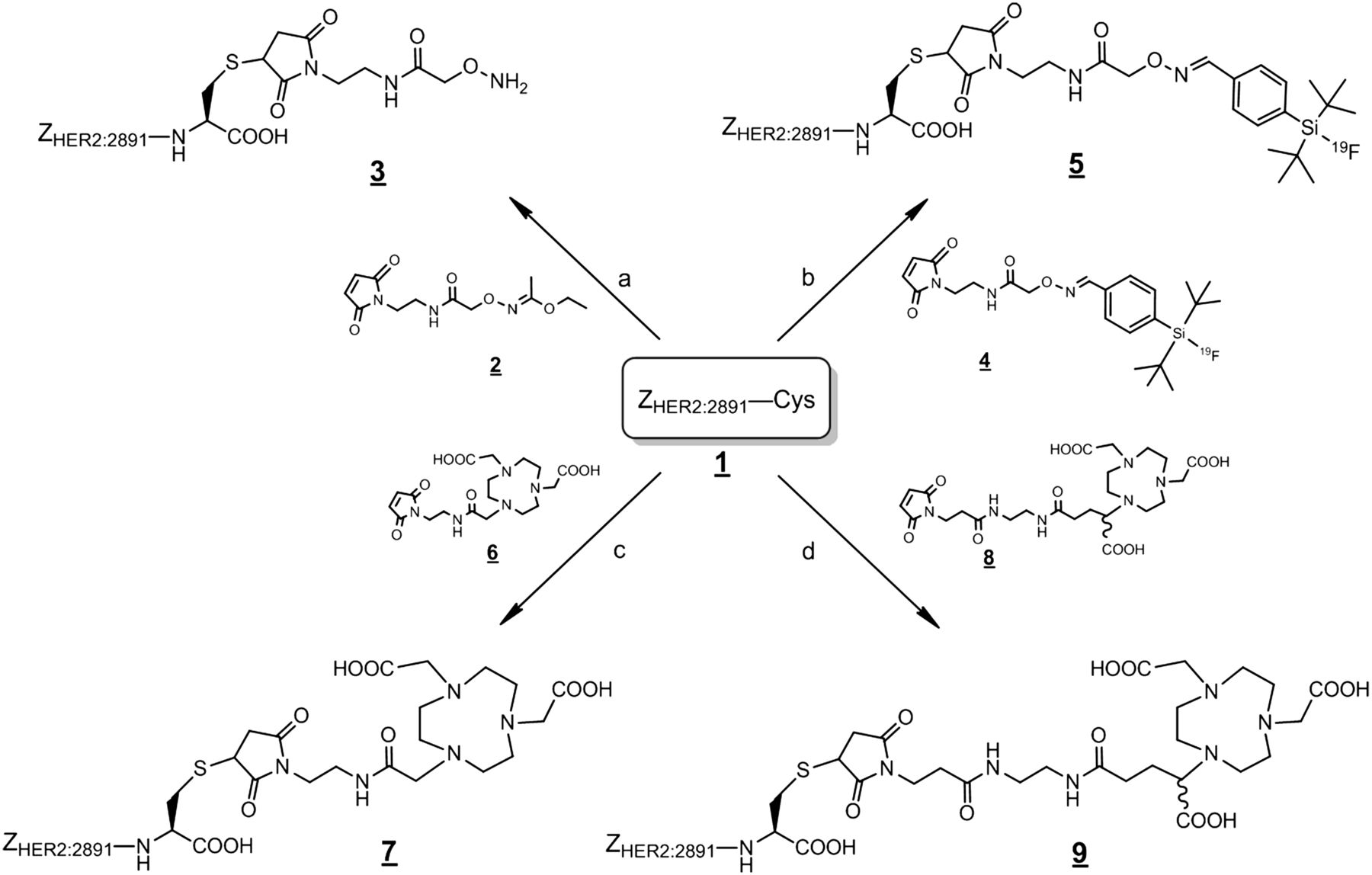
Preparation of ZHER2:2891 precursor compounds 3, 5, 7, and 9 for 18F labeling. Reaction conditions: (i) pH 6.0, 90 min r.t. (ii) water/2.5% TFA (v/v) (a); pH 6.0, 1 h r.t. (b); pH 6.0, 1 h r.t. (c); pH 6.0, 3 h r.t. (d). r.t. = room temperature.
Radiochemistry
For the synthesis of 18F-11, we used 18F-FBA following the established aminooxy peptide–labeling method (33). The non–decay-corrected RCY of FASTlab-produced 18F-FBA was 43% ± 5% (n = 5). An aliquot of the product batch of the 18F-FBA was used for manual conjugation experiments. The radioactive concentration of purified 18F-FBA was sufficient to produce 18F-11 from 3 in adequate quantities for preclinical studies, with an overall non–decay-corrected RCY of 13% ± 3% (Scheme 2; Table 1; Supplemental Figs. 1 and 2).

Preparation of 18F-labeled ZHER2:2891 compounds 18F-5 and 18F-11–13. Reaction conditions: dimethylsulfoxide, 9 min at 120°C (a); water, aniline hydrochloride, 20 min at 70°C (b); pH 4.0, 15 min at 95°C (c); pH 4.0, 15 min at 100°C (d).
Summary of Radiolabeling Data for 18F-Affibody Molecule 18F-5 and 18F-11–13
Following the single-step SiFA-labeling protocol by Schirrmacher et al. (23,24), we obtained Affibody molecule 18F-5 via isotopic 19F/18F exchange reaction in aqueous solvent, with 38% ± 2% RCY (HPLC analysis, Supplemental Fig. 3). The tracer was stable in saline at room temperature for at least 2 h.
The NOTA(COOH)2-modified Affibody molecule 7 was directly radiolabeled with 18F-fluoride/AlCl3 under literature conditions (27–29). The required 18F-fluoride was initially passed through a QMA cartridge with subsequent elution by saline, as suggested previously (27). However, no difference in labeling efficiencies was observed, compared with using nontreated 18F-fluoride (HPLC analysis, Supplemental Fig. 4).
A time course study revealed an optimum level of 18F incorporation of 49% after 15 min of heating at 100°C, with some decomposition afterward (Supplemental Fig. 5). Lowering the reaction temperature to 70°C reduced the prepeak but resulted in low reaction progress (Supplemental Table 2). Experimental data on the impact of the peptide/AlCl3 concentration showed a better labeling efficiency for the highest concentration but also a proportional increase of byproduct. This might have originated from thermal decomposition of the peptide (Supplemental Fig. 6).
The tris-acid NOTA Affibody molecule precursor 9 was also labeled with 18F-AlF according to Scheme 2. However, the analytic RCYs of 18F-13 (18%–20%) remained below the results of the bis-acid analog 7 (HPLC analysis, Supplemental Fig. 7).
Biacore Assay
Affibody analogs 5, 7, 9, and 11 have been tested for affinity toward human recombinant HER2 by Biacore surface plasmon resonance. The performed Biacore studies are based on interaction analysis using immobilized recombinant HER2 on a chip while the compounds were injected over the surface in different concentrations. The binding curves (Supplemental Fig. 8) of both compounds fitted well to a 1:1 binding model. The average KD values are shown in Table 2.
Binding Constants of Affibody Analogs 5, 7, 9, and 11 Toward Human HER2
Biodistribution in CD-1 Naïve Mice
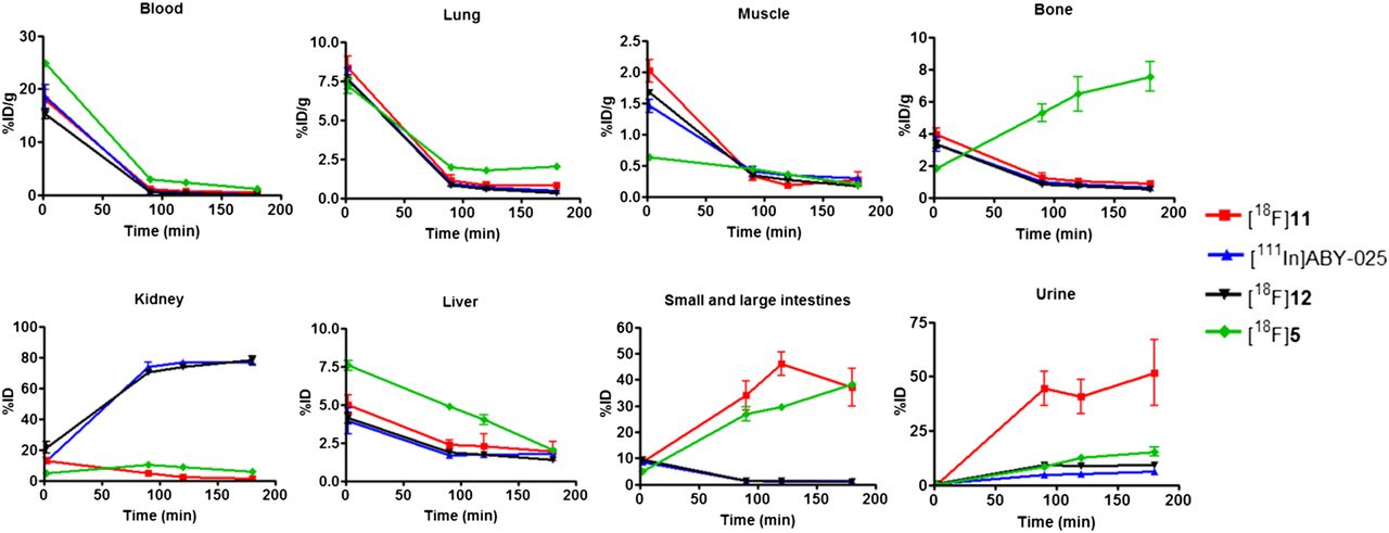
111In-ABY-025 showed high kidney retention, reaching 73.8 ± 3.0 and 76.9 ± 0.7 percentage injected dose (%ID) at 90 and 180 min, respectively (Fig. 1; Supplemental Table 3). The Affibody molecule 18F-12 gave a profile similar to 111In-ABY-025, with no reduction in kidney retention (Fig. 1; Supplemental Table 4). Tracers 18F-5 and 18F-11 demonstrated an improved excretion profile, characterized by reduced kidney retention and higher hepatobiliary excretion than with 111In-ABY-025 and 18F-12 (Fig. 1; Supplemental Tables 5 and 6). Furthermore, 18F-11 also revealed higher urinary excretion. In contrast to all other compounds assessed, the biodistribution of 18F-5 was characterized by increasing bone retention over time, which is indicative of tracer defluorination. Blood, muscle, lung, and liver profiles were comparable for all 4 agents, apart from a slight increase in blood, liver, and lung retention for 18F-5 (additional data in Supplemental Tables 3–6).

Biodistribution of Affibody molecules 111In-ABY-025, 18F-5, 18F-11, and 18F-12 in naïve CD-1 nude mice. Data are expressed as percentage administered activity (injected dose) per gram of tissue (%ID/g) for blood, lung, muscle, and bone and as %ID for kidney, liver, small and large intestine, and urine after intravenous injection of radiotracer at 2, 90, 120, and 180 min (n = 3).
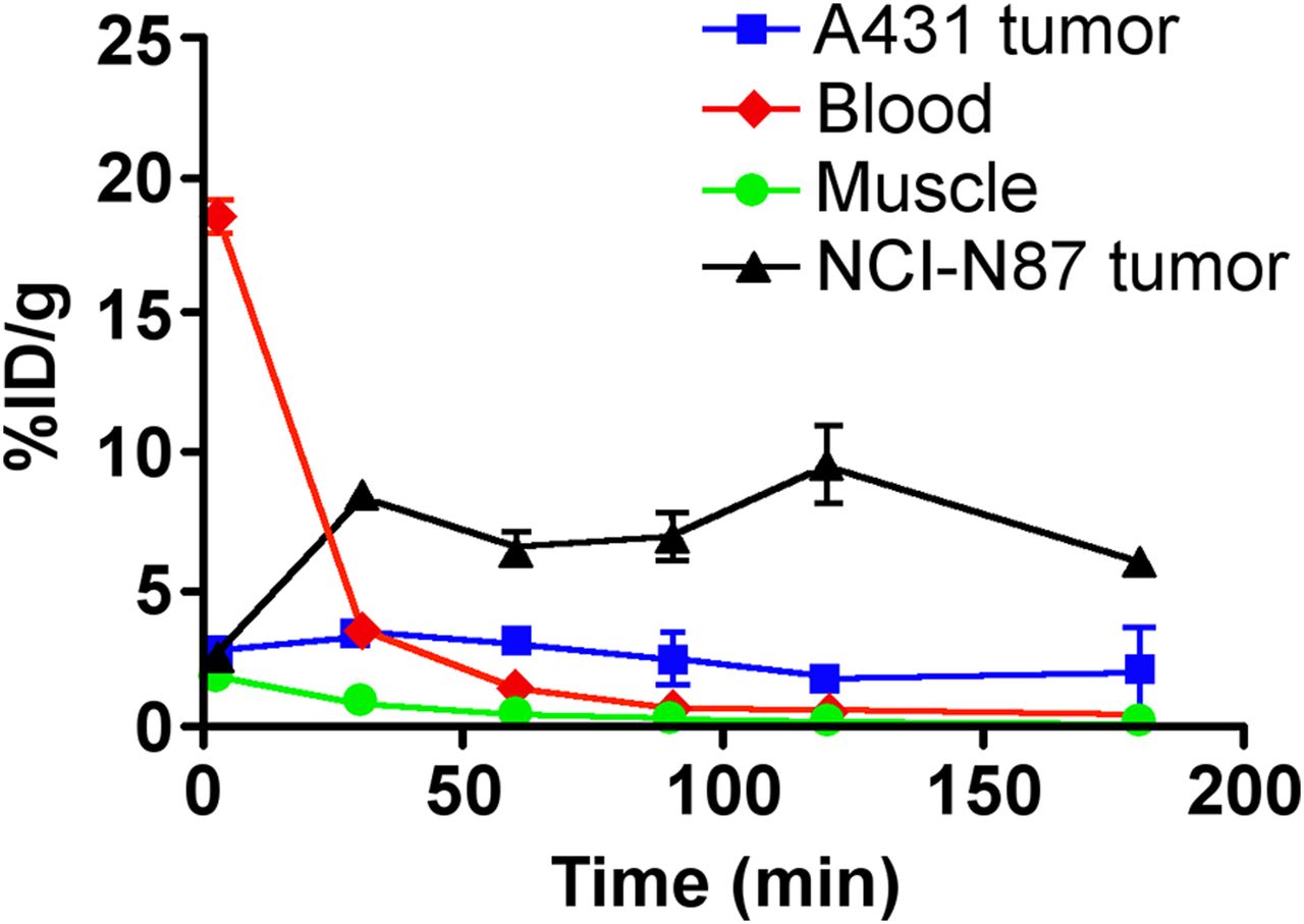
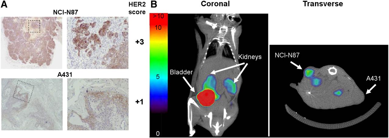
Biodistribution in A431/NCI-N87 Xenograft–Bearing Mice
HER2 expression in NCI-N87 (high HER2) and A431 (low HER2) xenografts was confirmed by immunohistochemistry on fixed tumor sections. Using the HercepTEST standard scoring scale (0 to +3), Figure 2A shows that NCI-N87 tumors stain strongly (+3), whereas A431 cells show a considerably weaker staining intensity (+1). In biodistribution studies, all radiolabeled Affibody molecules showed an increased retention in the high-HER2-expressing NCI-N87 tumors, compared with the low-HER2-expressing A431 tumors (Fig. 3). Of the 18F-radiolabeled Affibody molecules, 18F-11 demonstrated the highest retention in the NCI-N87 tumors and also the best differentiation between high- and low-HER2-expressing tumors (7.15 ± 0.69 %ID per gram [%ID/g] in NCI-N87 and 1.99 ± 0.73 %ID/g in A431 at 90 min after injection). It is followed by 18F-12 (4.79 ± 1.26 %ID/g in NCI-N87 and 1.37 ± 0.32 %ID/g in A431 at 90 min after injection), whereas 18F-5 had minimal differentiation between the 2 tumor types (3.49 ± 0.74 %ID/g in NCI-N87 and 2.07 ± 0.46 in A431 at 90 min after injection). Excluding 18F-5, all of the 18F-radiolabeled Affibody molecules tested had a significantly higher retention in NCI-N87 tumors than in A431 tumors at all time points (P < 0.05). High and moderate tumor–to–key organ ratios have been observed in the biodistributions of NCI-N87 and A431 tumor–bearing mice, respectively (Supplemental Tables 7 and 8). For the NCI-N87 biodistributions, the highest tumor-to-muscle and tumor-to-liver ratios observed were seen for 18F-11 at 28.89 and 2.83, respectively (90 min after injection).

PET/CT imaging of 18F-11 in dual A431/NCI- N87 tumor xenograft mouse model. (A) HER2 protein expression in tumor sections from NCI-N87 and A431 xenograft models by immunohistochemistry using HercepTEST by DAKO. Images on left are ×10 magnification; images on right are ×20 of highlighted square. With recommended intensity scale (0 to +3), NCI-N87 tumors stain strongly (+3), whereas A431 cells show considerably weaker staining intensity (+1). (B) Representative coronal and transverse PET/CT of dual-A431/NCI-N87 tumor xenograft mouse model imaged at 120–150 min after injection of 10 MBq of 18F-11. Color scale indicates %ID that accumulates per gram of tumor (%ID/g) ranging from 0% to 10 %ID/g (bladder exceeds 10 %ID/g indicated in scale bar).

Biodistribution of Affibody molecules 111In-ABY-025, 18F-5, 18F-11, and 18F-12 in NCI-N87- (high HER2) and A431 (low HER2)-expressing xenograft-bearing mice. Data are expressed as percentage administered activity (ID) per gram of tissue (%ID/g) after intravenous injection of radiotracer at 2, 90, 120, and 180 min (n = 3).
Dual-Flank A431/NCI-N87 Xenograft–Bearing Mice
Using the dual-flank A431/NCI-N87 xenograft model, Figure 4 shows that 18F-11 performance was comparable to that observed in the single-tumor animal studies, with consistent binder retention seen in the A431 and NCI-N87 tumors, starting from as early as 30 min after injection. As far as background tissue was concerned (Supplemental Table 10 provides key tissue ratios), blood levels at 60 min after injection were reduced significantly, providing an NCI-N87 tumor–to–blood ratio of 4.52, whereas at 30 min, reduced blood activity still gave a respectable ratio of 2.39, accompanied by a positive tumor-to-liver ratio of 1.39.
Biodistribution profile of 18F-11 in dual A431/NCI-N87 tumor xenograft mouse model. Data are expressed as percentage administered activity (ID) per gram of tissue (%ID/g) after intravenous injection of 18F-11 at 2, 30, 60, 90, 120, and 180 min (n = 3).
The dual-flank A431/NCI-N87 tumor mouse model was also used to perform a preliminary PET/CT study using Affibody molecule 18F-11. The image shown in Figure 2B demonstrated that elimination was through the kidneys and bladder, as previously observed in the biodistribution studies. The transverse image shows retention in both tumors, with the high-HER2-expressing NCI-N87 tumor showing considerably higher signal intensity than the low-HER2-expressing A431 tumor, in agreement with the dual-tumor biodistribution studies.
DISCUSSION
The Affibody molecule ZHER2:2891 is an advanced second-generation HER2-binding peptide (6). The aim of the study was to select a preferred compound on the basis of diagnostic efficacy potential and application of the radiochemistry for automation to provide 18F-labeled ZHER2:2891 for clinical evaluation. On the basis of the literature, we selected 3 of the currently most promising methods for radiolabeling biomacromolecules of approximately 7-kDa size: 18F-FBA/aminooxy (33), 18F-SiFA (24), and 18F-AlF/NOTA (27) coupling. Taking advantage of the C terminal cysteine modification of ZHER2:2891-Cys (1), we selectively attached the required reactive groups via the efficient maleimide-linking strategy. The aminooxy-labeled Affibody molecule 3 is stable under argon at −20°C for at least 2 mo and can be conveniently used for radiolabeling without the need of a protecting group. The desired precursor 5 was accessible in good yields via precoupled SiFA-maleimide 4.
Aniline-catalyzed coupling of 18F-FBA provided 18F-11 (GE226) in sufficient quantities for biologic evaluation. This use of a catalyst to generate the oxime-ligated molecule afforded significantly higher RCYs than with previous reports on Affibody molecule labeling using 18F-FBA (13%–18% decay-corrected RCY) (19). A less complex radiosynthesis using 18F-FBA/aniline proved to be beneficial with regards to labeling with 18F-FBEM (6.5% non–decay-corrected RCY (15) or 13%–18% decay-corrected RCY (17)). In an alternative approach avoiding the preparation of an indirect labeling reagent, the labeling protocol using isotopic 18F/19F exchange with SiFA precursor 5 was found to give best results in a mixed organic/aqueous solvent system. Another simple direct labeling method, the 18F-AlF/NOTA protocol, was subjected to some optimization. Two comparative experiments clearly demonstrated the NOTA(COOH)2 precursor 7 to be a more suitable precursor than the NOTA(COOH)3 derivative 9, with analytic labeling efficiencies of 42% and 15% for 18F-12 and 18F-13, respectively (Supplemental Fig. 7). It might be speculated that the third carboxylic group is not stoichiometrically required and can therefore cause some destabilization of the complex. Our labeling efficiencies from the NOTA(COOH)2-linked Affibody molecule precursor 7 appear to be comparable with a report by Heskamp et al. on a similar chelator-based ZHER2:2395 Affibody molecule (30). We subsequently applied 18F-12 for our biologic evaluation. In conclusion, when comparing the 3 labeling approaches, the 18F-SiFA protocol clearly gave the best RCYs, whereas the alternative direct labeling method using 18F-AlF and the 2-step 18F-FBA radiochemistry both were less efficient. However, all 3 approaches generated the desired Affibody molecule in high radiochemical purities and could be readily automated to provide a radiotracer of higher specific radioactivity.
For the in vivo biology assessment, NCI-N87 and A431 tumor xenograft models were chosen. The high and low HER2 expression levels for NCI-N87 and A431, respectively, were confirmed by immunohistochemistry using the Food and Drug Administration–approved HercepTest, indicating that the models are suitable for comparing the uptake of the different HER2-targeted Affibody molecules (Fig. 2). The biodistribution data show that 18F-5 and 18F-11 have a preferred biodistribution profile, compared with 18F-12, primarily due to the lower kidney retention (Fig. 1). All 4 compounds tested using Biacore 5, 7, 9, and 11 show a subnanomolar binding affinity to the HER2 (Table 2). This is in agreement with published data for ABY-025 in which subnanomolar affinity has also been demonstrated (6). Target selectivity of the ZHER2:2891-Cys pharmacophore has been previously reported by Feldwisch et al., where the compound bound only to HER2 and not to the other receptors in the HER family: EGFR (HER1), HER3, or HER4 (6).
The increased bone retention of 18F-5 with time may indicate defluorination of 18F-5 in vivo. Although a minor bone uptake could be preferential in some studies for more accurate mapping of internal organs, it may be unfavorable in metastatic breast cancer patients because it potentially increases the background signal for detection of bone metastasis. Furthermore, a higher bone uptake may result in a possibly higher radiation dose to the bone marrow, which could be critical for an oncology PET tracer. Additional disadvantages of 18F-5 arise from the higher blood, liver, and lung uptake but lower NCI-N87 tumor uptake. Taken together, these findings indicate that 18F-11 may be a preferable PET imaging agent in terms of in vivo background tissue pharmacokinetics. All of the 18F-radiolabeled Affibody molecules except 18F-5 at 120 min after injection demonstrated a significantly higher retention in NCI-N87 tumors than in A431 tumors at all time points, showing high specificity for HER2-expressing tumors. Ahlgren et al. performed biodistribution studies of the same reference tracer (111In-ABY-025) in a SKOV-3 tumor xenograft model, showing high tumor uptake levels (17 %ID/g of 111In-ABY-025 in SKOV-3 tumors at 60 min after injection) and high tumor-to-organ ratios. In comparison, the NCI-N87 tumor uptake levels and tumor-to-organ ratios of both tracers, 18F-11 and 111In-ABY-025, have been lower (∼7 %ID/g at 90 min after injection for 18F-11 and 111In-DOTA-ABY-025 in NCI-N87 tumors) (7). This might be due to the different tumor model (SKOV-3 vs. NCI-N87), with potential differences in the HER2 level or perfusion of the tumor type.
Biodistribution of our preferred tracer 18F-11 in a dual-flank NCI-N87/A431 xenograft model showed pharmacokinetics similar to the single-tumored mice. Small-animal PET imaging is in line with biodistribution data and suggests that the pharmacokinetics of 18F-11 is sufficient for imaging human subjects within a suitable imaging window.
The range of the specific radioactivities of the 18F-labeled Affibody molecules appeared to be suitable for the purpose of our study. However, controlling the specific radioactivity of such tracers is also important for potentially discriminating between tumors expressing high and low levels of HER2 (34).
Because the quality control data of formulated 18F-11 showed minor traces of stable side products (Supplemental Fig. 2), a toxicologic safety study will be required to enable a first-in-human study of the tracer.
CONCLUSION
We have established 3 simple routes to radiolabel the promising HER2-binding Affibody molecule ZHER2:2891 with 18F. On the basis of the favorable pharmacokinetic profile in normal mice and HER2-expressing xenograft mouse models, we selected the 18F-FBA/aminooxy–conjugated Affibody molecule as our lead candidate for further development of a fully automated protocol toward a first clinical study.
DISCLOSURE
The costs of publication of this article were defrayed in part by the payment of page charges. Therefore, and solely to indicate this fact, this article is hereby marked “advertisement” in accordance with 18 USC section 1734. No potential conflict of interest relevant to this article was reported.
Acknowledgments
The helpful comments and suggestions by Joachim Feldwisch, Affibody AB, and Brian Higley, GE Healthcare, on the manuscript are gratefully acknowledged. We also thank Joanne Nesbitt, Clare Durrant, Rochelle Lear, Mark Battle, Manisha Bapat, Roger Bjerke, and Astri Rogstad—all of GE Healthcare—for their technical support.
Footnotes
Published online Oct. 10, 2013.
- © 2013 by the Society of Nuclear Medicine and Molecular Imaging, Inc.
REFERENCES
- Received for publication March 1, 2013.
- Accepted for publication July 1, 2013.


Jump to section
Related Articles
Cited By...
- Site-Specific and Residualizing Linker for 18F Labeling with Enhanced Renal Clearance: Application to an Anti-HER2 Single-Domain Antibody Fragment
- 18F-AlF-Labeled Biomolecule Conjugates as Imaging Pharmaceuticals
- In Vivo Imaging of the Programmed Death Ligand 1 by 18F PET
- Preclinical Evaluation of 18F-Labeled Anti-HER2 Nanobody Conjugates for Imaging HER2 Receptor Expression by Immuno-PET
- Reply: Al18F Labeling of Affibody Molecules
- Al18F Labeling of Affibody Molecules
- Positron Emission Tomography Imaging with 18F-Labeled ZHER2:2891 Affibody for Detection of HER2 Expression and Pharmacodynamic Response to HER2-Modulating Therapies