


Abstract
The human growth factor receptor type 2 (HER2) is overexpressed in breast as well as other types of cancer. Immuno-PET, a noninvasive imaging procedure that could assess HER2 status in both primary and metastatic lesions simultaneously, could be a valuable tool for optimizing application of HER2-targeted therapies in individual patients. Herein, we have evaluated the tumor-targeting potential of the 5F7 anti-HER2 Nanobody (single-domain antibody fragment; ∼13 kDa) after 18F labeling by 2 methods. Methods: The 5F7 Nanobody was labeled with 18F using the novel residualizing label N-succinimidyl 3-((4-(4-18F-fluorobutyl)-1H-1,2,3-triazol-1-yl)methyl)-5-(guanidinomethyl)benzoate (18F-SFBTMGMB; 18F-RL-I) and also via the most commonly used 18F protein–labeling prosthetic agent N-succinimidyl 3-18F-fluorobenzoate (18F-SFB). For comparison, 5F7 Nanobody was also labeled using the residualizing radioiodination agent N-succinimidyl 4-guanidinomethyl-3-125I-iodobenzoate (125I-SGMIB). Paired-label (18F/125I) internalization assays and biodistribution studies were performed on HER2-expressing BT474M1 breast carcinoma cells and in mice with BT474M1 subcutaneous xenografts, respectively. Small-animal PET/CT imaging of 5F7 Nanobody labeled using 18F-RL-I also was performed. Results: Internalization assays indicated that intracellularly retained radioactivity for 18F-RL-I-5F7 was similar to that for coincubated 125I-SGMIB-5F7, whereas that for 18F-SFB-5F7 was lower than coincubated 125I-SGMIB-5F7 and decreased with time. BT474M1 tumor uptake of 18F-RL-I-5F7 was 28.97 ± 3.88 percentage injected dose per gram of tissue (%ID/g) at 1 h and 36.28 ± 14.10 %ID/g at 2 h, reduced by more than 90% on blocking with trastuzumab, indicating HER2 specificity of uptake, and was also 26%–28% higher (P < 0.05) than that of 18F-SFB-5F7. At 2 h, the tumor-to-blood ratio for 18F-RL-I-5F7 (47.4 ± 13.1) was significantly higher (P < 0.05) than for 18F-SFB-5F7 (25.4 ± 10.3); however, kidney uptake was 28–36-fold higher for 18F-RL-I-5F7. Conclusion: 18F-RL-I-5F7 is a promising tracer for evaluating HER2 status by immuno-PET; however, in settings in which renal background is problematic, strategies for reducing its kidney uptake may be needed.
Despite the introduction of molecularly targeted therapies, death rates in women from breast cancer remain higher than those from any other malignancy except lung cancer (1,2). Because the human epidermal growth factor receptor type 2 (HER2, ErbB2/neu) is associated with tumor aggressiveness and poor prognosis, a variety of HER2-targeted therapies have been developed (3). A notable example is trastuzumab, a monoclonal antibody reactive with the extracellular domain of HER2, which can significantly increase survival in 25%–30% of breast cancer patients with HER2-positive disease. As with other molecularly targeted therapies, those directed against HER2 are largely ineffective in patients who are HER2-negative at the time of treatment. Moreover, those patients not likely to benefit would be needlessly subjected to treatment-related side effects such as the cardiotoxicity associated with trastuzumab treatment (4), which could be avoided if their HER2 status was known. Thus, it is imperative to assess the HER2 levels in tumors of individual patients before administering trastuzumab or other HER2-targeted therapy. Indeed, evaluation of HER2 expression in every primary breast cancer has been recommended by both the American Society of Clinical Oncology and the European Group of Tumor Markers (5,6).
The 2 primary techniques for assaying HER2 levels, immunohistochemical staining and fluorescence in situ hybridization (7), are problematic because they are invasive and may not be representative because of heterogeneous HER2 expression within the primary tumor. Moreover, they are not informative about differences in HER2 levels between primary lesion and metastases or among different metastatic sites (8–10), leading to recommendations that a biopsy be obtained to evaluate target status in metastases before selecting an appropriate therapy (11). This need has provided motivation for the evaluation of HER2-specific antibodies, antibody fragments, and Affibody molecules labeled with positron emitters for assessment of global HER2 expression by immuno-PET (12–14).
Herein, we explore the feasibility of utilizing 18F-labeled Nanobodies as probes for evaluating HER2 status by immuno-PET. Nanobodies (a.k.a. VHH; 12–15 kDa) are antigen-binding fragments of heavy-chain-only antibodies from Camelidae (15,16) having biologic half-lives (1–2 h) that are ideal for labeling with 18F (1.8 h). Our 18F-labeling strategy is based on our previous studies with radioiodine labeling of the anti-HER2 Nanobody 5F7 using the residualizing label N-succinimidyl 4-guanidinomethyl-3-*I-iodobenzoate (*I-SGMIB) (16). The *I-SGMIB-5F7 conjugate exhibited substantially higher uptake in HER2-expressing xenografts than those reported previously for any combination of Nanobody, radionuclide, and tumor model. Herein, the 5F7 Nanobody was labeled with 18F using an agent conceptually analogous to *I-SGMIB, N-succinimidyl 3-((4-(4-18F-fluorobutyl)-1H-1,2,3-triazol-1-yl)methyl)-5-(guanidinomethyl)benzoate (18F-SFBTMGMB; 18F-RL-I) (Fig. 1) (17), and with 18F-SFB (18), and then evaluated in HER2-positive BT474M1 breast carcinoma cells and xenograft models.
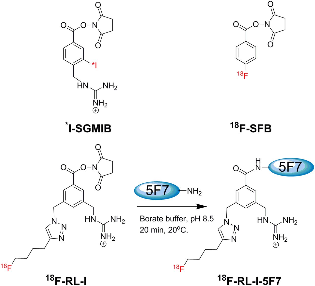
Structures of *I-SGMIB and 18F-SFB and scheme for labeling 5F7 Nanobody using 18F-RL-I.
MATERIALS AND METHODS
Nanobody, Cells, and Culture Conditions
The production, purification, and characteristics of anti-HER2 5F7 Nanobody, obtained from Ablynx, in the format lacking the GlyCysCys tail, have been described previously (19). HER2-expressing BT474M1 human breast carcinoma cells (20) were cultured in Dulbecco modified Eagle medium/F12 medium containing 10% fetal calf serum, streptomycin (100 μg/mL), and penicillin (100 IU/mL) (Sigma Aldrich). Cells were cultured at 37°C in a humidified incubator under 5% CO2 with medium changed every 2 d. When about 80% confluent, cells were subcultured by trypsinization (0.05% Trypsin-ethylenediaminetetraacetic acid).
Radiolabeling Nanobody 5F7
Details of the synthesis of 18F-RL-I-5F7, 18F-SFB-5F7, and 125I-SGMIB-5F7, as well as the affinity and immunoreactivity of these immunoconjugates, have been reported in a recent publication (17) (the supplemental materials provide additional synthetic details [supplemental materials are available at http://jnm.snmjournals.org]).
Internalization Assays
Two sets of internalization assays were performed on BT474M1 cells comparing the behavior of 18F-RL-I-5F7 or 18F-SFB-5F7 with coincubated 125I-SGMIB-5F7 as described previously (19) but only at 1, 2, and 4 h (details are provided in the supplemental materials).
Biodistribution Studies
Paired-label studies were performed in mice with BT474M1 subcutaneous xenografts (details are provided in the supplemental materials) following protocols approved by the Duke University Institutional Animal Care & Use Committee (16,19). In experiment 1, 2 groups of 5 mice were injected via the tail vein with 148 kBq (4 μCi; 0.9 μg) of 125I-SGMIB-5F7 and 370 kBq (10 μCi; 5.9 μg) of 18F-RL-I-5F7 in 100 μL of phosphate-buffered saline. In experiment 2, 2 groups of 5 mice received 148 kBq (4 μCi; 0. 5 μg) of 125I-SGMIB-5F7 and 555 kBq (15 μCi; 3.8 μg) of 18F-SFB-5F7. At 1 and 2 h after injection, 5 mice were killed with an overdose of isoflurane and dissected, and tissues and blood were harvested. Tissues, blood, and urine were weighed and counted for 125I and 18F activity in an automated γ-counter. From these data, percentage injected dose per gram of tissue (%ID/g) and tumor–to–normal-tissue ratios were calculated.
Small-Animal PET/CT Imaging
Imaging was performed on an Inveon microPET/CT system (Siemens) in groups of 4 mice with BT474M1 xenografts with and without HER2 blocking. For HER2 blocking, mice were injected intravenously with trastuzumab in phosphate buffer (4.4 mg in 200 μL; 220 mg/kg) 24 h before injection of 3.0–4.5 MBq (80–120 μCi; ∼10 μg) of 18F-RL-I-5F7 in 100 μL of phosphate-buffered saline. Mice were anesthetized using 2%–3% isoflurane in oxygen and placed prone in the scanner gantry for a 5-min PET acquisition followed by a 5-min CT scan. Control mice were imaged at 1 and 2 h while HER2-blocked mice were imaged at 1 h. List-mode PET data were histogram-processed and the images reconstructed using standard 3-dimensional ordered-subset expectation maximization/maximum a posteriori algorithm—two 3-dimensional ordered-subset expectation maximization iterations, and eighteen maximum a posteriori iterations—with a cutoff (Nyquist) of 0.5. Images were corrected for attenuation (CT-based) and radioactive decay. Image analysis was performed using Inveon Research Workplace software (Siemens). Regions of interest were drawn around tumors on the coregistered PET and CT images, and 18F uptake was expressed as SUV and %ID/g.
Statistical Analysis
Results are presented as mean ± SD. The statistical significance of differences in uptake between 2 tracers that were coinjected (18F vs. 125I) was calculated with a 2-tailed, paired Student t test using Microsoft Excel, whereas a 2-tailed unpaired Student t test was used to compare the results obtained for the 2 18F-labeling methods in different groups of animals. A P value of less than 0.05 was considered statistically significant.
RESULTS
Internalization Assays
In the first study (Fig. 2A), intracellularly trapped 18F-RL-I-5F7 activity was 49.3% ± 1.6%, 49.9% ± 2.1%, and 47.5% ± 2.1%, of initially cell-bound levels, at 1, 2, and 4 h, respectively, values that were slightly lower than those for coincubated 125I-SGMIB-5F7 (53.4% ± 0.8%, 55.0% ± 1.2%, and 52.1% ± 0.3%, respectively). In contrast, intracellular counts from 18F-SFB-5F7 decreased from 39.9% ± 0.3% at 1 h to 24.5% ± 1.1% at 4 h (Fig. 2B), values that were significantly lower (P < 0.05) than those for coincubated 125I-SGMIB-5F7 (1 h, 51.2% ± 0.7%; 4 h, 51.3% ± 2.6%). Normalizing to coadministered 125I-SGMIB-5F7 was performed for the 2 experiments, and the resultant 18F-to-125I ratios shown in Figure 2C further demonstrate the advantage of 18F-RL-I-5F7 over 18F-SFB-5F7 with regard to intracellular trapping of 18F activity. These results suggest that RL-I, like SGMIB, helps retain radioactivity in BT474M1 cells in vitro after internalization of labeled Nanobody.

Paired-label internalization of 125I-SGMIB-5F7 (blue bars) versus 18F-RL-I-5F7 (magenta bars) (A) and 125I-SGMIB-5F7 (blue bars) versus 18F-SFB-5F7 (green bars) (B) in BT-471M1 breast cancer cells in vitro. (C) Ratio of 18F to 125I obtained from 2 experiments.
Biodistribution Studies
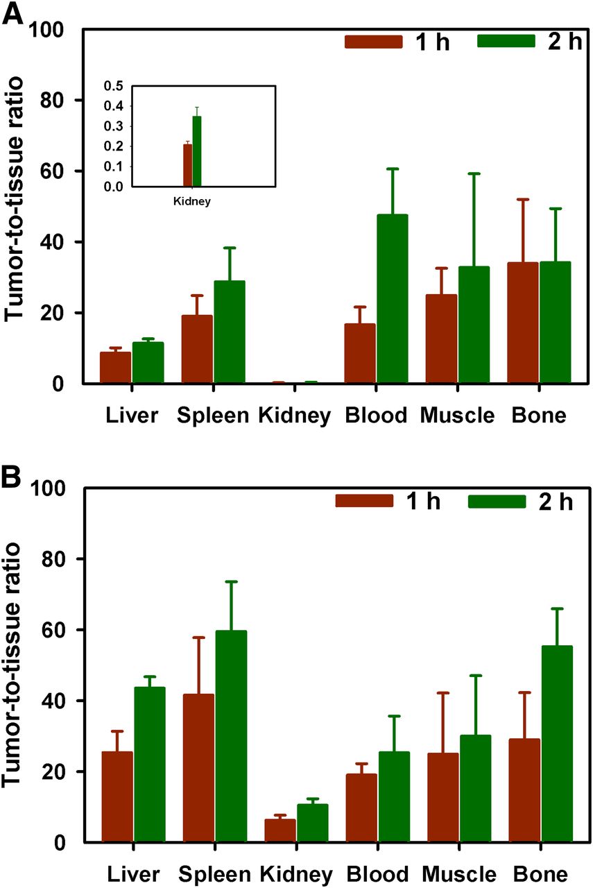
The tissue distribution of 18F-RL-I-5F7 and 18F-SFB-5F7 were compared with that of coadministered 125I-SGMIB-5F7 in mice with BT474M1 xenografts at 1 and 2 h, and the results are presented in Tables 1 and 2, respectively. Tumor uptake of 18F-RL-I-5F7 increased from 29.0 ± 3.9 %ID/g at 1 h to 36.3 ± 14.1 %ID/g at 2 h and was significantly higher (P < 0.05) than that of coadministered 125I-SGMIB-5F7. In contrast, tumor uptake of 18F-SFB-5F7 was also more than 20 %ID/g but significantly lower (P < 0.05) than that of coadministered 125I-SGMIB-5F7 at all time points. When normalized to 125I-SGMIB-5F7 levels (Fig. 3), the tumor uptake of 18F-RL-I-5F7 was 26%–28% higher (P < 0.05) than that of 18F-SFB-5F7, consistent with the hypothesized effects of charged guanidine (17) and polar triazole (21) moieties in 18F-RL-I on trapping of labeled catabolites, thereby enhancing tumor uptake. Generally, uptake of the 2 18F-5F7 conjugates in normal tissues was more than an order of magnitude lower than that in tumor, with the exception of renal levels for 18F-RL-I-5F7, which were comparable to those for 125I-SGMIB-5F7. Tumor–to–normal-tissue ratios for both 18F-labeled 5F7 conjugates increased from 1 to 2 h after injection (Fig. 4). With 18F-RL-I-5F7, the tumor-to-blood and tumor-to-muscle ratios at 2 h reached 47 ± 13 and 38 ± 21, respectively, values that were higher than those observed for 18F-SFB-5F7 (25 ± 10; P < 0.02 and 30 ± 17; P > 0.05, respectively). In contrast, tumor-to-tissue ratios for 18F-SFB-5F7 were higher than those for 18F-RL-I-5F7 in the liver, spleen, bone, and most notably, kidneys (P < 0.04–0.001).
Paired-Label Biodistribution of 18F-RL-I-5F7 and 125I-SGMIB-5F7 in Severe Combined Immunodeficiency Mice Bearing BT474M1 Xenografts
Paired Label Biodistribution of 18F-SFB-5F7 and 125I-SGMIB-5F7 in Severe Combined Immunodeficiency Mice Bearing BT474M1 Xenografts

18F-to-125I ratio in tumor from paired-label biodistribution of 18F-RL-I5F7 and 125I-SGMIB-5F7 and 18F-SFB-5F7 and 125I-SGMIB-5F7 in severe combined immunodeficiency mice bearing BT474M1 xenografts. Green bars = 18F-RL-I-5F7; magenta bars = 18F-SFB-5F7.

Tumor-to-tissue ratios for selected tissues obtained from biodistribution of 18F-RL-I-5F7 (A) and 18F-SFB-5F7 (B).
Small-Animal PET/CT Imaging
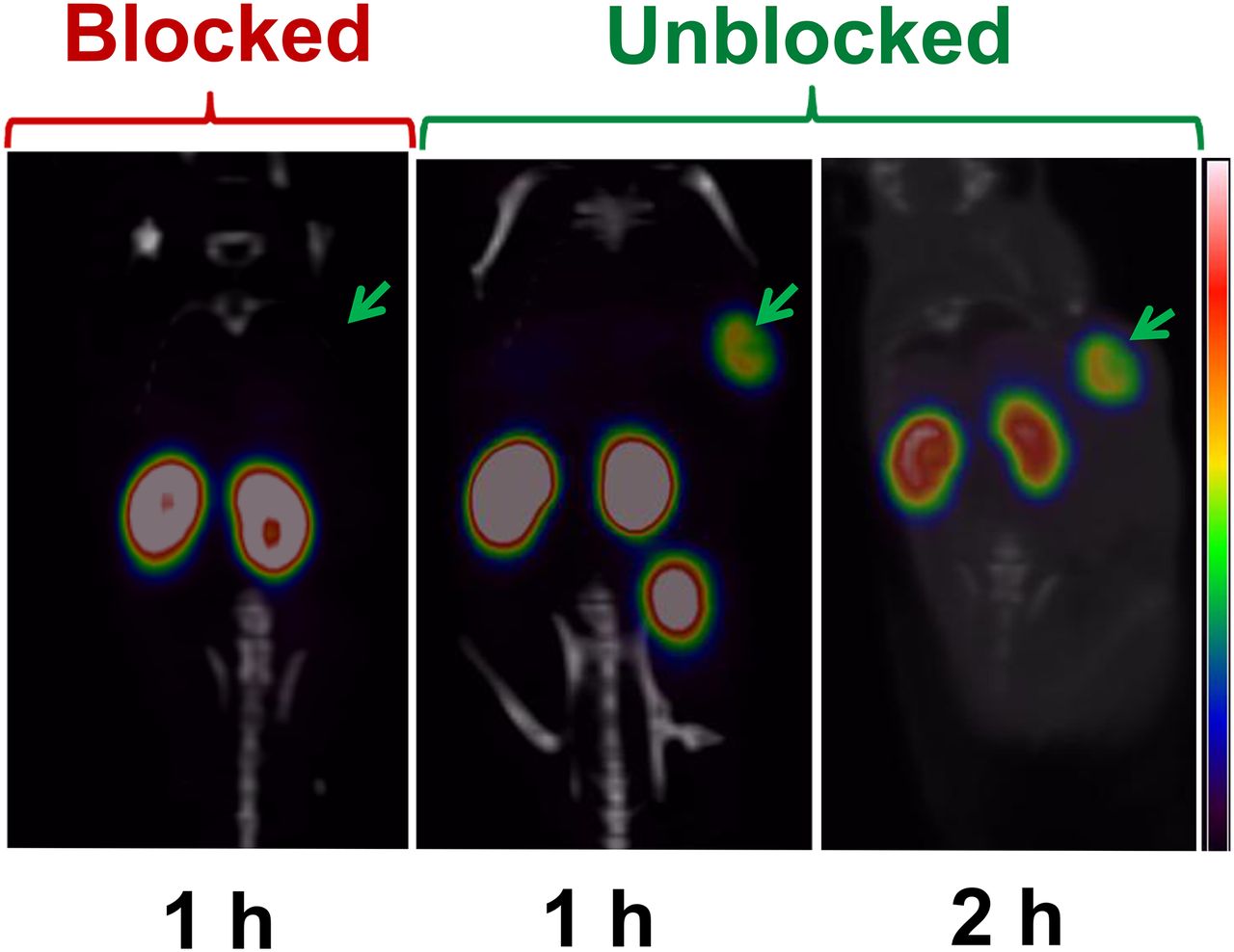
Representative small-animal PET/CT whole-body coronal images of the mice with BT474M1 xenografts obtained 1 and 2 h after injection of 18F-RL-I-5F7 as well as for a mouse receiving a blocking dose of trastuzumab 24 h before tracer injection are shown in Figure 5. SUV and %ID/g values calculated from the imaging data are presented in Table 3; consistent with the necropsy experiments, high tumor uptake was observed at both time points. No significant uptake was seen in normal organs other than the kidneys and bladder, resulting in high-contrast images. Preinjection of trastuzumab reduced tumor accumulation of 18F-RL-I-5F7 by more than 90%, confirming that tumor localization was HER2-specific.

PET/CT images of mice bearing BT474M1 xenografts after injection of 18F-RL-I-5F7. Images were obtained at 1 and 2 h without and at 1 h with blocking of HER2 receptors by preadministration of trastuzumab.
Tumor SUV and %ID/g from Small-Animal PET/CT Imaging of Severe Combined Immunodeficiency Mice Bearing BT474M1 Xenografts after Injection of 18F-RL-I-5F7
DISCUSSION
Nanobodies are an attractive platform for use in tandem with short-lived positron emitters because of their rapid tumor uptake and normal-tissue clearance (15). A recent phase-1 clinical study with a 68Ga-labeled Nanobody (68Ga-NOTA-2Rs15 d) demonstrated the feasibility of evaluating HER2 status in patients with breast carcinoma metastases by immuno-PET (22). Although encouraging results were reported, 18F might be an even more attractive radionuclide for labeling Nanobodies for several reasons. Compared with 68Ga, 18F has a more than 3-fold-lower energy and tissue range, resulting in improved spatial resolution. Moreover, the longer physical half-life of 18F provides the option for delayed imaging in circumstances in which background activity may be problematic and also allows radiopharmaceutical distribution from regional production sites, facilitating widespread use. In the present study, we have evaluated 2 approaches for labeling Nanobodies with 18F—a novel residualizing label that we developed for 18F labeling of proteins and peptides targeting internalizing receptors such as HER2 (17) as well as 18F-SFB, the most widely used protein/peptide radiofluorination agent for which several automated procedures have already been developed (23).
Our previous studies with radioiodinated anti-HER2 5F7 Nanobody documented the importance of using a residualizing labeling approach for maximizing retention of radioactivity in HER2-expressing tumors (16,19). Because peak and cumulative tumor radioactivity levels with 131I-SGMIB-5F7 were considerably higher than previously reported for any Nanobody radionuclide combination (15), SGMIB was selected as the design template for creating an 18F-labeled residualizing label. 18F-RL-I was synthesized and used to label the 5F7 Nanobody in reasonable radiochemical yield, with preservation of immunoreactivity (62%–80%) and affinity (4.7 ± 0.9 nM) for HER2 after labeling (17).
The potential advantage of the residualizing labeling agent was first evaluated in internalization assays performed with HER2-expressing BT474M1 breast carcinoma cells. Because there is no suitable fluorine radionuclide to use in tandem with 18F, direct paired-label comparisons of Nanobody labeled with 18F-RL-I and 18F-SFB cannot be performed. Instead, indirect comparison was made by performing 2 paired-label studies with 125I-SGMIB-5F7 serving as a common reference. Intracellularly trapped radioactivity levels for 5F7 labeled with 18F-RL-I remained constant at more than 47% of initial cell-bound radioactivity over the 4-h experiment and were similar to those for coincubated 125I-SGMIB-5F7. In contrast, intracellular radioactivity levels for 18F-SFB-5F7 were lower and decreased with time, and exhibited behavior on this cell line similar to 5F7-GGC Nanobody labeled using IODO-GEN (16), demonstrating the residualizing capability of the 18F-RL-I moiety.
Biodistribution and small-animal PET imaging experiments in severe combined immunodeficiency mice with HER2-expressing BT474M1 xenografts demonstrated rapid tumor accumulation and blood-pool clearance of 18F-RL-I-5F7. Pretreatment with trastuzumab reduced tumor levels more than 10-fold, confirming that uptake was HER2-specific. When normalized to coadministered 125I-SGMIB-5F7, tumor accumulation of 18F-RL-I-5F7 was 26%–28% higher than that observed with 18F-SFB-5F7 at 1 and 2 h, consistent with the 16%–24% higher normalized intracellular activity measured with BT474M1 cells in vitro for 18F-RL-I-5F7. By 4 h, the intracellular retention advantage increased to 47%, suggesting that the residualizing ability of the RL-I prosthetic group might be even more pronounced in vivo at later time points.
It is worth noting that the tumor accumulation of 5F7 after labeling with both 18F-labeled prosthetic groups was higher than that observed in this xenograft model when this Nanobody was radioiodinated using either the IODO-GEN or the IB-Mal-d-GEEEK methods (16,19) and considerably higher than that reported for any other combination of Nanobody, radionuclide, and xenograft model (15,24,25). With regard to other studies with 18F, tumor accumulation of Nanobodies labeled using 18F-SFB and targeting the macrophage mannose receptor (26) and HER2 (27) were reported to be 2.40 ± 0.46 %ID/g (3 h) and 3.09 ± 0.02 %ID/g (1 h), respectively, about 10-fold lower than observed in the current study. Use of a sortase-based site-specific method involving a click reaction for labeling Nanobodies with 18F also has been reported (28); however, the goal was imaging immune response to tumor, not a cancer cell surface molecular target.
It is also relevant to compare the tumor targeting of these 18F-labeled 5F7 conjugates with 18F-labeled anti-HER2 Affibody molecules because of the similarity in molecular weight (6.5 vs. 12–15 kDa) and intended clinical application for these labeled proteins. In studies with HER2-specific ZHER2:342 Affibody labeled via N-2-(4-18F-fluorobenzamido)ethyl]maleimide performed in mice with xenografts expressing high levels of HER2, peak tumor uptake occurred at 1 h and ranged from about 10–22 %ID/g (29,30). A second-generation Affibody, ZHER2:2891 (GE-226) with improved HER2 affinity (76 pM), was evaluated in mice with HER2-expressing NCI-N87 xenografts after labeling with 18F by 3 methods; optimal tumor accumulation was obtained (7.15 ± 0.69 %ID/g at 90 min) when labeling was performed using 4-18F-fluorobenzaldehyde (FBA) (31). In a subsequent PET imaging study with 18F-FBA-GE-226, peak tumor uptake in 3 high-HER2-expressing xenografts ranged from 10.9 ± 1.5 %ID/mL for MCF7-HER2 to 18.7 ± 2.4 %ID/mL for SKOV-3 (14). Although differences in variables such as animal model, protein dose, and internalization rate could play a role (32), the results obtained in the current study with 18F-labeled anti-HER2 5F7 Nanobody compare favorably with those reported for 18F-labeled Affibody molecules.
Normal-tissue clearance of the labeled Nanobody conjugates was quite rapid except from the kidneys for 18F-RL-I-5F7 and 125I-SGMIB-5F7. This behavior is consistent with the high degree of renal retention observed with other proteins with molecular weights less than 60 kDa (33) as well as Nanobodies labeled with radiometals (15), other residualizing radiohalgen moieties (19), and those containing polar amino acid residues at the C-terminal (24,25). Exceptions to this behavior are Nanobodies labeled with radioiodine using IODO-GEN (16,19), presumably reflecting their rapid dehalogenation in vivo and the about 30-fold-lower kidney uptake observed in the current study for 18F-SFB-5F7, compared with 18F-RL-I-5F7 and 125I-SGMIB-5F7. A factor that could contribute to the low renal activity levels seen with 18F-SFB-5F7 is the formation of 4-18F-fluorohippuric acid, the primary metabolite reported from other proteins labeled using the 18F-SFB method (34). On the other hand, the polar triazole (21) and especially the guanidine moieties in 18F-RL-I might have contributed to its high renal retention as seen with Nanobodies bearing other polar functionalities (24,25). Future studies are planned to determine whether the radioactivity retained in the kidneys is due to intact 18F-RL-I-5F7 or trapping of lower-molecular-weight catabolites generated by its lysosomal proteolysis (33,35).
From an imaging perspective, high renal activity levels should not interfere with lesion detection both for primary and the most common sites of metastases for HER2-positive cancers, as was demonstrated in a recent study with a 68Ga-NOTA anti-HER2 Nanobody (22). If necessary, a significant reduction in kidney uptake of radiolabeled Nanobodies can be achieved through the use of positively charged amino acids or the plasma expander Gelofusine (Braun Medical) (24). It also may be possible to decrease kidney uptake by introducing brush border enzyme-cleavable linkers in the prosthetic moiety (35,36), and efforts in this direction are under way in our laboratories. In addition, these 18F-labeled 5F7 conjugates are not retained in the liver, a frequent site of metastases for HER2-positive breast cancers. This is a potential advantage compared with other HER2-specific immuno-PET agents such as 89Zr-DFO-trastuzumab that exhibit significant accumulation in the liver (37).
CONCLUSION
The results of this study demonstrate the feasibility of using 18F-labeled anti-HER2 Nanobodies for the evaluation of HER2-expressing cancers. Excellent tumor targeting was observed with both reagents; however, use of the recently developed residualizing agent 18F-RL-I resulted accumulation and retention of significantly higher 18F levels in BT474M1 human breast carcinoma cells and xenografts compared with 18F-SFB. As has been observed previously with Nanobodies labeled with residualizing radiometals, renal uptake of 18F-RL-I-5F7 was high, which if problematic, might require compensatory strategies such as Gelofusine administration. Both 18F-RL-I-5F7 and 18F-SFB-5F7 warrant further evaluation as tracers for the evaluation of HER2-expressing cancers using immuno-PET.
DISCLOSURE
The costs of publication of this article were defrayed in part by the payment of page charges. Therefore, and solely to indicate this fact, this article is hereby marked “advertisement” in accordance with 18 USC section 1734. This work was supported in part by National Institutes of Health grants CA188177 and CA42324 and for small-animal PET imaging, by S10RR31792. No other potential conflict of interest relevant to this article was reported.
Acknowledgments
We thank Hilde Revets (Ablynx, Belgium) for providing the 5F7 Nanobody, Xiao-Guang Zhao for biodistribution studies, and Thomas Hawk for help with small-animal PET imaging studies.
Footnotes
Published online Feb. 18, 2016.
- © 2016 by the Society of Nuclear Medicine and Molecular Imaging, Inc.
REFERENCES
- Received for publication December 14, 2015.
- Accepted for publication February 3, 2016.


Jump to section
Related Articles
Cited By...
- Single-Domain Antibody Theranostics on the Horizon
- Site-Specific and Residualizing Linker for 18F Labeling with Enhanced Renal Clearance: Application to an Anti-HER2 Single-Domain Antibody Fragment
- PET Imaging of Receptor Tyrosine Kinases in Cancer
- Same-Day Imaging Using Small Proteins: Clinical Experience and Translational Prospects in Oncology
- Preclinical and clinical applications of specific molecular imaging for HER2-positive breast cancer