Main menu

  • Home
  • Content
    • Current
    • Ahead of print
    • Past Issues
    • JNM Supplement
    • SNMMI Annual Meeting Abstracts
    • Continuing Education
    • JNM Podcasts
  • Subscriptions
    • Subscribers
    • Institutional and Non-member
    • Rates
    • Journal Claims
    • Corporate & Special Sales
  • Authors
    • Submit to JNM
    • Information for Authors
    • Assignment of Copyright
    • AQARA requirements
  • Info
    • Reviewers
    • Permissions
    • Advertisers
  • About
    • About Us
    • Editorial Board
    • Contact Information
  • More
    • Alerts
    • Feedback
    • Help
    • SNMMI Journals
  • SNMMI
    • JNM
    • JNMT
    • SNMMI Journals
    • SNMMI

User menu

  • Subscribe
  • My alerts
  • Log in
  • My Cart

Search

  • Advanced search
Journal of Nuclear Medicine
  • SNMMI
    • JNM
    • JNMT
    • SNMMI Journals
    • SNMMI
  • Subscribe
  • My alerts
  • Log in
  • My Cart
Journal of Nuclear Medicine

Advanced Search

  • Home
  • Content
    • Current
    • Ahead of print
    • Past Issues
    • JNM Supplement
    • SNMMI Annual Meeting Abstracts
    • Continuing Education
    • JNM Podcasts
  • Subscriptions
    • Subscribers
    • Institutional and Non-member
    • Rates
    • Journal Claims
    • Corporate & Special Sales
  • Authors
    • Submit to JNM
    • Information for Authors
    • Assignment of Copyright
    • AQARA requirements
  • Info
    • Reviewers
    • Permissions
    • Advertisers
  • About
    • About Us
    • Editorial Board
    • Contact Information
  • More
    • Alerts
    • Feedback
    • Help
    • SNMMI Journals
  • View or Listen to JNM Podcast
  • Visit JNM on Facebook
  • Join JNM on LinkedIn
  • Follow JNM on Twitter
  • Subscribe to our RSS feeds
OtherThis Month in JNM

This Month in JNM

Journal of Nuclear Medicine May 2008, 49 (5) 11A-12A;
  • Article
  • Info & Metrics
  • PDF
Loading

MRI in tumor microenvironments: Penet and colleagues review the potential for MRI of cancer cells within the complex environment formed by the extracellular matrix, blood and lymphatic vessels, infiltrating leukocytes, and other cells.
Page 687

131I therapy risks: Boreham and Dolling survey current challenges in assessing the effects of therapeutic exposure to radioiodine during pregnancy and preview an article on this topic in this issue of JNM.
Page 691

11C-MET PET of irradiated brain tumors: Terakawa and colleagues evaluate the diagnostic accuracy of 11C-MET PET for the differentiation of recurrence from radiation necrosis in patients previously treated for metastatic brain tumors or glioma.
Page 694

Figure

18F-fluoride PET accuracy: Frost and colleagues compare the precision of 18F-fluoride PET with that of biochemical markers of bone turnover in the assessment of regional bone disease over a 6-mo period.
Page 700

PET in uterine smooth muscle tumors: Yoshida and colleagues investigate whether combined MRI and 18F-FDG PET imaging is more accurate than MRI alone in differentiating nonbenign uterine smooth muscle tumors from leiomyomas.
Page 708

Figure

New HSV1-tk mutant: Likar and colleagues describe the development of a reporter gene with specificity and high phosphorylation activity for acycloguanosine derivatives and potential for PET imaging of suicidal gene therapy protocols.
Page 713

Metabolic imaging of human gliomas: Stadlbauer and colleagues report on research supporting multimodal evaluation of metabolic changes in cerebral gliomas, correlating 18F-FET uptake on PET and concentrations of choline, creatine, and N-acetyl-aspartate as determined with proton MR spectroscopic imaging.
Page 721

PET and MRI for NILs: Floeth and colleagues describe characteristic MRI growth patterns and 18F-FET uptake in nonspecific incidental brain lesions as predictors of eventual development of low- and high-grade gliomas.
Page 730

Figure

Radiation dose in cardiac PET/CT: Gould and colleagues test the quantitative accuracy of a single poststress cine CT attenuation scan for reconstructing cardiac rest perfusion images, thus eliminating resting CT attenuation scans and reducing cumulative radiation dose.
Page 738

Figure

Gated SPECT for diastolic function: Patel and colleagues explore the benefits of combining data on perfusion defects with data on diastolic impairment to predict left ventricular end-diastolic pressure at subsequent cardiac catheterization.
Page 746

SPECT/CT and brown adipose tissue: Goetze and colleagues assess the frequency with which uptake of 99mTc-MIBI is present in brown adipose tissue in an adult patient population.
Page 752

Figure

SPECT and the pathophysiology of anxiety: van der Wee and colleagues use SPECT to examine 123I-β-CIT binding potentials for serotonin and dopamine transporters in individuals with and without a generalized social anxiety disorder.
Page 757

rhTSH-stimulated ablation and recurrence: Tuttle and colleagues report on rates of disease recurrence with recombinant human thyroid-stimulating hormone administration after radioiodine therapy and compare these rates with those from conventional thyroid hormone withdrawal.
Page 764

Xenon gas index for emphysema: Mathews and colleagues investigate the potential of a new quantitative gas trapping index derived from a 133Xe ventilation scan to assess the severity of emphysema and provide valuable data for lung volume reduction procedures.
Page 771

Figure

Lower gastrointestinal tract evaluation: Mariani and colleagues provide an educational overview of the technical aspects and diagnostic performance parameters of nuclear medicine procedures used in patients with disorders of the lower gastrointestinal tract.
Page 776

In vivo evaluation of nanobodies: Gainkam and colleagues use SPECT/CT to compare in vivo tumor uptake and biodistribution of two 99mTc-labeled anti–epidermal growth factor receptor nanobodies with potential for radioimmunodetection of specific targets early after therapy.
Page 788

Figure

Galectin-3 targeting and breast tumors: Kumar and Deutscher evaluate the tumor cell–targeting and SPECT properties of a 111In-labeled galectin-3–avid peptide in human breast carcinoma cells and human breast tumor–bearing mice.
Page 796

Figure

HER2 PET with 18F-labeled affibody: Cheng and colleagues explore the potential of 2 radiofluorinated anti–human epidermal growth factor receptor type 2 protein scaffold molecules as potential molecular probes for small-animal PET.
Page 804

Figure

18F-DPA-714 for imaging TSPO: James and colleagues detail the synthesis, radiofluorination, and pharmacologic evaluation of a new translocator protein–specific pyrazolopyrimidine.
Page 814

Figure

Imaging probe for melanoma: Miao and colleagues evaluate the potential utility of an imaging surrogate for an experimentally successful peptide-targeted α-therapy for melanoma and discuss the potential for patient-specific dosimetry and monitoring of tumor response.
Page 823

Novel ligand for angiogenesis imaging: Jeong and colleagues describe the conjugation and 68Ga labeling of an RGD derivative for angiogenesis imaging in ischemic tissue and detail the results of initial evaluations and PET studies.
Page 830

Dosimetry in myeloablative RIT: Rajendran and colleagues review the records of 100 patients who underwent tracer infusion of 131I-tositumomab before radioimmunotherapy for B-cell non-Hodgkin's lymphoma for data supporting an optimal approach to dosimetry.
Page 837

Radioiodine therapy and pregnancy: Garsi and colleagues update a 10-y-old study about pregnancy outcomes and the health of offspring of women exposed to 131I during thyroid carcinoma treatment.
Page 845

Internal dose uncertainties: Stabin presents a systematic analysis of inherent uncertainty in internal dose calculations for radiopharmaceuticals and discusses the level of risk and concern associated with this uncertainty in clinical practice.
Page 853

Figure

ON THE COVER

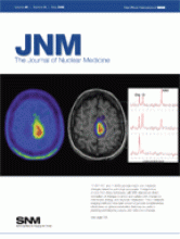
18F-FET PET and 1H MRSI provide insight into metabolic changes related to pathologic processes. Coregistration of data from these techniques with MRI data allows direct correlation of changes in amino acid uptake with changes in membrane, energy, and neuronal metabolism. The 2 metabolic imaging methods have been shown to provide complementary information on glioma metabolism that may be useful in planning and targeting surgery and radiochemotherapy.

Figure

See page 724.

PreviousNext
Back to top

In this issue

Journal of Nuclear Medicine: 49 (5)
Journal of Nuclear Medicine
Vol. 49, Issue 5
May 2008
  • Table of Contents
  • Table of Contents (PDF)
  • About the Cover
  • Index by author
Print
Download PDF
Article Alerts
Sign In to Email Alerts with your Email Address
Email Article

Thank you for your interest in spreading the word on Journal of Nuclear Medicine.

NOTE: We only request your email address so that the person you are recommending the page to knows that you wanted them to see it, and that it is not junk mail. We do not capture any email address.

Enter multiple addresses on separate lines or separate them with commas.
This Month in JNM
(Your Name) has sent you a message from Journal of Nuclear Medicine
(Your Name) thought you would like to see the Journal of Nuclear Medicine web site.
Citation Tools
This Month in JNM
Journal of Nuclear Medicine May 2008, 49 (5) 11A-12A;

Citation Manager Formats

  • BibTeX
  • Bookends
  • EasyBib
  • EndNote (tagged)
  • EndNote 8 (xml)
  • Medlars
  • Mendeley
  • Papers
  • RefWorks Tagged
  • Ref Manager
  • RIS
  • Zotero
Share
This Month in JNM
Journal of Nuclear Medicine May 2008, 49 (5) 11A-12A;
Twitter logo Facebook logo LinkedIn logo Mendeley logo
  • Tweet Widget
  • Facebook Like
  • Google Plus One
Bookmark this article

Jump to section

  • Article
  • Info & Metrics
  • PDF

Related Articles

  • Molecular and Functional MRI of the Tumor Microenvironment
  • Risks Associated with Therapeutic 131I Radiation Exposure
  • Diagnostic Accuracy of 11C-Methionine PET for Differentiation of Recurrent Brain Tumors from Radiation Necrosis After Radiotherapy
  • Long-Term Precision of 18F-Fluoride PET Skeletal Kinetic Studies in the Assessment of Bone Metabolism
  • Comparison of 18F-FDG PET and MRI in Assessment of Uterine Smooth Muscle Tumors
  • A New Acycloguanosine-Specific Supermutant of Herpes Simplex Virus Type 1 Thymidine Kinase Suitable for PET Imaging and Suicide Gene Therapy for Potential Use in Patients Treated with Pyrimidine-Based Cytotoxic Drugs
  • Metabolic Imaging of Cerebral Gliomas: Spatial Correlation of Changes in O-(2-18F-Fluoroethyl)-l-Tyrosine PET and Proton Magnetic Resonance Spectroscopic Imaging
  • Prognostic Value of 18F-Fluoroethyl-l-Tyrosine PET and MRI in Small Nonspecific Incidental Brain Lesions
  • Reducing Radiation Dose in Rest–Stress Cardiac PET/CT by Single Poststress Cine CT for Attenuation Correction: Quantitative Validation
  • Diastolic Filling Parameters Derived from Myocardial Perfusion Imaging Can Predict Left Ventricular End-Diastolic Pressure at Subsequent Cardiac Catheterization
  • Visualization of Brown Adipose Tissue with 99mTc-Methoxyisobutylisonitrile on SPECT/CT
  • Increased Serotonin and Dopamine Transporter Binding in Psychotropic Medication–Naïve Patients with Generalized Social Anxiety Disorder Shown by 123I-β-(4-Iodophenyl)-Tropane SPECT
  • Recombinant Human TSH–Assisted Radioactive Iodine Remnant Ablation Achieves Short-Term Clinical Recurrence Rates Similar to Those of Traditional Thyroid Hormone Withdrawal
  • New 133Xe Gas Trapping Index for Quantifying Severe Emphysema Before Partial Lung Volume Reduction
  • Radionuclide Evaluation of the Lower Gastrointestinal Tract
  • Comparison of the Biodistribution and Tumor Targeting of Two 99mTc-Labeled Anti-EGFR Nanobodies in Mice, Using Pinhole SPECT/Micro-CT
  • 111In-Labeled Galectin-3–Targeting Peptide as a SPECT Agent for Imaging Breast Tumors
  • Small-Animal PET Imaging of Human Epidermal Growth Factor Receptor Type 2 Expression with Site-Specific 18F-Labeled Protein Scaffold Molecules
  • DPA-714, a New Translocator Protein–Specific Ligand: Synthesis, Radiofluorination, and Pharmacologic Characterization
  • 203Pb-Labeled α-Melanocyte–Stimulating Hormone Peptide as an Imaging Probe for Melanoma Detection
  • Preparation of a Promising Angiogenesis PET Imaging Agent: 68Ga-Labeled c(RGDyK)–Isothiocyanatobenzyl-1,4,7-Triazacyclononane-1,4,7-Triacetic Acid and Feasibility Studies in Mice
  • Myeloablative 131I-Tositumomab Radioimmunotherapy in Treating Non-Hodgkin's Lymphoma: Comparison of Dosimetry Based on Whole-Body Retention and Dose to Critical Organ Receiving the Highest Dose
  • Therapeutic Administration of 131I for Differentiated Thyroid Cancer: Radiation Dose to Ovaries and Outcome of Pregnancies
  • Uncertainties in Internal Dose Calculations for Radiopharmaceuticals
  • Google Scholar

Cited By...

  • No citing articles found.
  • Google Scholar

More in this TOC Section

  • This Month in JNM
  • This Month in JNM
  • This Month in JNM
Show more This Month in JNM

Similar Articles

SNMMI

© 2025 SNMMI

Powered by HighWire