
Main menu

  • Home
  • Content
    • Current
    • Ahead of print
    • Past Issues
    • JNM Supplement
    • SNMMI Annual Meeting Abstracts
    • Continuing Education
    • JNM Podcasts
  • Subscriptions
    • Subscribers
    • Institutional and Non-member
    • Rates
    • Journal Claims
    • Corporate & Special Sales
  • Authors
    • Submit to JNM
    • Information for Authors
    • Assignment of Copyright
    • AQARA requirements
  • Info
    • Reviewers
    • Permissions
    • Advertisers
  • About
    • About Us
    • Editorial Board
    • Contact Information
  • More
    • Alerts
    • Feedback
    • Help
    • SNMMI Journals
  • SNMMI
    • JNM
    • JNMT
    • SNMMI Journals
    • SNMMI

User menu

  • Subscribe
  • My alerts
  • Log in
  • My Cart

Search

  • Advanced search
Journal of Nuclear Medicine
  • SNMMI
    • JNM
    • JNMT
    • SNMMI Journals
    • SNMMI
  • Subscribe
  • My alerts
  • Log in
  • My Cart
Journal of Nuclear Medicine

Advanced Search

  • Home
  • Content
    • Current
    • Ahead of print
    • Past Issues
    • JNM Supplement
    • SNMMI Annual Meeting Abstracts
    • Continuing Education
    • JNM Podcasts
  • Subscriptions
    • Subscribers
    • Institutional and Non-member
    • Rates
    • Journal Claims
    • Corporate & Special Sales
  • Authors
    • Submit to JNM
    • Information for Authors
    • Assignment of Copyright
    • AQARA requirements
  • Info
    • Reviewers
    • Permissions
    • Advertisers
  • About
    • About Us
    • Editorial Board
    • Contact Information
  • More
    • Alerts
    • Feedback
    • Help
    • SNMMI Journals
  • View or Listen to JNM Podcast
  • Visit JNM on Facebook
  • Join JNM on LinkedIn
  • Follow JNM on Twitter
  • Subscribe to our RSS feeds
Research ArticleCLINICAL INVESTIGATIONS

Comparison of 18F-FDG PET and MRI in Assessment of Uterine Smooth Muscle Tumors

Yoshio Yoshida, Tetsuji Kurokawa, Yoko Sawamura, Akiko Shinagawa, Tetsuya Tsujikawa, Hidehiko Okazawa, Tatsuro Tsuchida, Yoshiaki Imamura, Narufumi Suganuma and Fumikazu Kotsuji
Journal of Nuclear Medicine May 2008, 49 (5) 708-712; DOI: https://doi.org/10.2967/jnumed.107.047142
Yoshio Yoshida
  • Find this author on Google Scholar
  • Find this author on PubMed
  • Search for this author on this site
Tetsuji Kurokawa
  • Find this author on Google Scholar
  • Find this author on PubMed
  • Search for this author on this site
Yoko Sawamura
  • Find this author on Google Scholar
  • Find this author on PubMed
  • Search for this author on this site
Akiko Shinagawa
  • Find this author on Google Scholar
  • Find this author on PubMed
  • Search for this author on this site
Tetsuya Tsujikawa
  • Find this author on Google Scholar
  • Find this author on PubMed
  • Search for this author on this site
Hidehiko Okazawa
  • Find this author on Google Scholar
  • Find this author on PubMed
  • Search for this author on this site
Tatsuro Tsuchida
  • Find this author on Google Scholar
  • Find this author on PubMed
  • Search for this author on this site
Yoshiaki Imamura
  • Find this author on Google Scholar
  • Find this author on PubMed
  • Search for this author on this site
Narufumi Suganuma
  • Find this author on Google Scholar
  • Find this author on PubMed
  • Search for this author on this site
Fumikazu Kotsuji
  • Find this author on Google Scholar
  • Find this author on PubMed
  • Search for this author on this site
  • Article
  • Figures & Data
  • Info & Metrics
  • PDF
Loading

Article Figures & Data

Figures

  • Tables
  • FIGURE 1. 
    • Download figure
    • Open in new tab
    • Download powerpoint
    FIGURE 1. 

    Results of ROC analysis with histologic results used as reference standard. ROC curve generated for presence of nonbenign USMT demonstrates improved accuracy for MRI with 18F-FDG PET when compared with MRI alone.
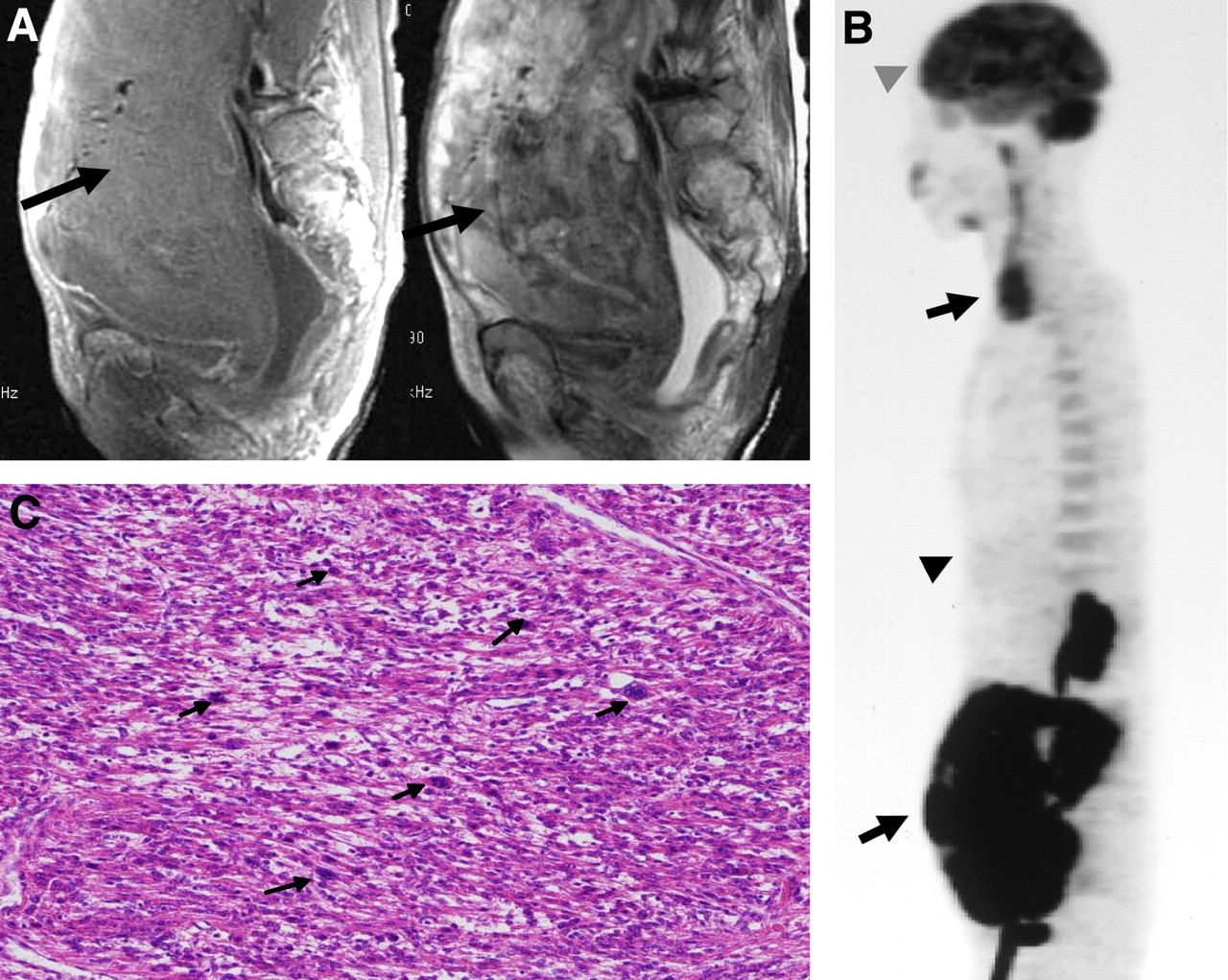
  • FIGURE 2. 
    • Download figure
    • Open in new tab
    • Download powerpoint
    FIGURE 2. 

    A 66-y-old woman with LMS. (A) Sagittal MRI shows large tumor with heterogeneous high signal intensity (arrow) on T1-weighted (left) and T2-weighted (right) images (MRI score, 3). (B) Sagittal 18F-FDG PET shows 18F-FDG uptake (top arrow indicates physiologic uptake in vocal cords, bottom arrow indicates USMT, bottom arrowhead indicates liver, and top arrowhead indicates brain) equivalent to that in brain (PET score, 3). Consensus score was “nonbenign.” (C) Histopathologic section of this tumor demonstrates LMS (hematoxylin-eosin stain, ×40. Arrows indicate mitotic figures).
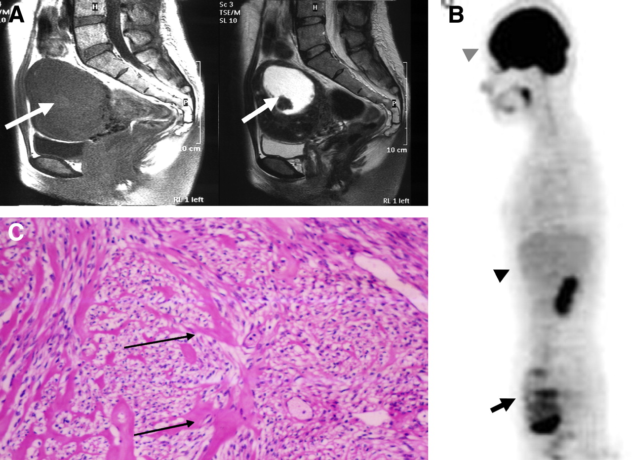
  • FIGURE 3. 
    • Download figure
    • Open in new tab
    • Download powerpoint
    FIGURE 3. 

    A 38-y-old woman with myxoid variant leiomyoma. (A) Sagittal MRI shows uterine mass with spotty pocket of high (arrow) signal intensity on T1-weighted (left) images and background of low signal intensity and a mass with central high (arrow) signal intensity on T2-weighted (right) images (MRI score, 3). (B) Sagittal 18F-FDG PET shows diffuse high (arrow) 18F-FDG uptake (arrow indicates USMT, bottom arrowhead indicates liver, and top arrowhead indicates brain) with multispots (PET score, 2). Consensus score was “probably nonbenign.” (C) Histopathologic section of this tumor reveals myxoid variant leiomyoma (hematoxylin-eosin stain, ×100. Arrows show myxoid degeneration).

  • FIGURE 4. 
    • Download figure
    • Open in new tab
    • Download powerpoint
    FIGURE 4. 

    A 38-y-old woman with USMTs of uncertain malignant potential. (A) Sagittal MRI shows uterine mass with low signal intensity (arrow) on T1-weighted (left) and T2-weighted (right) images (MRI score, 0). (B) Sagittal 18F-FDG PET shows equivalent-to-liver 18F-FDG uptake (arrow indicates USMT, bottom arrowhead indicates liver, and top arrowhead indicates brain) with multispots (PET score, 1). Consensus score was “benign.” (C) Histopathologic section of the tumor confirms uncertain malignant potential (hematoxylin-eosin stain, ×100. Arrows indicate severe cytologic atypia).

Tables

  • Figures
    • View popup
    TABLE 1

    Clinical and Histopathologic Findings in 70 Patients Suspected of Having Nonbenign USMTs

    Findingn
    Histopathologic
     Nonbenign USMTs
      LMS10
      Smooth muscle tumors of uncertain malignant potential (USMTsUMP)5
     Benign USMTs
      Uncomplicated26
      Mitotically active variant1
      Cellular variant1
      Hemorrhagic cellular variant1
      Myxoid variant20
      Atypical variant1
      Lipoleiomyoma variant3
      Uncomplicated plus adenomyosis2
    Clinical
     Total number of patients
      Nonbenign USMTs15
      Benign USMTs55
     Average age (y)
      Nonbenign USMTs53.6 ± 15.1 (28–77)*
      Benign USMTs49.5 ± 8.2 (30–64)*
     Total number of postmenopausal patients
      Nonbenign USMTs10
      Benign USMTs23
     Average tumor size (cm)
      Nonbenign USMTs10.7 ± 5.76 (4–20)†
      Benign USMTs7.82 ± 2.65 (5–15)†
     Number of metastasis
      Nonbenign USMTs0
      Benign USMTs0
    • ↵* P > 0.05 (no significant difference in average age of patients between nonbenign USMT and benign USMT groups).

    • ↵† P > 0.05 (no significant difference in tumor size between nonbenign USMT and benign USMT groups).

    • Data for average age and tumor size are mean ± SD, with minimum and maximum values in parentheses.

    • View popup
    TABLE 2

    Analysis of Accuracy of 18F-FDG PET, MRI, and MRI Plus 18F-FDG PET

    SensitivitySpecificity
    Imaging techniquePercentageCIPercentageCIAccuracy (%)
    MRI (%)73.3 (11/15)0.45–0.9185.5 (47/55)0.73–0.9382.9 (58/70)
    18F-FDG PET (%)86.7 (13/15)0.58–0.9892.7 (51/55)0.82–0.9891.4 (64/70)
    MRI plus 18F-FDG PET (%)93.3 (14/15)0.66–0.9992.7 (51/55)0.82–0.9892.9 (65/70)
    • Probable nonbenign results were considered positive for the purpose of analysis.

PreviousNext
Back to top

In this issue

Journal of Nuclear Medicine: 49 (5)
Journal of Nuclear Medicine
Vol. 49, Issue 5
May 2008
  • Table of Contents
  • Table of Contents (PDF)
  • About the Cover
  • Index by author
Print
Download PDF
Article Alerts
Sign In to Email Alerts with your Email Address
Email Article

Thank you for your interest in spreading the word on Journal of Nuclear Medicine.

NOTE: We only request your email address so that the person you are recommending the page to knows that you wanted them to see it, and that it is not junk mail. We do not capture any email address.

Enter multiple addresses on separate lines or separate them with commas.
Comparison of 18F-FDG PET and MRI in Assessment of Uterine Smooth Muscle Tumors
(Your Name) has sent you a message from Journal of Nuclear Medicine
(Your Name) thought you would like to see the Journal of Nuclear Medicine web site.
Citation Tools
Comparison of 18F-FDG PET and MRI in Assessment of Uterine Smooth Muscle Tumors
Yoshio Yoshida, Tetsuji Kurokawa, Yoko Sawamura, Akiko Shinagawa, Tetsuya Tsujikawa, Hidehiko Okazawa, Tatsuro Tsuchida, Yoshiaki Imamura, Narufumi Suganuma, Fumikazu Kotsuji
Journal of Nuclear Medicine May 2008, 49 (5) 708-712; DOI: 10.2967/jnumed.107.047142

Citation Manager Formats

  • BibTeX
  • Bookends
  • EasyBib
  • EndNote (tagged)
  • EndNote 8 (xml)
  • Medlars
  • Mendeley
  • Papers
  • RefWorks Tagged
  • Ref Manager
  • RIS
  • Zotero
Share
Comparison of 18F-FDG PET and MRI in Assessment of Uterine Smooth Muscle Tumors
Yoshio Yoshida, Tetsuji Kurokawa, Yoko Sawamura, Akiko Shinagawa, Tetsuya Tsujikawa, Hidehiko Okazawa, Tatsuro Tsuchida, Yoshiaki Imamura, Narufumi Suganuma, Fumikazu Kotsuji
Journal of Nuclear Medicine May 2008, 49 (5) 708-712; DOI: 10.2967/jnumed.107.047142
Twitter logo Facebook logo LinkedIn logo Mendeley logo
  • Tweet Widget
  • Facebook Like
  • Google Plus One
Bookmark this article

Jump to section

  • Article
    • Abstract
    • MATERIALS AND METHODS
    • RESULTS
    • DISCUSSION
    • CONCLUSION
    • Acknowledgments
    • Footnotes
    • References
  • Figures & Data
  • Info & Metrics
  • PDF

Related Articles

  • This Month in JNM
  • PubMed
  • Google Scholar

Cited By...

  • The Effectiveness of 18F-FDG PET/CT Combined with STIR MRI for Diagnosing Nodal Involvement in the Thorax
  • Google Scholar

More in this TOC Section

  • Feasibility of Ultra-Low-Activity 18F-FDG PET/CT Imaging Using a Long–Axial-Field-of-View PET/CT System
  • Cardiac Presynaptic Sympathetic Nervous Function Evaluated by Cardiac PET in Patients with Chronotropic Incompetence Without Heart Failure
  • Validation and Evaluation of a Vendor-Provided Head Motion Correction Algorithm on the uMI Panorama PET/CT System
Show more Clinical Investigations

Similar Articles

SNMMI

© 2025 SNMMI

Powered by HighWire