
Main menu

  • Home
  • Content
    • Current
    • Ahead of print
    • Past Issues
    • JNM Supplement
    • SNMMI Annual Meeting Abstracts
    • Continuing Education
    • JNM Podcasts
  • Subscriptions
    • Subscribers
    • Institutional and Non-member
    • Rates
    • Journal Claims
    • Corporate & Special Sales
  • Authors
    • Submit to JNM
    • Information for Authors
    • Assignment of Copyright
    • AQARA requirements
  • Info
    • Reviewers
    • Permissions
    • Advertisers
  • About
    • About Us
    • Editorial Board
    • Contact Information
  • More
    • Alerts
    • Feedback
    • Help
    • SNMMI Journals
  • SNMMI
    • JNM
    • JNMT
    • SNMMI Journals
    • SNMMI

User menu

  • Subscribe
  • My alerts
  • Log in
  • My Cart

Search

  • Advanced search
Journal of Nuclear Medicine
  • SNMMI
    • JNM
    • JNMT
    • SNMMI Journals
    • SNMMI
  • Subscribe
  • My alerts
  • Log in
  • My Cart
Journal of Nuclear Medicine

Advanced Search

  • Home
  • Content
    • Current
    • Ahead of print
    • Past Issues
    • JNM Supplement
    • SNMMI Annual Meeting Abstracts
    • Continuing Education
    • JNM Podcasts
  • Subscriptions
    • Subscribers
    • Institutional and Non-member
    • Rates
    • Journal Claims
    • Corporate & Special Sales
  • Authors
    • Submit to JNM
    • Information for Authors
    • Assignment of Copyright
    • AQARA requirements
  • Info
    • Reviewers
    • Permissions
    • Advertisers
  • About
    • About Us
    • Editorial Board
    • Contact Information
  • More
    • Alerts
    • Feedback
    • Help
    • SNMMI Journals
  • View or Listen to JNM Podcast
  • Visit JNM on Facebook
  • Join JNM on LinkedIn
  • Follow JNM on Twitter
  • Subscribe to our RSS feeds
OtherBASIC SCIENCE INVESTIGATIONS

A New 18F-Labeled Myocardial PET Tracer: Myocardial Uptake After Permanent and Transient Coronary Occlusion in Rats

Takahiro Higuchi, Stephan G. Nekolla, Marc M. Huisman, Sybille Reder, Thorsten Poethko, Ming Yu, Hans-Jurgen Wester, David S. Casebier, Simon P. Robinson, Rene M. Botnar and Markus Schwaiger
Journal of Nuclear Medicine October 2008, 49 (10) 1715-1722; DOI: https://doi.org/10.2967/jnumed.108.053967
Takahiro Higuchi
  • Find this author on Google Scholar
  • Find this author on PubMed
  • Search for this author on this site
Stephan G. Nekolla
  • Find this author on Google Scholar
  • Find this author on PubMed
  • Search for this author on this site
Marc M. Huisman
  • Find this author on Google Scholar
  • Find this author on PubMed
  • Search for this author on this site
Sybille Reder
  • Find this author on Google Scholar
  • Find this author on PubMed
  • Search for this author on this site
Thorsten Poethko
  • Find this author on Google Scholar
  • Find this author on PubMed
  • Search for this author on this site
Ming Yu
  • Find this author on Google Scholar
  • Find this author on PubMed
  • Search for this author on this site
Hans-Jurgen Wester
  • Find this author on Google Scholar
  • Find this author on PubMed
  • Search for this author on this site
David S. Casebier
  • Find this author on Google Scholar
  • Find this author on PubMed
  • Search for this author on this site
Simon P. Robinson
  • Find this author on Google Scholar
  • Find this author on PubMed
  • Search for this author on this site
Rene M. Botnar
  • Find this author on Google Scholar
  • Find this author on PubMed
  • Search for this author on this site
Markus Schwaiger
  • Find this author on Google Scholar
  • Find this author on PubMed
  • Search for this author on this site
  • Article
  • Figures & Data
  • Info & Metrics
  • PDF
Loading

Article Figures & Data

Figures

  • FIGURE 1. 
    • Download figure
    • Open in new tab
    • Download powerpoint
    FIGURE 1. 

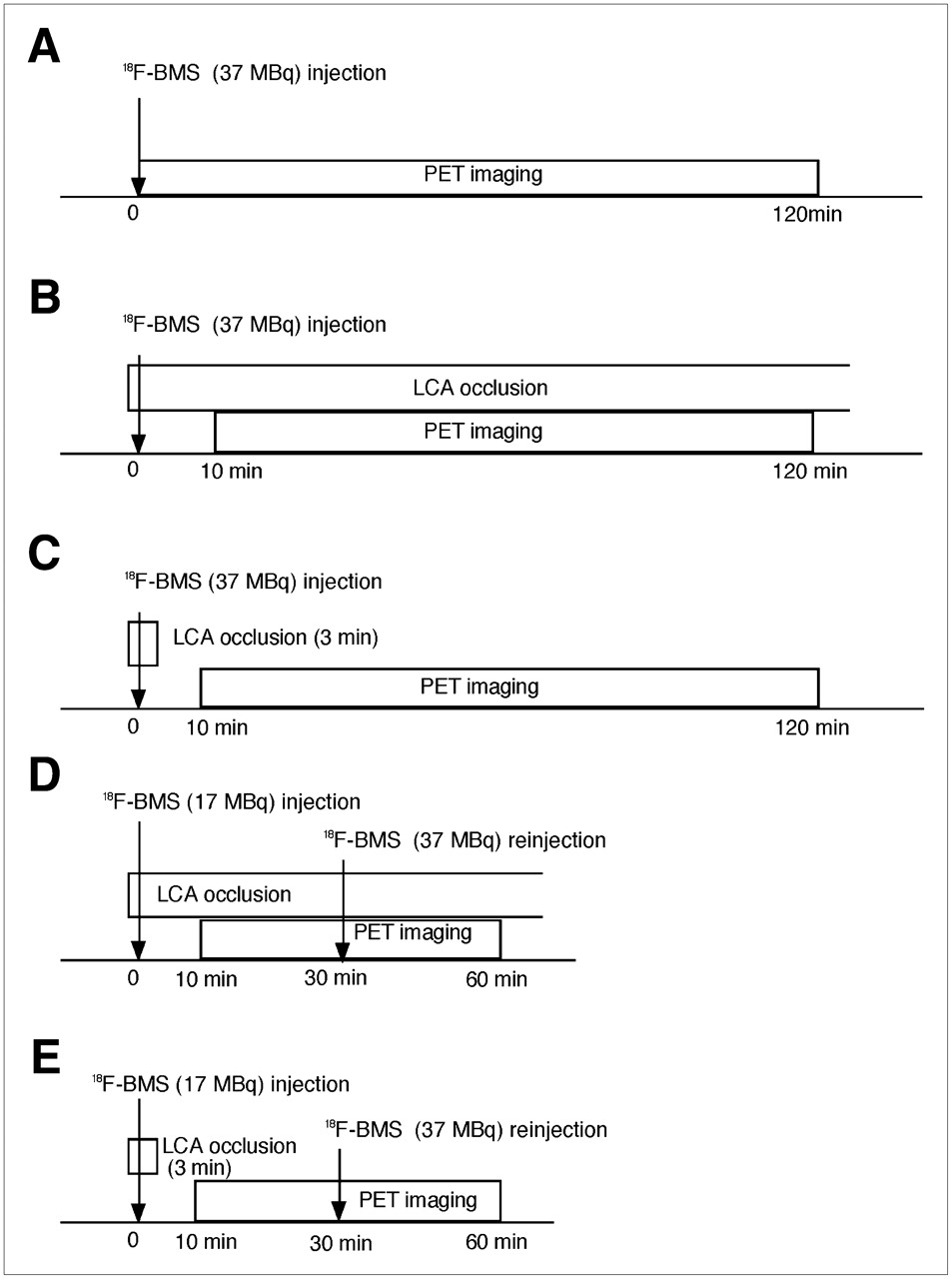
    Schemas of experimental protocols of permanent and transient coronary occlusion and imaging: normal heart (A), permanent LCA occlusion (B), transient LCA occlusion (C), reinjection after permanent LCA occlusion (D), and reinjection after transient LCA occlusion (E). 18F-BMS = 18F-BMS-747158-02.

  • FIGURE 2. 
    • Download figure
    • Open in new tab
    • Download powerpoint
    FIGURE 2. 

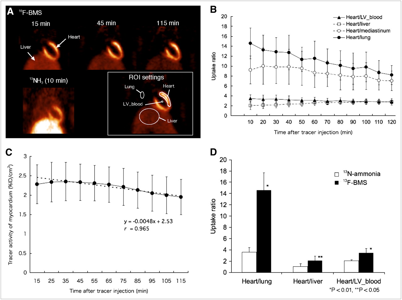
    (A) Examples of 18F-BMS-747158-02 (18F-BMS) PET images of chest of healthy rat at 15, 45, and 115 min after tracer injection and 13N-ammonia PET image at 10 min in coronal view. Example of regions of interest for measurement of tracer activity is displayed in white box. (B and C) Ratio of 18F-BMS uptake between myocardium and surrounding organs (B) and absolute 18F-BMS activity of myocardium (%ID/cm3) (C). (D) Comparison of 18F-BMS uptake ratios at 15 min after injection and 13N-ammonia at 10 min.

  • FIGURE 3. 
    • Download figure
    • Open in new tab
    • Download powerpoint
    FIGURE 3. 

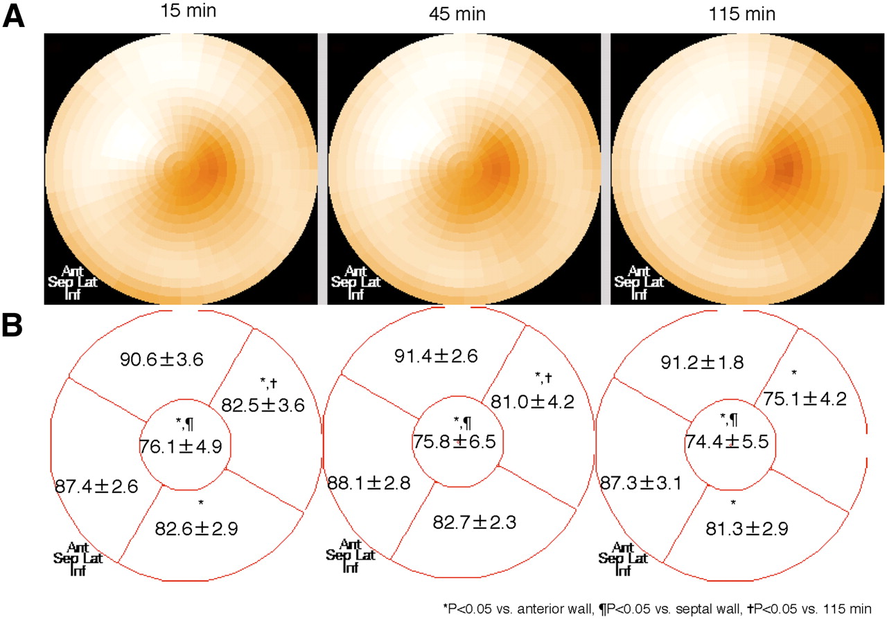
    Normal pattern of left ventricular 18F-BMS-747158-02 (18F-BMS) distribution at 15, 45, and 115 min after tracer injection as shown in mean polar maps (A) and as mean uptake values (% left ventricle maximum) with SD (B). Uptake was nearly homogeneous throughout left ventricle, with slightly lower uptake in apical segment.

  • FIGURE 4. 
    • Download figure
    • Open in new tab
    • Download powerpoint
    FIGURE 4. 

    Short-axis images of rat heart 1 wk after coronary occlusion using 18F-BMS-747158-02 (18F-BMS) PET and 13N-ammonia PET. Arrows indicate localization of myocardial infarction.
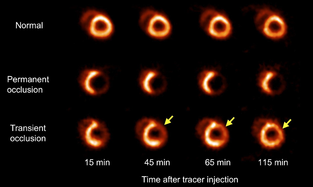
  • FIGURE 5. 
    • Download figure
    • Open in new tab
    • Download powerpoint
    FIGURE 5. 

    Representative short-axis images of rat hearts without coronary occlusion, permanent coronary occlusion, and transient coronary occlusion at different time points after tracer administration. Tracer redistribution is seen in induced defect after reperfusion (arrows).

  • FIGURE 6. 
    • Download figure
    • Open in new tab
    • Download powerpoint
    FIGURE 6. 

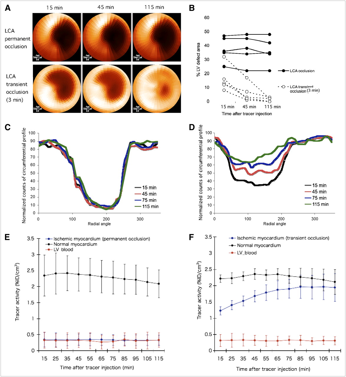
    Changes in myocardial 18F-BMS-747158-02 (18F-BMS) distribution after both permanent and transient coronary occlusion are shown in representative polar maps (A) and as defect area expressed as percentage of total left ventricular myocardium (B). Normalized counts of circumferential profiles after permanent occlusion (C) or transient occlusion (D) are also shown. Mean values are shown for tracer activity in induced defect and normally perfused myocardium after permanent (E) and transient occlusion (F). Tracer activity in defect increased over time after reperfusion in transient coronary occlusion.
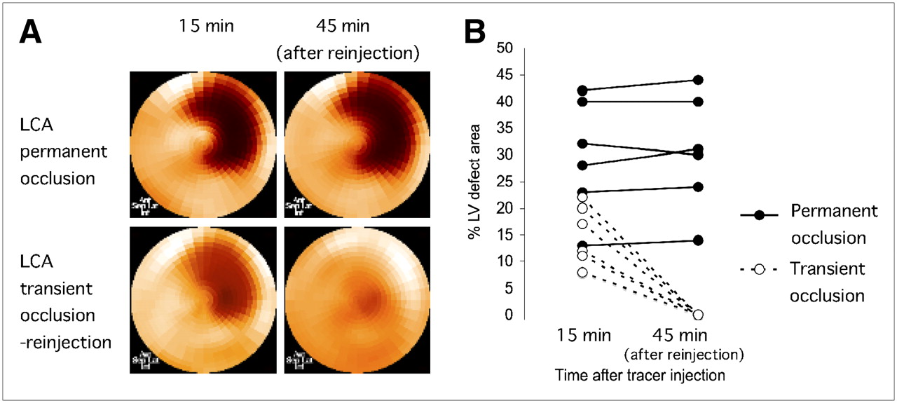
  • FIGURE 7. 
    • Download figure
    • Open in new tab
    • Download powerpoint
    FIGURE 7. 

    Myocardial 18F-BMS-747158-02 (18F-BMS) distribution patterns before and after tracer reinjection in rats after permanent and transient coronary occlusion. Representative polar maps (A) and plots of defect area (B) are shown. Tracer uptake defects resolved after tracer reinjection in rats after transient coronary occlusion but not in permanent coronary occlusion.

PreviousNext
Back to top

In this issue

Journal of Nuclear Medicine: 49 (10)
Journal of Nuclear Medicine
Vol. 49, Issue 10
October 2008
  • Table of Contents
  • Table of Contents (PDF)
  • About the Cover
  • Index by author
Print
Download PDF
Article Alerts
Sign In to Email Alerts with your Email Address
Email Article

Thank you for your interest in spreading the word on Journal of Nuclear Medicine.

NOTE: We only request your email address so that the person you are recommending the page to knows that you wanted them to see it, and that it is not junk mail. We do not capture any email address.

Enter multiple addresses on separate lines or separate them with commas.
A New 18F-Labeled Myocardial PET Tracer: Myocardial Uptake After Permanent and Transient Coronary Occlusion in Rats
(Your Name) has sent you a message from Journal of Nuclear Medicine
(Your Name) thought you would like to see the Journal of Nuclear Medicine web site.
Citation Tools
A New 18F-Labeled Myocardial PET Tracer: Myocardial Uptake After Permanent and Transient Coronary Occlusion in Rats
Takahiro Higuchi, Stephan G. Nekolla, Marc M. Huisman, Sybille Reder, Thorsten Poethko, Ming Yu, Hans-Jurgen Wester, David S. Casebier, Simon P. Robinson, Rene M. Botnar, Markus Schwaiger
Journal of Nuclear Medicine Oct 2008, 49 (10) 1715-1722; DOI: 10.2967/jnumed.108.053967

Citation Manager Formats

  • BibTeX
  • Bookends
  • EasyBib
  • EndNote (tagged)
  • EndNote 8 (xml)
  • Medlars
  • Mendeley
  • Papers
  • RefWorks Tagged
  • Ref Manager
  • RIS
  • Zotero
Share
A New 18F-Labeled Myocardial PET Tracer: Myocardial Uptake After Permanent and Transient Coronary Occlusion in Rats
Takahiro Higuchi, Stephan G. Nekolla, Marc M. Huisman, Sybille Reder, Thorsten Poethko, Ming Yu, Hans-Jurgen Wester, David S. Casebier, Simon P. Robinson, Rene M. Botnar, Markus Schwaiger
Journal of Nuclear Medicine Oct 2008, 49 (10) 1715-1722; DOI: 10.2967/jnumed.108.053967
Twitter logo Facebook logo LinkedIn logo Mendeley logo
  • Tweet Widget
  • Facebook Like
  • Google Plus One
Bookmark this article

Jump to section

  • Article
    • Abstract
    • MATERIALS AND METHODS
    • RESULTS
    • DISCUSSION
    • CONCLUSION
    • Acknowledgments
    • Footnotes
    • References
  • Figures & Data
  • Info & Metrics
  • PDF

Related Articles

  • This Month in JNM
  • PubMed
  • Google Scholar

Cited By...

  • Assessment of the 18F-Labeled PET Tracer LMI1195 for Imaging Norepinephrine Handling in Rat Hearts
  • Myocardial Uptake of 7'-(Z)-[123I]Iodorotenone During Vasodilator Stress in Dogs With Critical Coronary Stenoses
  • Stable Delineation of the Ischemic Area by the PET Perfusion Tracer 18F-Fluorobenzyl Triphenyl Phosphonium After Transient Coronary Occlusion
  • Radionuclide Imaging of Angiotensin II Type 1 Receptor Upregulation After Myocardial Ischemia-Reperfusion Injury
  • Evaluation of the Novel Myocardial Perfusion Positron-Emission Tomography Tracer 18F-BMS-747158-02: Comparison to 13N-Ammonia and Validation With Microspheres in a Pig Model
  • Evaluation of a Novel 18F-Labeled Positron-Emission Tomography Perfusion Tracer for the Assessment of Myocardial Infarct Size in Rats
  • Google Scholar

More in this TOC Section

  • PET/MRI of Hypoxic Atherosclerosis Using 64Cu-ATSM in a Rabbit Model
  • Tumor Uptake of Anti-CD20 Fabs Depends on Tumor Perfusion
  • How Sensitive Is the Upper Gastrointestinal Tract to 90Y Radioembolization? A Histologic and Dosimetric Analysis in a Porcine Model
Show more Basic Science Investigations

Similar Articles

SNMMI

© 2025 SNMMI

Powered by HighWire