
Main menu

  • Home
  • Content
    • Current
    • Ahead of print
    • Past Issues
    • JNM Supplement
    • SNMMI Annual Meeting Abstracts
  • Subscriptions
    • Subscribers
    • Institutional and Non-member
    • Rates
    • Corporate & Special Sales
    • Journal Claims
  • Authors
    • Submit to JNM
    • Information for Authors
    • Assignment of Copyright
    • AQARA requirements
  • Info
    • Continuing Education
    • Reviewers
    • Permissions
    • Advertisers
  • About
    • About Us
    • Editorial Board
    • Contact Information
  • More
    • Alerts
    • Feedback
    • Help
    • SNMMI Journals
  • SNMMI
    • JNM
    • JNMT
    • SNMMI Journals
    • SNMMI

User menu

  • Subscribe
  • My alerts
  • Log in
  • My Cart

Search

  • Advanced search
Journal of Nuclear Medicine
  • SNMMI
    • JNM
    • JNMT
    • SNMMI Journals
    • SNMMI
  • Subscribe
  • My alerts
  • Log in
  • My Cart
Journal of Nuclear Medicine

Advanced Search

  • Home
  • Content
    • Current
    • Ahead of print
    • Past Issues
    • JNM Supplement
    • SNMMI Annual Meeting Abstracts
  • Subscriptions
    • Subscribers
    • Institutional and Non-member
    • Rates
    • Corporate & Special Sales
    • Journal Claims
  • Authors
    • Submit to JNM
    • Information for Authors
    • Assignment of Copyright
    • AQARA requirements
  • Info
    • Continuing Education
    • Reviewers
    • Permissions
    • Advertisers
  • About
    • About Us
    • Editorial Board
    • Contact Information
  • More
    • Alerts
    • Feedback
    • Help
    • SNMMI Journals
  • Follow JNM on Twitter
  • Visit JNM on Facebook
  • Join JNM on LinkedIn
  • Subscribe to our RSS feeds

About the Cover

October 2008; Volume 49,Issue 10

Cover image

Cover image expansion

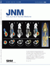
The need to study dynamic biologic processes in intact small-animal models has stimulated the development of high-resolution nuclear imaging methods. Above, small-animal SPECT/CT is applied to oncology research. In a prostate cancer xenograft model, radiolabeled antibodies for prostate-specific membrane antigen are used to monitor expression of the antigen. CT images and 3D rendering of fused images help define the tumor boundaries for more accurate image quantification.
See page 1651.

Back to top
PreviousNext

In this issue

Journal of Nuclear Medicine: 49 (10)
Journal of Nuclear Medicine
Vol. 49, Issue 10
October 2008
  • Table of Contents
  • Table of Contents (PDF)
  • About the Cover
  • Index by author
Sign up for alerts

Jump to

  • This Month in JNM
  • SNM Newsline
  • Focus on Molecular Imaging
  • Invited Perspectives
  • Clinical Investigations
  • Continuing Education
  • Erratum
  • Basic Science Investigations
  • Letters to the Editor
  • Book Reviews
  • Most Cited
  • Most Read
Loading
  • Initial Clinical Experience with 90Y-FAPI-46 Radioligand Therapy for Advanced-Stage Solid Tumors: A Case Series of 9 Patients
  • Total-Body PET Multiparametric Imaging of Cancer Using a Voxelwise Strategy of Compartmental Modeling
  • Prospective phase 2 trial of PSMA-targeted molecular RadiothErapy with 177Lu-PSMA-617 for metastatic castration-reSISTant Prostate Cancer (RESIST-PC): efficacy results of the UCLA cohort
  • Why We Did What We Did: PSMA PET/CT Selection Criteria for the VISION Trial
  • 68Ga-FAPI as a Diagnostic Tool in Sarcoma: Data from the 68Ga-FAPI PET Prospective Observational Trial
More...
SNMMI

© 2023 Journal of Nuclear Medicine

Powered by HighWire