


Article Figures & Data
Figures
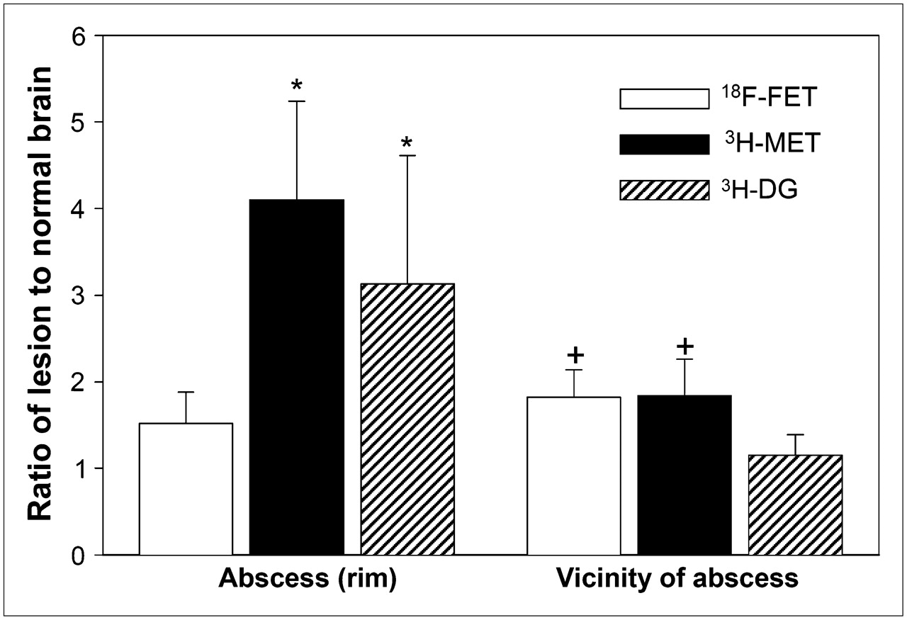
- FIGURE 1.
Comparison of lesion-to-normal brain ratios of 18F-FET, 3H-MET, and 3H-DG. (Left) Data for abscess rim (macrophage infiltration). (Right) Data for vicinity of abscess. Uptake of 3H-MET and 3H-DG is significantly increased in macrophages (*P < 0.01 vs. 18F-FET), whereas uptake of 18F-FET is close to that in normal brain tissue. In vicinity of abscess, slightly increased uptake of 18F-FET and 3H-MET is noted (+P < 0.01 vs. 3H-DG), whereas 3H-DG uptake is unchanged.
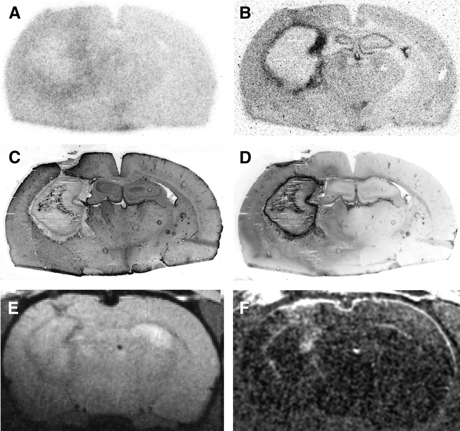
- FIGURE 2.
Coronal rat brain slices 5 d after abscess induction (rat 4). (A) 18F-FET autoradiography. (B) 3H-MET autoradiography. (C) Immunofluorescent imaging for GFAP. (D) Immunofluorescent imaging for CD68. (E) T1-weighted MRI. (F) Contrast-enhanced T1-weighted MRI after intravenous injection of Gd-DTPA (subtraction). Increased 3H-MET uptake in abscess rim is congruent with macrophage infiltration. 18F-FET is negative in that area. MRI shows typical ring-enhancing lesion.
- FIGURE 3.
Coronal rat brain slices 5 d after abscess induction (rat 9). (A) 18F-FET autoradiography. (B) 3H-DG autoradiography. (C) Immunofluorescent imaging for GFAP. (D) Immunofluorescent imaging for CD68. Increased 3H-DG uptake in abscess rim is congruent with macrophage infiltration. Again, 18F-FET is negative in that area. However, diffuse 18F-FET uptake in vicinity of abscess shows similarity to reactive astrocytosis (GFAP).
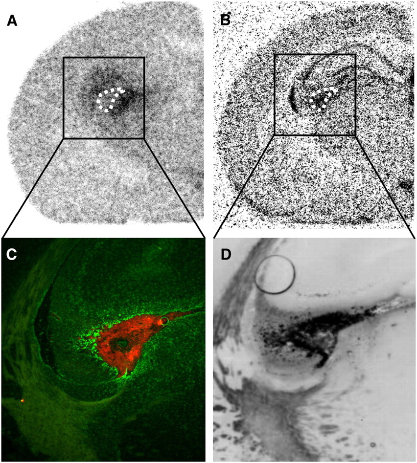
- FIGURE 4.
Coronal rat brain slices 5 d after abscess induction (rat 1). (A) 18F-FET autoradiography. (B) 3H-MET autoradiography. (C) Overlay of double immunofluorescent imaging for astrocytosis (GFAP, green) and microglia (CD11b, red). (D) Immunofluorescent imaging for CD68. C and D are same area at higher magnification. Dotted line indicates position of area with increased GFAP and CD68 binding in autoradiograms. Increased 18F-FET uptake appears to be colocalized with reactive astrocytosis in surrounding of this area (green cells), whereas increased 3H-MET uptake is found in area of activated microglia (red cells) that also shows immunoreactivity for macrophages (D).
- FIGURE 5.
Slices of calf muscle 5 d after abscess induction (rat 11). (A) 18F-FET autoradiography. (B) 3H-MET autoradiography. (C) Immunofluorescent imaging for cell nuclei (DAPI). (D) Immunofluorescent imaging for macrophages (CD68). Increased 3H-MET uptake in abscess rim is congruent with macrophage infiltration. 18F-FET is negative in that area.


Tables
18F-FET uptake in abscess wall and vicinity 3H-MET uptake in abscess wall and vicinity 3H-DG uptake in abscess wall and vicinity No. SUVmax abscess SUV vicinity L/B abscess L/B vicinity SUVmax abscess SUV vicinity L/B abscess L/B vicinity SUVmax abscess SUV vicinity L/B abscess L/B vicinity 1 1.3 1.6 1.9 2.2 6.1 3.7 4.1 2.5 2 1.1 1.4 1.5 1.9 2.3 1.0 3.3 1.4 3 1.1 1.5 1.3 1.7 4.2 2.8 2.7 1.8 4 1.8 1.8 2.1 2.2 7.7 2.6 5.5 1.9 5 1.6 1.4 1.5 1.7 8.5 2.7 4.9 1.6 6 0.5 0.6 1.7 2.2 7.4 2.5 2.8 1.5 7 2.1 1.5 1.1 1.4 10.3 4.4 2.1 1.0 8 1.9 3.0 1.0 1.5 18.5 8.6 2.3 1.1 9 1.6 2.1 1.4 1.8 15.1 2.8 5.3 1.0 Mean 1.4 1.7 1.5 1.8* 5.8† 2.6 4.1† 1.8* 12.8† 4.6 3.1† 1.2 SD 0.5 0.6 0.4 0.3 2.5 1.0 1.1 0.4 4.9 2.8 1.5 0.2 18F-FET uptake in abscess wall 3H-MET uptake in abscess wall No. SUVmax abscess SUV muscle L/B abscess SUVmax abscess SUV muscle L/B abscess 10 1.7 1.6 1.1 5.1 1.3 4.0 11 1.7 1.5 1.1 6.7 1.7 4.6 12 1.5 1.5 1.0 9.3 2.0 4.6 13 1.9 2.1 0.9 7.9 1.7 4.6 14 1.5 1.8 0.9 3.7 1.2 3.2 Mean 1.7 1.7 1.0 6.5 1.6 4.2* SD 0.2 0.3 0.1 2.2 0.3 0.6 ↵* P < 0.01 vs. L/B of FET.


Jump to section
Related Articles
Cited By...
- Distinguishing Progression from Pseudoprogression in Glioblastoma Using 18F-Fluciclovine PET
- Amino Acid PET in Neurooncology
- Utility of Amino Acid PET in the Differential Diagnosis of Recurrent Brain Metastases and Treatment-Related Changes: A Meta-analysis
- Amino Acid PET in Neurooncology
- Diagnostic value of PET with different radiotracers and MRI for recurrent glioma: a Bayesian network meta-analysis
- Treatment-Related Uptake of O-(2-18F-Fluoroethyl)-L-Tyrosine and L-[Methyl-3H]-Methionine After Tumor Resection in Rat Glioma Models
- Epileptic Activity Increases Cerebral Amino Acid Transport Assessed by 18F-Fluoroethyl-L-Tyrosine Amino Acid PET: A Potential Brain Tumor Mimic
- TSPO Imaging in Glioblastoma Multiforme: A Direct Comparison Between 123I-CLINDE SPECT, 18F-FET PET, and Gadolinium-Enhanced MR Imaging
- The Usefulness of Dynamic O-(2-18F-Fluoroethyl)-L-Tyrosine PET in the Clinical Evaluation of Brain Tumors in Children and Adolescents
- Replay: Being Sensitive: to Specify When Amino Acid Tracers Accumulate in a Brain Lesion
- Role of O-(2-18F-Fluoroethyl)-L-Tyrosine PET for Differentiation of Local Recurrent Brain Metastasis from Radiation Necrosis
- Transport of 3-Fluoro-L-{alpha}-Methyl-Tyrosine by Tumor-Upregulated L-Type Amino Acid Transporter 1: A Cause of the Tumor Uptake in PET
- Performance of 18F-Fluoro-Ethyl-Tyrosine (18F-FET) PET for the Differential Diagnosis of Primary Brain Tumor: A Systematic Review and Metaanalysis
- Classification of Peritumoral Fiber Tract Alterations in Gliomas Using Metabolic and Structural Neuroimaging
- Comparison of O-(2-18F-Fluoroethyl)-L-Tyrosine and L-3H-Methionine Uptake in Cerebral Hematomas
- Comparison of 99mTc- and 18F-Ubiquicidin Autoradiography to Anti-Staphylococcus aureus Immunofluorescence in Rat Muscle Abscesses
- Metabolic Imaging of Cerebral Gliomas: Spatial Correlation of Changes in O-(2-18F-Fluoroethyl)-L-Tyrosine PET and Proton Magnetic Resonance Spectroscopic Imaging