


Article Figures & Data
Figures
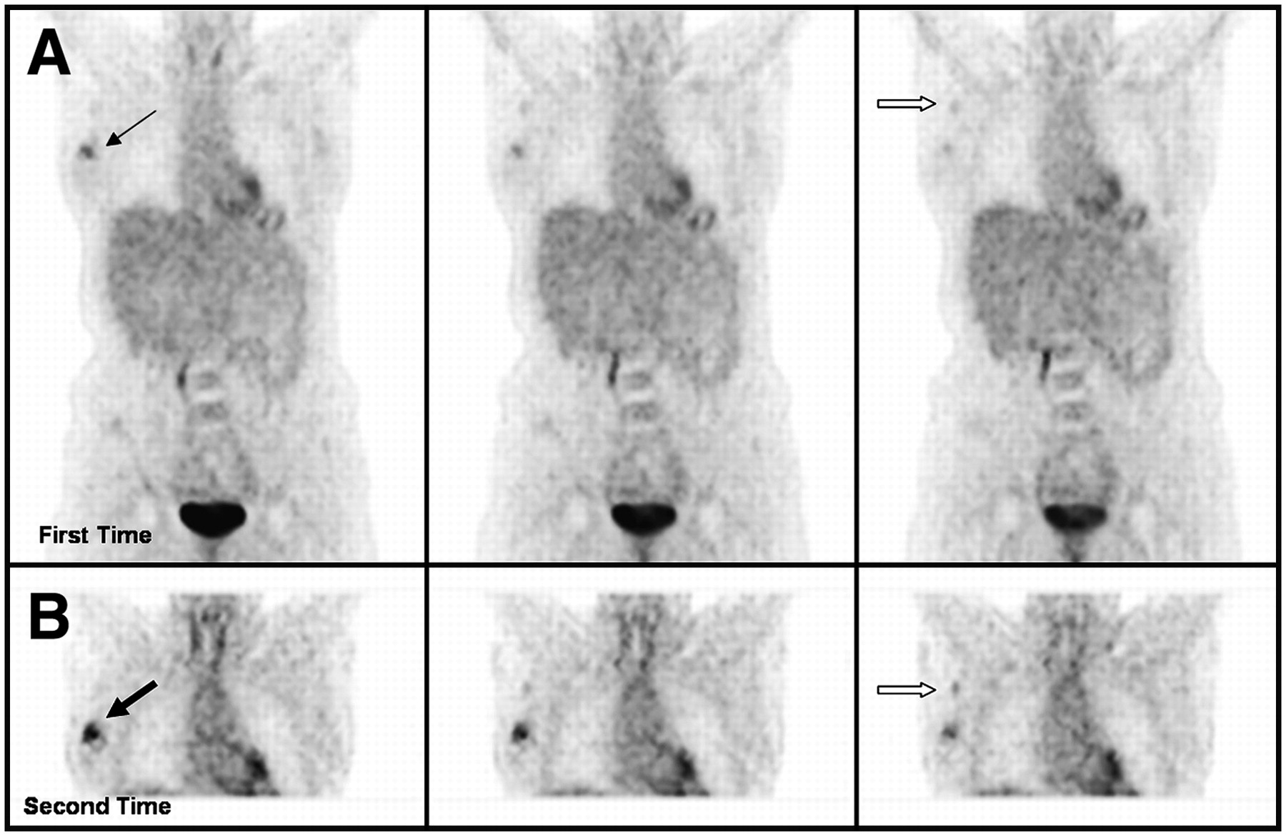
- FIGURE 1.
Patient with history of invasive ductal cancer of right breast had dual time point PET. (A) Coronal slices in top row were obtained at first time point. (B) Corresponding scans in bottom row were acquired at second time point. Measured SUVmax1 of lesion in first image set was 4.3 (thin arrow), whereas that of second set was 4.8 (thick arrow). Percent increase in SUV of the lesion was 11.6%. Measured SUVmax1 of normal contralateral glandular breast tissue was 1.1 in first image set, whereas that of second set was 0.9. Therefore, increase in T/B ratios between first image set and second image set was 36.4%. Surgical pathology confirmed 2-cm invasive ductal cancer.
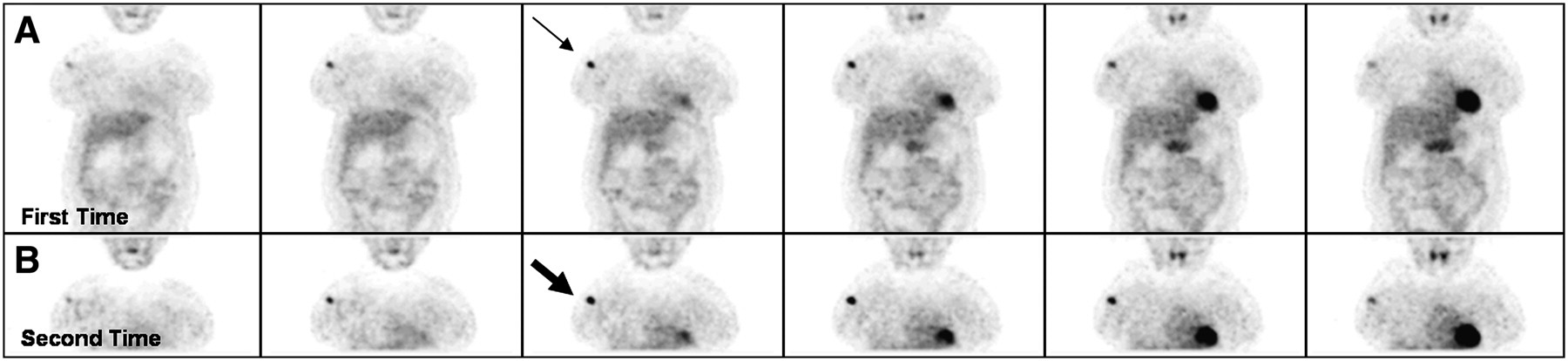
- FIGURE 2.
Patient with invasive ductal carcinoma of right breast was examined with dual time point PET. (A) Coronal slices in top row were obtained at first time point. (B) Corresponding images in bottom row were acquired at second time point. Images in both sets clearly show primary lesion. However, intensity of uptake was substantially higher on delayed images. In addition, axillary lymph node metastasis was faintly visualized on first set but was clearly demonstrated on second set. Measured SUVmax1 of lesion in first image set was 2.2 (thin black arrow), whereas that of second set was 2.6 (thick black arrow). Percent increase in SUV of the lesion was 18.2%. SUVmax1 of metastatic right axillary lymph node was 1.0 in first set (open arrow in A) and increased to 1.1 in second set (open arrow in B). Surgical pathology confirmed 2.5-cm invasive ductal carcinoma with axillary metastasis.
- FIGURE 3.
Patient with history of noninvasive carcinoma of right breast underwent dual time point 18F-FDG PET for preoperative staging. (A) Coronal slices in top row were obtained at first time point. (B) Corresponding scans in bottom row were acquired at second time point. Lesion is questionable in first set, whereas it is clearly visualized in second set. Measured SUVmax1 of lesion in first image set (thin arrow) was 1.4, whereas that of second set was 1.9 (thick arrow). Percent change from first to second time point of this measurement was 35.7%. Surgical pathology confirmed noninvasive breast tumor.


Tables
- TABLE 1
SUVmax Measurements and Changes over Time in Normal Breast, Invasive Cancer, Noninvasive Cancer, and T/B Ratios
Histopathology SUVmax1 SUVmax2 Δ%SUVmax Δ% in ratio 1 and ratio 2 (T/B ratio) Group A (n = 82) 3.9 ± 3.7 4.3 ± 4.0 8.3 ± 11.5 22.0 ± 26.8 Group B (n = 24) 2.0 ± 0.6 2.1 ± 0.6 3.4 ± 13.0 15.7 ± 18.6 Group C (n = 120) 1.2 ± 0.3 1.1 ± 0.2 −10.0 ± 10.8 Group A = invasive cancer; group B = noninvasive cancer; group C = contralateral breast; ratio 1 = T/B ratios of SUVmax at first time point; ratio 2 = T/B ratios of SUVmax at second time point; Δ% = percent change.
Data are presented as mean ± SD.
- TABLE 2
SUVmax Measurements and Changes over Time in Invasive Cancers According to Subtypes
Group A (n = 82) SUVmax1 SUVmax2 Δ% SUVmax Invasive ductal (n = 66) 4.3 ± 3.9* 4.7 ± 4.3* 8.1 ± 10.6* Invasive lobular (n = 7) 2.7 ± 1.8* 3.1 ± 2.3* 10.5 ± 14.0* Invasive mixed (n = 7) 2.0 ± 0.9* 2.2 ± 1.1* 9.3 ± 14.6* Medullary (n = 1) 7.2 8.6 19.4 Mucinous (n = 1) 1.0 1.0 0 ↵* Data are presented as mean ± SD.
Group A = invasive cancers; Δ% = percent change.
- TABLE 3
SUVmax Measurements and Changes over Time According to Tumor Size in Invasive Cancers with Increase in T/B Ratios
Group A (n = 82) SUVmax1 SUVmax2 Δ%SUVmax Δ% in ratio 1 and ratio 2 (T/B ratio) Tumors >10 mm (n = 57) 4.8 ± 4.1 5.3 ± 4.4 8.6 ± 12.2 23.1 ± 28.5 Tumors 4−10 mm (n = 25) 1.9 ± 0.8 2.0 ± 0.7 6.5 ± 9.9 18.8 ± 23.5 Group A = invasive cancer; ratio 1 = T/B ratios of SUVmax at first time point; ratio 2 = T/B ratios of SUVmax at second time point; Δ% = percent change.
Data are presented as mean ± SD.


Jump to section
Related Articles
Cited By...
- Assessing PET Parameters in Oncologic 18F-FDG Studies
- Multicenter Clinical Trials Using 18F-FDG PET to Measure Early Response to Oncologic Therapy: Effects of Injection-to-Acquisition Time Variability on Required Sample Size
- Voxel-Based Analysis of Dual-Time-Point 18F-FDG PET Images for Brain Tumor Identification and Delineation
- PET Tumor Metabolism in Locally Advanced Breast Cancer Patients Undergoing Neoadjuvant Chemotherapy: Value of Static versus Kinetic Measures of Fluorodeoxyglucose Uptake
- 18F-FDG PET of Locally Invasive Breast Cancer and Association of Estrogen Receptor Status with Standardized Uptake Value: Microarray and Immunohistochemical Analysis
- The Effect of Age, Menopausal State, and Breast Density on 18F-FDG Uptake in Normal Glandular Breast Tissue
- 18F-FDG Uptake in Lung, Breast, and Colon Cancers: Molecular Biology Correlates and Disease Characterization
- Reproducibility of Standardized Uptake Value Measurements Determined by 18F-FDG PET in Malignant Tumors
- Time and Again, Children Resemble Their Parents
- Breast Cancer Staging in a Single Session: Whole-Body PET/CT Mammography