


Article Figures & Data
Figures
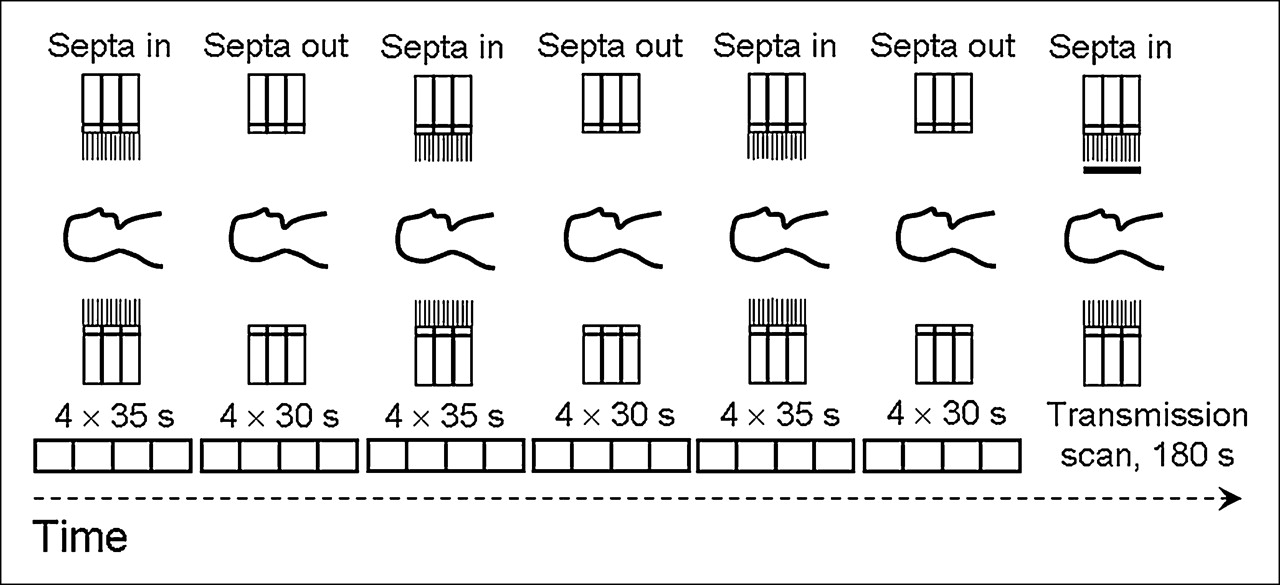
- FIGURE 1.
Research data acquisition occurred over a single-bed position using an interleaved septa-in/septa-out acquisition protocol. At each position of the septa, emission data were acquired in a dynamic mode (4 frames of either 30- or 35-s duration). Six dynamic emission scans were acquired followed by a 3-min transmission scan.
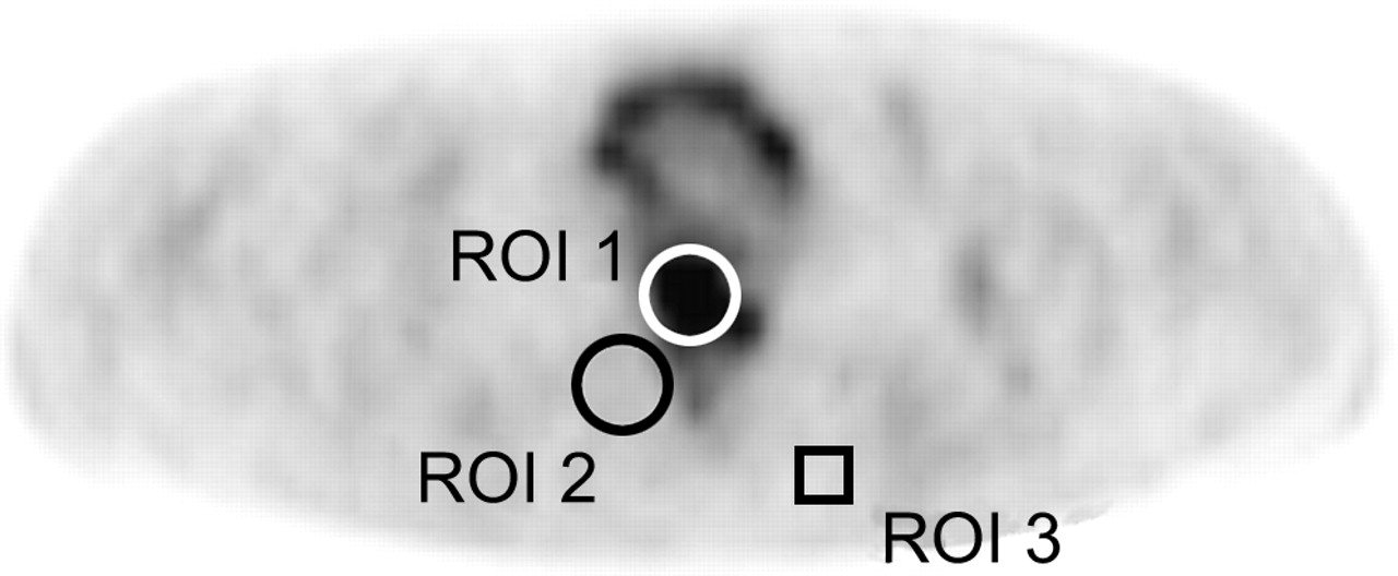
- FIGURE 2.
Lesion contrast was estimated by dividing the maximum pixel value in a target region (ROI 1), by the mean pixel value in a background region (ROI 2). Image noise was estimated by placing a 1.5 × 1.5 cm square region (ROI 3) in a background area of the central slice. Note that in this example the lesion was present in the central slice, although this was not typically the case.
- FIGURE 3.
Target-to-background ratios for the 420-s 2D and 360-s 3D images. (A) Both 2D and 3D images were smoothed with a 6-mm FWHM gaussian filter after OSEM reconstruction. Mean target-to-background ratios for 2D and 3D images were 6.0 ± 3.3 and 5.5 ± 2.8 (P = 0.005), respectively. (B) 2D images were smoothed with a 6-mm FWHM gaussian filter; and 3D images were smoothed with a 5-mm gaussian filter. Mean target-to-background ratios for 2D and 3D images were 6.0 ± 3.3 and 5.8 ± 3.0 (P = 0.16), respectively.
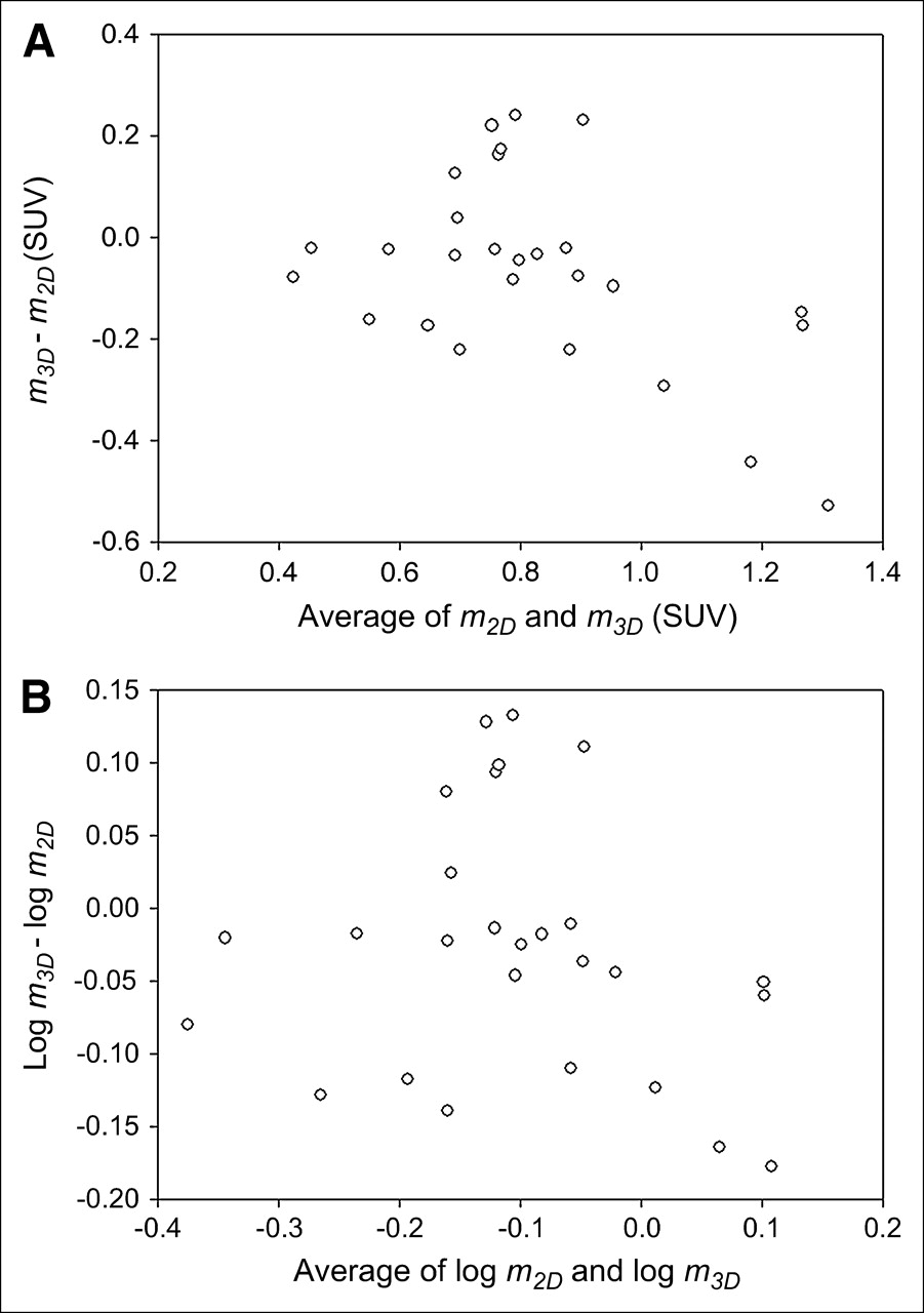
- FIGURE 4.
Difference between 2D and 3D measurements, d, as a function of the average of the 2D and 3D measurements, a. (A) Data are shown in original units (SUV = standardized uptake value). (B) Data are shown after logarithmic transformation.
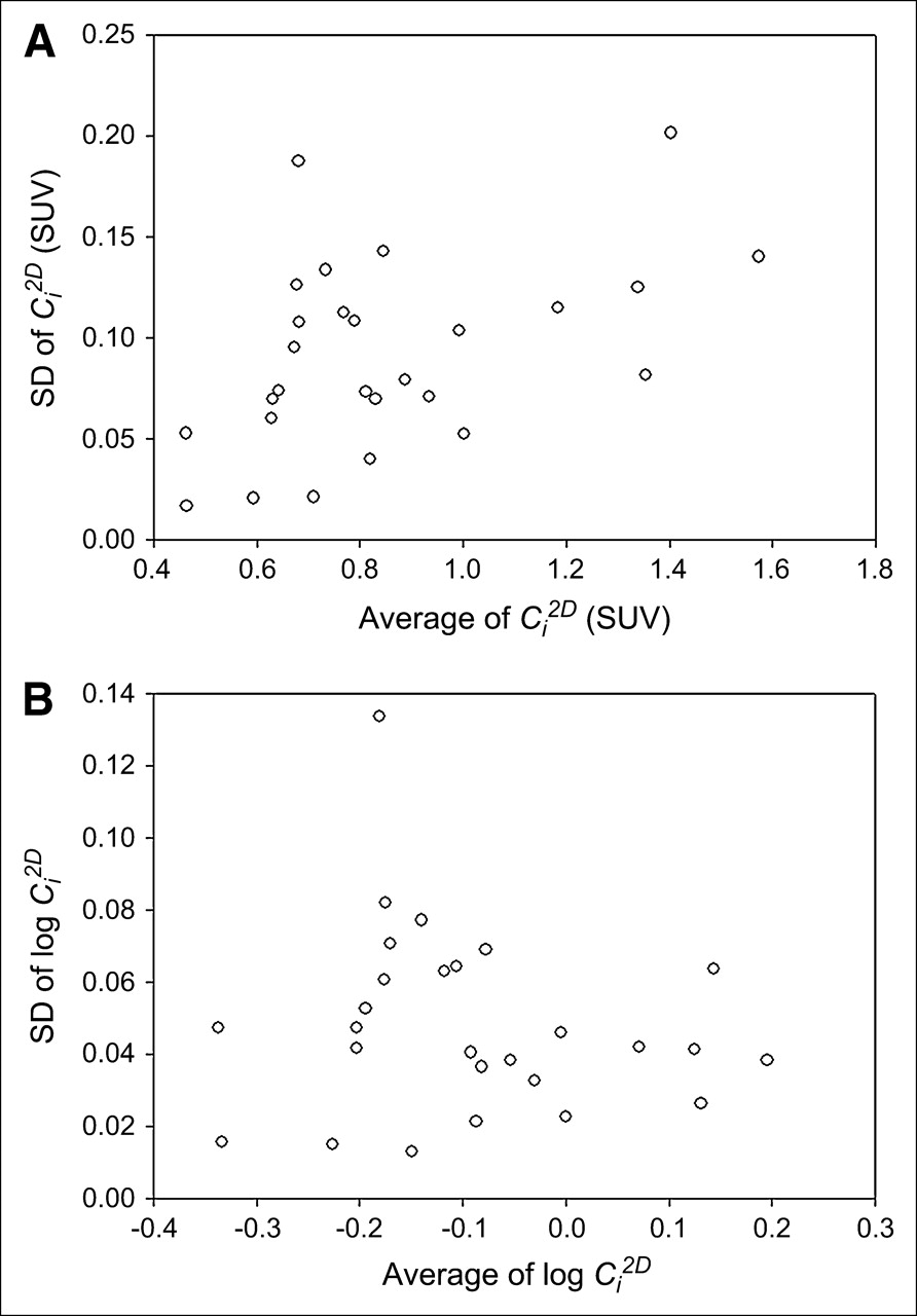
- FIGURE 5.
SD of 2D ROI data
 as a function of their average. (A) Data are shown in original units (SUV = standardized uptake value). (B) Data are shown after logarithmic transformation.
 - FIGURE 6.
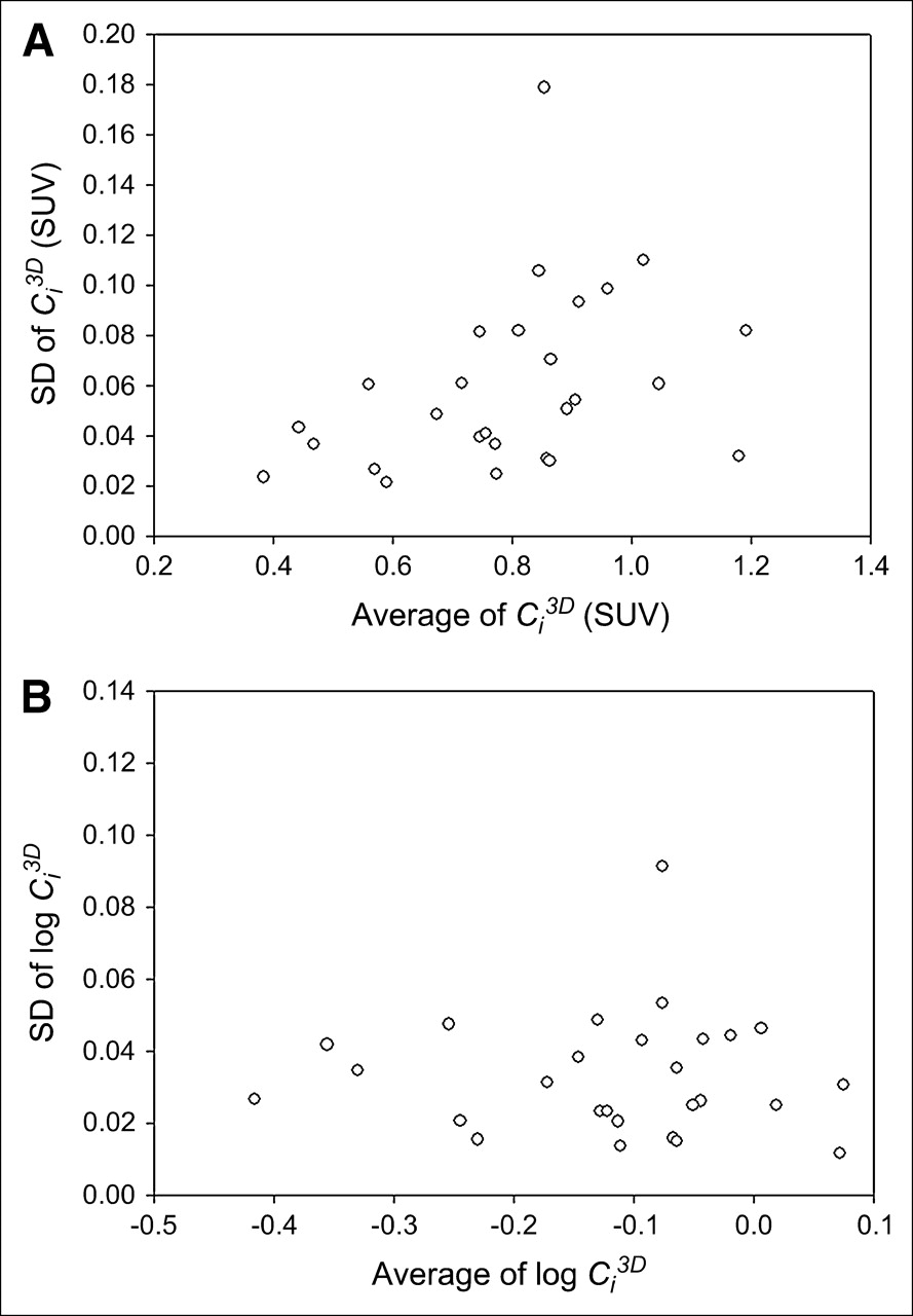
SD of 3D ROI data
as a function of their average. (A) Data are shown in original units (SUV = standardized uptake value). (B) Data are shown after logarithmic transformation. - FIGURE 7.
Example images from 3 patient studies. Images A, C, and E were acquired in 2D (105 s, 6-mm gaussian); images B, D, and F are the corresponding image slices acquired in 3D (90 s, 5-mm gaussian). The lower coefficient of variation in the 3D images can be perceived in images B, D, and F.


Jump to section
Related Articles
Cited By...
- High Reproducibility of Tumor Hypoxia Evaluated by 18F-Fluoromisonidazole PET for Head and Neck Cancer
- Scan-Time Reduction Using Noise-Matched Images in 2- and 3-Dimensional Bismuth Germanate PET/CT: Clinical Study in Head and Neck Cancer
- Deep-Inspiration Breath-Hold PET/CT of Lung Cancer: Maximum Standardized Uptake Value Analysis of 108 Patients
- Dual-Modality Imaging: Combining Anatomy and Function
- Impact of Acquisition Geometry, Image Processing, and Patient Size on Lesion Detection in Whole-Body 18F-FDG PET
- Absolute Quantification of Myocardial Blood Flow with 13N-Ammonia and 3-Dimensional PET