


Article Figures & Data
Figures
- FIGURE 1.
18F-FDG PET images of healthy control subject (A and B) and RA patient with active disease (C and D). (A) 3D projection image of normal tracer distribution in knee. (B) Normal distribution in hand and wrist. (C) Rheumatoid knee. (D) Rheumatoid hand and wrist.
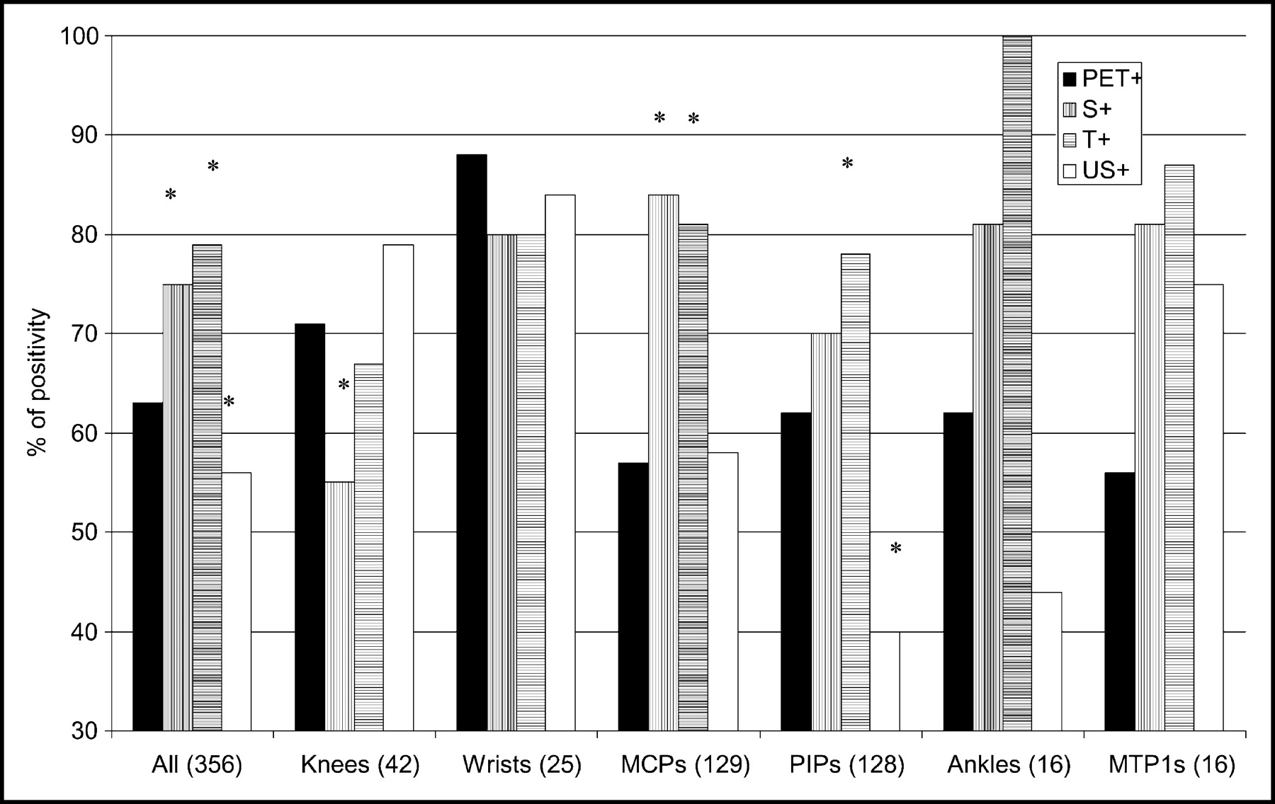
- FIGURE 2.
Percentage of PET positivity of different joints studied. *P < 0.05 compared with PET positivity (χ2 test). In ankles and MTP-1 joints, cohorts were too small to allow valid statistical analysis. S+ = swollen joints; T+ = tender joints; US+ = joints with synovial thickness ≥ 1 mm.
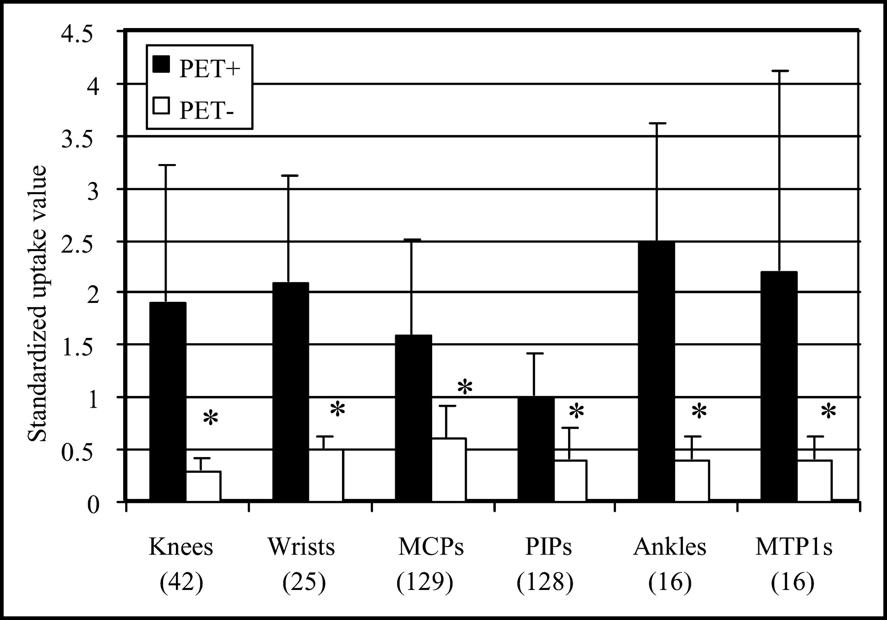
- FIGURE 3.
SUVs (mean ± SD) in joints in which synovitis is observed (PET+) or not observed (PET−) by 18F-FDG PET analysis. *P < 0.05 compared with PET-positive joints.
- FIGURE 4.
PET positivity (A) and SUVs (mean ± SD) (B) in different types of joints studied as joints were not swollen, not tender, and also US negative (0; n = 43) or were positive for 1 (n = 41), 2 (n = 110), or 3 (n = 162) parameters studied. *P < 0.05 compared with PET-positive joints.
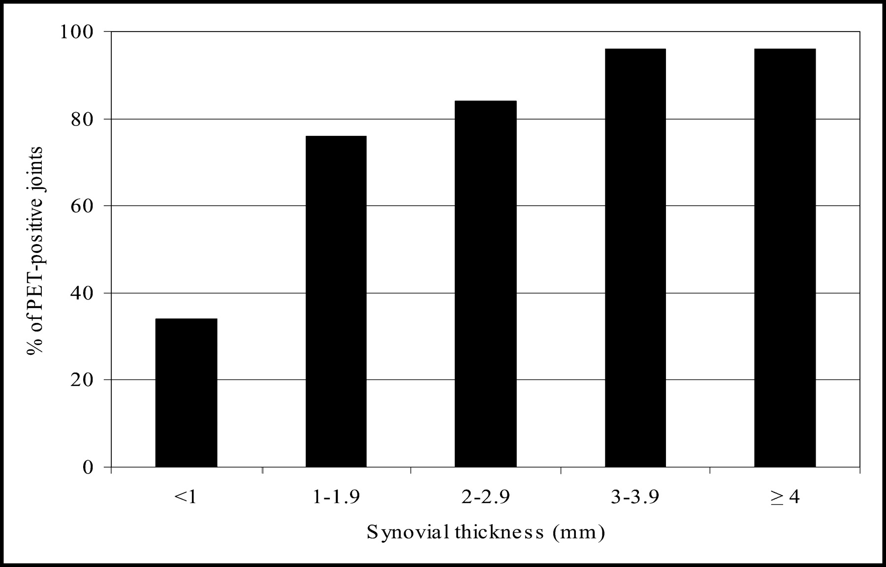
- FIGURE 5.
Percentage of PET-positive joints according to synovial thickness measured by US.


Tables
- TABLE 1
Comparison of PET Positivity According to Visual Analysis and Clinical or US Positivity
Joints (n) Odd ratio* (95% CI) Swelling Tenderness US positivity All (356) 4.8 (2.9–8) 8.6 (4.8–15.6) 11.7 (7–19.7) Knees (42) 3.4 (0.8–13.4) 4.6 (2.2–18.5) 9.0 (1.8–45) Wrists† (25) 10.0 (0.9–187) 10.0 (0.9–187) 10.0 (0.6–181) MCP (129) 20 (4.5–89) 57 (2.7–162) 2.6 (1.7–3.4) PIP (128) 5.1 (2.3–11.5) 13.6 (4.7–40) 22.5 (6.3–78) Ankles‡ (16) 0.8 (0.3–2.1) NA NA MTP-1§ (16) NA NA 6.1 (0.5–78) ↵* Odd ratios in bold type are statistically significant.
↵† Of 22 PET-positive wrists, 19 were swollen or tender and 20 were US positive.
↵‡ Of 10 PET-positive ankles, 8 were swollen, 10 were tender, and 7 were US positive.
↵§ Of 9 PET-positive MTP-1 joints, 9 were swollen, 7 were tender, and 8 were US positive.
NA = nonapplicable because of no value in some categories.
Joints (n) Location of US scanning r P All (356) 0.56 <0.0001 Knees (42) Lateral patellar recess 0.56 <0.0001 Suprapatellar recess 0.78 <0.0001 Medial patellar recess 0.73 <0.0001 Sum 0.80 <0.0001 Wrists (25) Radiocarpal (top of lunate) 0.44 0.02 Intercarpal (top of capitate) 0.40 0.04 Sum 0.50 0.01 MCP (129) Dorsal surface 0.50 <0.0001 PIP (128) Dorsal surface 0.39 <0.0001 Ankles (16) Top of talar neck 0.64 0.008 MTP-1 (16) Dorsal surface 0.36 0.2 Sum = combined thickness resulting from multiple scans (3 sites for knees and 2 sites for wrists).
P values ≤ 0.05 are significant.
- TABLE 3
Individual Parameters of 13 Patients Who Had PET Studies of Knees, Wrists, and MCP and PIP Joints
Patient Parameters studied Clinical Biologic US Composite indices 18F-FDG PET DD (y) S (n) T (n) PGA (mm) PhyGA (mm) MS (min) HAQ ESR (mm/h)* CRP (mg/L) US (n)† CST (mm) Doppler‡ Doppler score§ DAS28 SDAI PET¶ CSUV 1 8 14 24 90 88 150 2.50 53 81 17 66.1 9 21 8.1 60.9 23 61.9 2 7 13 17 82 79 90 1.87 33 5 6 14.5 3 5 7.2 50.5 11 10.5 3 7 20 15 87 84 180 1.25 26 22 18 63.1 9 23 7.2 58.3 19 31.1 4 3 10 11 63 49 120 1.62 14 23 3 9.9 0 0 5.6 36.5 4 8.3 5 7 24 24 96 94 300 2.50 40 35 18 44.6 5 15 8.3 75.5 20 27.3 6 10 24 24 58 93 15 2.12 69 29 15 42.6 0 0 8.2 73.0 20 24.6 7 18 22 22 88 87 240 1.25 31 60 22 46.8 8 16 7.8 71.5 24 42.3 8 18 20 21 93 91 240 2.25 84 41 14 41.0 0 0 8.5 69.5 19 23.4 9 9 17 17 92 94 300 2.75 46 97 17 51.3 2 5 7.7 67.3 15 23.0 10 1 12 8 58 70 60 1.62 12 11 7 10.5 0 0 5.2 34.9 0 6.0 11 14 16 20 94 94 90 2.25 14 36 9 30.3 3 10 6.9 62.4 14 26.1 12 1 21 21 65 87 120 1.62 36 55 4 20.0 0 0 7.5 67.7 13 26.8 13 16 17 17 88 84 150 2.00 54 70 18 61.3 1 2 7.6 59.2 16 26.9 ↵* Millimeters per first hour.
↵† No. of joints with synovial thickness ≥1 mm measured by US.
↵‡ No. of joints with at least 1 power Doppler signal.
↵§ Sum of all scores attributed to all detected signals.
↵¶ No. of joints where synovitis is observed on 18F-FDG PET analysis.
DD = disease duration; S = swollen joints; T = tender joints; MS = morning stiffness.
The clinical parameters swelling and tenderness, sonographic, and PET parameters were determined on the same 24 joints (knees, wrists, and MCP and PIP joints), whereas indices DAS28 and SDAI were calculated using a 28-joint count (the same 24, plus elbows and shoulders).
- TABLE 4
Correlations Between PET Parameters and Other Parameters of Disease Activity in RA
Parameters studied No. of PET-positive joints log CSUV r P r P Clinical No. of swollen joints 0.71 0.007 0.58 0.04 No. of tender joints 0.86 0.0002 0.80 0.0009 PGA 0.60 0.03 0.58 0.04 PhyGA 0.73 0.004 0.70 0.007 Duration of morning stiffness 0.45 0.1 0.40 0.2 HAQ 0.23 0.4 0.23 0.4 Biologic log ESR 0.68 0.01 0.56 0.049 log CRP 0.56 0.049 0.72 0.006 US No. of US-positive joints 0.80 0.0009 0.71 0.006 log cumulative synovial thickness 0.88 <0.0001 0.88 <0.0001 No. of Doppler-positive joints 0.61 0.03 0.63 0.02 Doppler score 0.59 0.03 0.63 0.02 Composite indices DAS28 0.90 <0.0001 0.78 0.002 SDAI 0.90 <0.0001 0.85 0.0002 P values ≤ 0.05 are significant.
In this issue


Jump to section
Related Articles
Cited By...
- Oncologic Staging with 68Ga-FAPI PET/CT Demonstrates a Lower Rate of Nonspecific Lymph Node Findings Than 18F-FDG PET/CT
- Total-Body 18F-FDG PET/CT in Autoimmune Inflammatory Arthritis at Ultra-Low Dose: Initial Observations
- Whole-Body Macrophage Positron Emission Tomography Imaging for Disease Activity Assessment in Early Rheumatoid Arthritis
- Early prediction of treatment response in rheumatoid arthritis by quantitative macrophage PET
- A model of chronic enthesitis and new bone formation characterized by multimodal imaging
- 18F-FEDAC as a Targeting Agent for Activated Macrophages in DBA/1 Mice with Collagen-Induced Arthritis: Comparison with 18F-FDG
- In Vivo Imaging of Cell Proliferation Enables the Detection of the Extent of Experimental Rheumatoid Arthritis by 3'-Deoxy-3'-18F-Fluorothymidine and Small-Animal PET
- 18F-FDG PET as a Tool to Predict the Clinical Outcome of Infliximab Treatment of Rheumatoid Arthritis: An Explorative Study
- Contrast-enhanced Coded Phase-inversion Harmonic Sonography of Knee Synovitis Correlates with Histological Vessel Density: 2 Automated Digital Quantifications
- Inflammatory Cytokines and Hypoxia Contribute to 18F-FDG Uptake by Cells Involved in Pannus Formation in Rheumatoid Arthritis
- Positron emission tomography with computed tomography imaging of neuroinflammation in experimental autoimmune encephalomyelitis
- Increased 18F-FDG Uptake in Degenerative Disease of the Spine: Characterization with 18F-FDG PET/CT
- FDG-PET/CT scan of inflammatory spondylodiscitis lesions in ankylosing spondylitis, and short term evolution during anti-tumour necrosis factor treatment
- 18F-FDG PET in Rheumatoid Arthritis: There Still Is a Long Way to Go