


Abstract
Activated macrophages have been known to play pivotal roles in the pathogenesis of rheumatoid arthritis (RA). 18F-FEDAC (N-benzyl-N-methyl-2-[7,8-dihydro-7-(2-18F-fluoroethyl)-8-oxo-2-phenyl-9H-purin-9-yl]acetamide) is a radiolabeled ligand for the 18-kDa translocator protein (TSPO), which is abundant in activated macrophages. We evaluated the feasibility of using 18F-FEDAC in a murine RA model. Methods: RAW 264.7 mouse macrophages were activated by lipopolysaccharide. TSPO expression levels in activated and inactivated macrophages were measured by quantitative polymerase chain reaction and Western blotting. The cellular uptake and specific binding of 18F-FEDAC were measured using a γ-counter. For the in vivo study, collagen-induced arthritis (CIA) was developed in DBA/1 mice, and the clinical score for arthritis was measured regularly. 18F-FEDAC and 18F-FDG PET images were acquired on days 23 and 37 after the first immunization. Histologic examinations were performed to evaluate macrophages and TSPO expression. Results: We found increased TSPO messenger RNA and protein expression in activated macrophages. Uptake of 18F-FEDAC in activated macrophages was higher than that in nonactivated cells and was successfully blocked by the competitor, PK11195. In CIA mice, joint swelling was apparent on day 26 after the first immunization, and the condition worsened by day 37. 18F-FEDAC uptake by arthritic joints increased early on (day 23), whereas 18F-FDG uptake did not. However, 18F-FDG uptake by arthritic joints markedly increased at later stages (day 37) to a higher level than 18F-FEDAC uptake. The 18F-FEDAC uptake correlated weakly with summed severity score (P = 0.019, r = 0.313), whereas the 18F-FDG uptake correlated strongly with summed severity score (P < 0.001, r = 0.897). Histologic sections of arthritic joints demonstrated an influx of macrophages compared with that in normal joints. Conclusion: 18F-FEDAC enabled the visualization of active inflammation sites in arthritic joints in a CIA model by targeting TSPO expression in activated macrophages. The results suggest the potential usefulness of 18F-FEDAC imaging in the early phase of RA.
Rheumatoid arthritis (RA) is a common autoimmune disease characterized by chronic synovial inflammation, which ultimately leads to joint deformity and dysfunction (1). Once the immune system is triggered by genetic and environmental factors, subclinical inflammation is induced by activation of T cells, B cells, and macrophages. After this, symptoms related with RA are detectable and clinical diagnoses can be made.
Current imaging modalities for diagnosis and monitoring of RA are confined exclusively to the anatomic level. Radiography and CT effectively help visualize morphologic changes such as articular bony erosion or joint space narrowing. However, such changes cannot be reliably detected earlier than 6–12 mo after RA onset. Ultrasonography, MRI, and 99mTc-disphosphonate SPECT help visualize abnormal findings in the early stages of RA; however, they are still limited by a lack of specificity regarding inflammation (2–6).
18F-FDG PET has been clinically used for tumor imaging, because glucose metabolism increases in cancer cells. Recently, use of 18F-FDG PET has expanded for inflammatory diseases, including RA, because glucose metabolism also increases in neutrophils and activated macrophages; however, 18F-FDG has limited value in subclinical inflammation (7–11).
Besides 18F-FDG, more specific PET tracers based on RA pathogenesis have been developed. Activated macrophages are central effectors of synovial inflammation in early stages of RA and release proinflammatory cytokines, including tumor necrosis factor-α, interleukin-1, and interleukin-6 (1,12,13). Therefore, targeting and visualization of macrophages in subclinically inflamed synovium enable earlier diagnosis and therapeutic intervention.
The 18-kDa translocator protein (TSPO) is overexpressed in activated macrophages (14–17). 11C-PK11195 ((R)-N-methyl-N-(1-methylpropyl)-1-(2-chlorophenyl)-isoquinoline-3-carboxamide) is a prototypical radioligand for PET imaging and has recently been applied to the study of RA (17). However, its clinical applications are limited by a relatively low specific-to-nonspecific uptake ratio, high plasma protein binding, very high lipophilicity, and considerable periarticular background activity (15). 18F-FEDAC (N-benzyl-N-methyl-2-[7,8-dihydro-7-(2-18F-fluoroethyl)-8-oxo-2-phenyl-9H-purin-9-yl]acetamide) is a second-generation TSPO ligand with high selectivity. The feasibility of using 18F-FEDAC for RA research has not been studied, although it has been used to study neuroinflammation, brain infarction, and lung inflammation models by reflecting increased TSPO expression (18–20).
The aim of the present study was to apply the new TSPO radioligand 18F-FEDAC to visualize synovial inflammation associated with RA. We performed in vitro assays and in vivo PET to evaluate TSPO expression in activated macrophage and inflamed joints of collagen-induced arthritis (CIA) models. In addition, for the detection of subclinical arthritis, we evaluated the value of 18F-FEDAC compared with 18F-FDG, which is the most widely used radioligand for inflammation via glucose metabolism of diverse inflammatory cells.
MATERIALS AND METHODS
Synthesis of 18F-FEDAC
18F-fluoride was produced in-house using a cyclotron (PETtrace; GE Healthcare) and captured on a Chromafix cartridge (PS-HCO3; ABX), which was preconditioned using ethanol (1 mL) and water (1 mL). 18F-fluoride on the cartridge was eluted into a reaction vial containing a mixture (1 mL) of acetonitrile (83.8%) and tetrabutylammonium bicarbonate (2.3%). Two azeotropic evaporations were conducted at 110°C, after the addition of 1 mL of acetonitrile each time, under a gentle stream of nitrogen gas. The precursor of 18F-FEDAC, TosOEt-DAC (N-benzyl-N-methyl-2-[7,8-dihydro-7-(2-tosyloxyethyl)-8-oxo-2-phenyl-9H-purin-9-yl]acetamide, 1.3 mg), in dimethylformamide (0.6 mL) was added to the reaction vial and heated to 95°C for 15 min. The reaction mixture was diluted using 40% EtOH and subjected to semipreparative high-performance liquid chromatography (Xterra Prep RP18, 10 μm, 10 × 250 mm; 40% EtOH for 20 min; 5 mL/min; Waters). The radiochemical purity was more than 99% on analytic high-performance liquid chromatography (Xterra RP18, 10 μm, 4.6 × 100 mm; 40% EtOH for 20 min, 1 mL/min; Waters), and the radiochemical yield of 18F-FEDAC was 24.8% ± 1.4% in a total synthesis time of 50 min. The specific activity of 18F-FEDAC was 265.1 ± 93.1 GBq/μmol (n = 3) (21). All the technical support for radiosynthesis, including the precursor and cold standard of 18F-FEDAC, was kindly provided by Lin Xie and Ming-Rong Zhang (National Institute of Radiologic Sciences and National Institutes for Quantum and Radiologic Science and Technology, Chiba, Japan).
Cell Culture
The mouse macrophage cell line RAW 264.7 was obtained from the American Type Culture Collection. RAW 264.7 cells were grown using Dulbecco modified Eagle medium (WelGene Inc.) supplemented with 10% fetal bovine serum (Invitrogen) and 1% antibiotics (Invitrogen) at 37°C in a humidified atmosphere containing 5% CO2. For activation, RAW 264.7 cells were treated with a 1 μg/mL concentration of lipopolysaccharide (Sigma-Aldrich) for 24 h (22).
In Vitro Real-Time Polymerase Chain Reaction (PCR) Analysis and Western Blot Analysis
Total RNA was extracted from cells using TRIzol reagent (Invitrogen) and converted into complementary DNA using a complementary DNA synthesis kit (GenDEPOT). TSPO in macrophages was quantified using SYBR Green real-time PCR Master Mix (TaKaRa) with 2 ng of complementary DNA and 0.2 μM primers. Expression of the housekeeping gene 18S rRNA was used to standardize TSPO expression. Real-time PCR was performed using an Applied Biosystems 7500 Sequence Detection System instrument with the following parameters: 50°C for 2 min, 95°C for 10 min, and 40 cycles of 95°C for 15 s and 60°C for 1 min.
Total protein was isolated from the cells using radioimmunoprecipitation assay buffer (Sigma-Aldrich). The lysate of each sample was loaded onto NuPAGE 4%–12% Bis-Tris Gel (Invitrogen). After electrophoresis, the gels were blotted onto polyvinylidene difluoride membranes (Millipore), which were subsequently blocked with 5% skim milk for 1 h at room temperature. The membranes were incubated overnight at 4°C with primary antibodies targeting TSPO (Abcam; diluted 1:10,000) and β-actin (Sigma-Aldrich; diluted 1:5,000). Membranes were then probed with horseradish peroxidase–conjugated antirabbit or antimouse IgG (Cell Signaling Technology). The signal intensity was measured using an LAS-3000 imaging system (Fujifilm).
In Vitro Cellular Uptake of TSPO Radioligand
18F-FEDAC uptake in vitro was examined using a 0.37–55.5 kBq/mL concentration of 18F-FEDAC in assay medium (Hanks balanced salt solution, 0.5% bovine serum albumin [w/v], 0.24% 4-(2-hydroxyethyl)-1-piperazineethanesulfonic acid [w/v], pH 7.4). Incubation was performed for 1 h at room temperature based on the pretest results (Supplemental Fig. 1; supplemental materials are available at http://jnm.snmjournals.org). RAW 264.7 cells were incubated in culture medium with or without lipopolysaccharide stimulation for 24 h, and the culture medium was then replaced with 1 mL of assay medium. After incubation with 18F-FEDAC, uptake was terminated by 2 washes using cold phosphate-buffered saline. Then, the cells were solubilized with 500 μL of 1% sodium dodecyl sulfate buffer. The radioactivity of the nonactivated control and the activated macrophages was measured using a γ-counter (PerkinElmer). The uptake of 18F-FEDAC was calculated from the ratio of 18F-FEDAC associated with the cells to the initial dose. To verify in vitro specific TSPO binding of 18F-FEDAC, an excess of cold-form PK11195 (1,000-fold molar excess) was used to compete with 18F-FEDAC.
Induction and Assessment of CIA Mouse Model
All experimental procedures were approved by the Institutional Animal Care and Use Committee of the Seoul National University Hospital. All specific pathogen-free male DBA/1 (22.8 ± 2.5 g, 6-wk-old) mice were purchased from Japan SLC Inc. The CIA mouse model was induced as described previously (23). Bovine type II collagen (Chondrex, Inc.) dissolved in 0.1 M acetic acid was emulsified with an equal volume of complete Freund adjuvant and then injected subcutaneously at the base of the tail (50 μL per mouse). Three weeks after the first immunization, type II collagen emulsified with incomplete Freund adjuvant was injected subcutaneously as a booster immunization (50 μL per mouse).
From the day of the second immunization, the mice were clinically examined every 2 d. The clinical severity scoring of arthritis in the paw was performed as described previously (23): score 0, no macroscopic evidence of erythema or swelling; score 1, erythema and mild swelling confined to the tarsals; score 2, erythema and mild swelling extending from the ankle to the tarsals; score 3, erythema and moderate swelling extending from the ankle to the tarsals; and score 4, erythema and severe swelling encompassing the ankle, foot, and digits, or ankylosis of the limb. To obtain the summed severity score per animal, the results from all paws of that animal were added.
18F-FEDAC and 18F-FDG PET Imaging
PET was performed using a small-animal PET/CT scanner (eXplore Vista; GE Healthcare). We measured 18F-FEDAC uptake in all paws of mice with CIA (n = 10) at 2 different time points of disease (days 23 and 37) and compared it with the uptake of the control group (n = 4). We measured 18F-FDG uptake in all paws of mice with CIA (n = 5) at 2 different time points of disease (days 23 and 37) and compared it with the uptake of the control group (n = 2).
The mice were anesthetized with 1.5% isoflurane, and the radiotracers were intravenously injected (8.7 ± 0.1 MBq/100 μL for 18F-FEDAC; 9.5 ± 0.7 MBq/100 μL for 18F-FDG). PET/CT images were acquired 2 and 1 h after injection of 18F-FEDAC and 18F-FDG, respectively. Images were reconstructed using a 3-dimensional ordered-subset expectation maximization algorithm. To verify specific TSPO binding, CIA mice were pretreated with a more than 1,000-fold molar excess of the nonradioactive PK11195 before the injection of 18F-FEDAC and followed by PET imaging.
PET Image Analysis
Quantitative analysis was performed by an experienced nuclear medicine physician using PMOD software, version 3.3. The SUVmax was measured by placing a 3-dimensional volume of interest encasing each paw with a margin threshold set as 40% SUVmax. The SUV in a pixel was calculated as tissue radioactivity concentration/(injected radioactivity/body weight). Next, the SUVmax of the paw was divided by the blood-pool SUVmax measured from the aorta for normalization. Finally, after these calculations, a target-to-background ratio (TBR) was acquired.
Histopathology and Immunostaining
The paws were amputated and fixed in 10% paraformaldehyde for 7 d at 4°C. Bone tissue was decalcified for about 1 wk at 4°C in a solution with 14% ethylenediaminetetraacetic acid (pH 7.2; Sigma-Aldrich). After decalcification, the joints were washed and prepared for paraffin embedding. Sagittal sections (4 μm) from the center of the joint were used for staining. The sections were stained with hematoxylin and eosin for histologic evaluation.
Immunofluorescence staining of the macrophages was performed using anti-CD68 antibody (diluted 1:100; Abcam), and TSPO receptor staining was performed using anti-TSPO antibody (diluted 1:400; Abcam) incubated overnight at 4°C. After washing with phosphate-buffered saline, Alexa 488– and Alexa 647–conjugated secondary antibodies were incubated for 1 h at room temperature. After staining, the slides were mounted using Prolong Gold reagent with 4′,6-diamidino-2-phenylindole (Invitrogen). Fluorescence images were obtained using an LSM 800 confocal imaging system (Carl Zeiss).
Statistics
All statistical analyses were performed using MedCalc software. Mann–Whitney tests were performed to analyze differences in PET uptake between arthritic joints of CIA mice and normal joints of control mice. Kruskal–Wallis testing was performed to evaluate the PET uptake according to the clinical severity score. The clinical severity score was reclassified as 3 grades by merging scores 2–4, for which arthritis extended from the ankle to the tarsals (score 0, grade 0; score 1, grade 1; scores 2–4, grade 2), and then was referred for further statistical analysis. Rank correlation testing was performed to evaluate the relationship of PET uptake with summed severity score. A Wilcoxon signed rank test was performed for paired observations, such as nonblocking versus blocking subjects. P values of less than 0.05 were regarded as statistically significant. In addition, receiver-operating-characteristic curves were used to evaluate the predictive performance of PET uptake for the development of clinical arthritis. The optimal cutoff—the point that has the maximal sum of sensitivity and specificity—was determined using the Youden index.
RESULTS
Expression Level of TSPO in Activated Macrophages
After 24 h of lipopolysaccharide stimulation, RAW 264.7 cells were activated, as observed under a microscope (Supplemental Fig. 2). Real-time PCR demonstrated increased RNA expression of TSPO in activated macrophages compared with nonactivated controls (Fig. 1A). Western blot analysis demonstrated greater TSPO protein expression in activated macrophages than in nonactivated controls (Fig. 1B).

(A) Time-dependent TSPO messenger RNA level after lipopolysaccharide (LPS) stimulation. (B) TSPO protein level after 24-h stimulation. (C) Dose-dependent 18F-FEDAC uptake of RAW 264.7 cells (blue). Stimulation for 24 h enhanced 18F-FEDAC uptake (red), whereas cold-form PK11195 blocked 18F-FEDAC uptake (black).
In Vitro Cell Uptake and Specific Binding of 18F-FEDAC in Activated Macrophages
Uptake of 18F-FEDAC in activated macrophage was higher than that in nonactivated controls (Fig. 1C). In addition, specific TSPO binding of 18F-FEDAC was demonstrated, and an approximately 0.3-fold decrease in 18F-FEDAC uptake was observed in activated macrophages by the specific competitor, PK11195.
Clinical Course of CIA
Among the 26 mice used for CIA modeling, 24 successfully developed clinical arthritis (92.3%). Joint swelling was apparent on day 26 after the first immunization (day 0), and the condition worsened by day 37 (Supplemental Fig. 3).
PET Imaging Analysis
Quantification of Joint Inflammation Using 18F-FEDAC and 18F-FDG PET/CT
Joint uptake of 18F-FEDAC in CIA mice was observed on day 23 but did not increase by day 37. The TBR in CIA mice was significantly higher than that in the control group on day 23 (P < 0.001, Supplemental Figs. 4A–4C and Fig. 2A), although no CIA mice developed clinical arthritis. On day 37, all CIA mice developed clinical arthritis, and the TBR in CIA mice was still higher than that in control mice (P < 0.001, Supplemental Figs. 4D–4F and Fig. 2A). Despite the deterioration of clinical arthritis as compared with that on day 23, the TBR in CIA mice on day 37 was no different from 18F-FEDAC uptake on day 23 (P = 0.183, Supplemental Figs. 4G–4I).
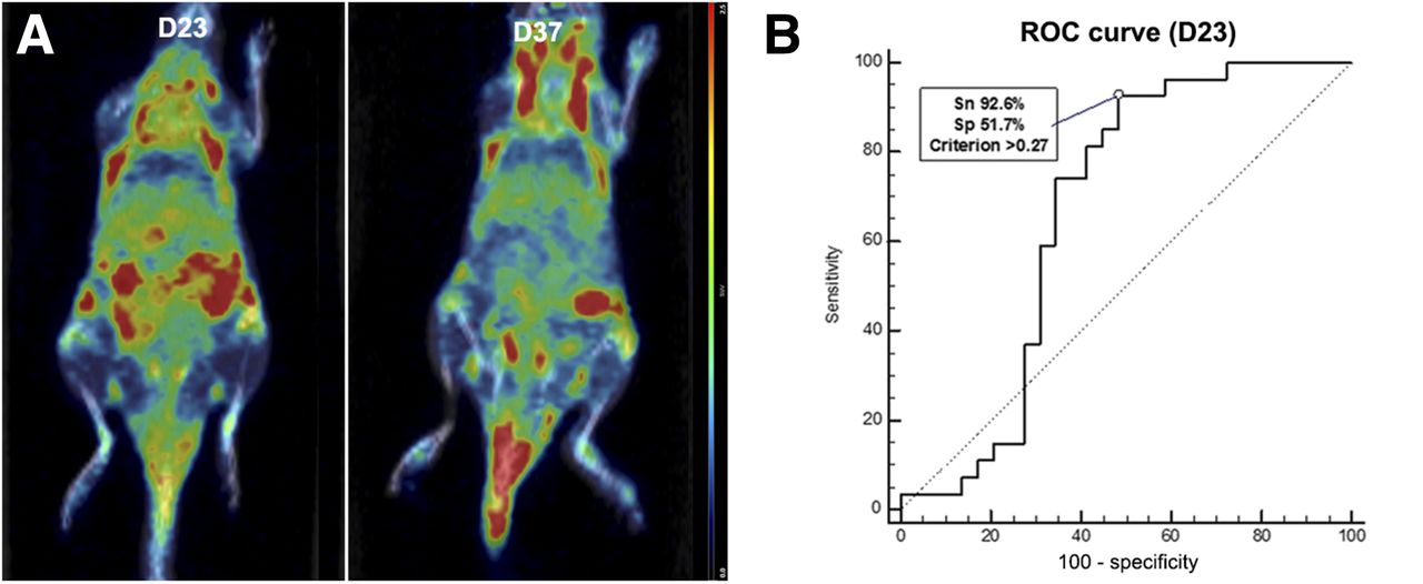
(A) Coronal 18F-FEDAC PET/CT section of same CIA mouse on days 23 and 37, showing increased uptake in front and hind paws. (B) Predictive performance of day 23 18F-FEDAC uptake for development of clinical arthritis. ROC = receiver operating characteristic; Sn = sensitivity; Sp = specificity.
Joint uptake of 18F-FDG in CIA mice was detected only on day 37. The TBR in CIA mice was not significantly different from that in control mice on day 23 (P = 0.838, Supplemental Figs. 5A–5C and Fig. 3). On day 37, all CIA mice showed prominent signs of clinical arthritis, and the TBR in CIA mice was significantly higher than that in control mice (P < 0.001, Supplemental Figs. 5D–5F and Fig. 3). With deterioration of clinical arthritis, the TBR in CIA mice on day 37 was significantly higher than that on day 23 (P < 0.001, Supplemental Figs. 5G–5I). Detailed data for 18F-FEDAC and 18F-FDG are summarized in Table 1.
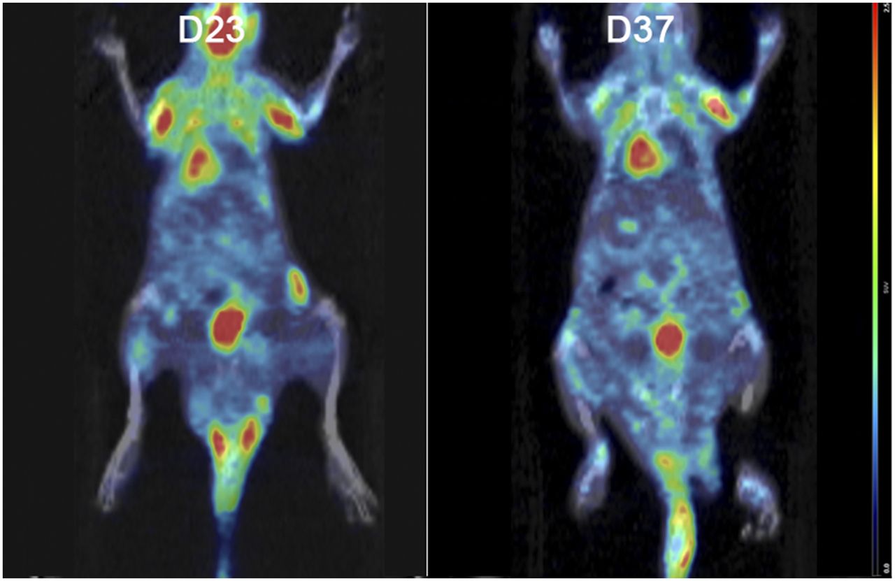
Coronal 18F-FDG PET/CT section of same mouse on days 23 and 37. On day 23, before onset of arthritis, uptake by joints was not observed. On day 37, increased 18F-FDG uptake was observed for all 4 arthritic joints.
TBR in Normal and CIA Mice
Relationship of Clinical Score with 18F-FEDAC and 18F-FDG Uptake
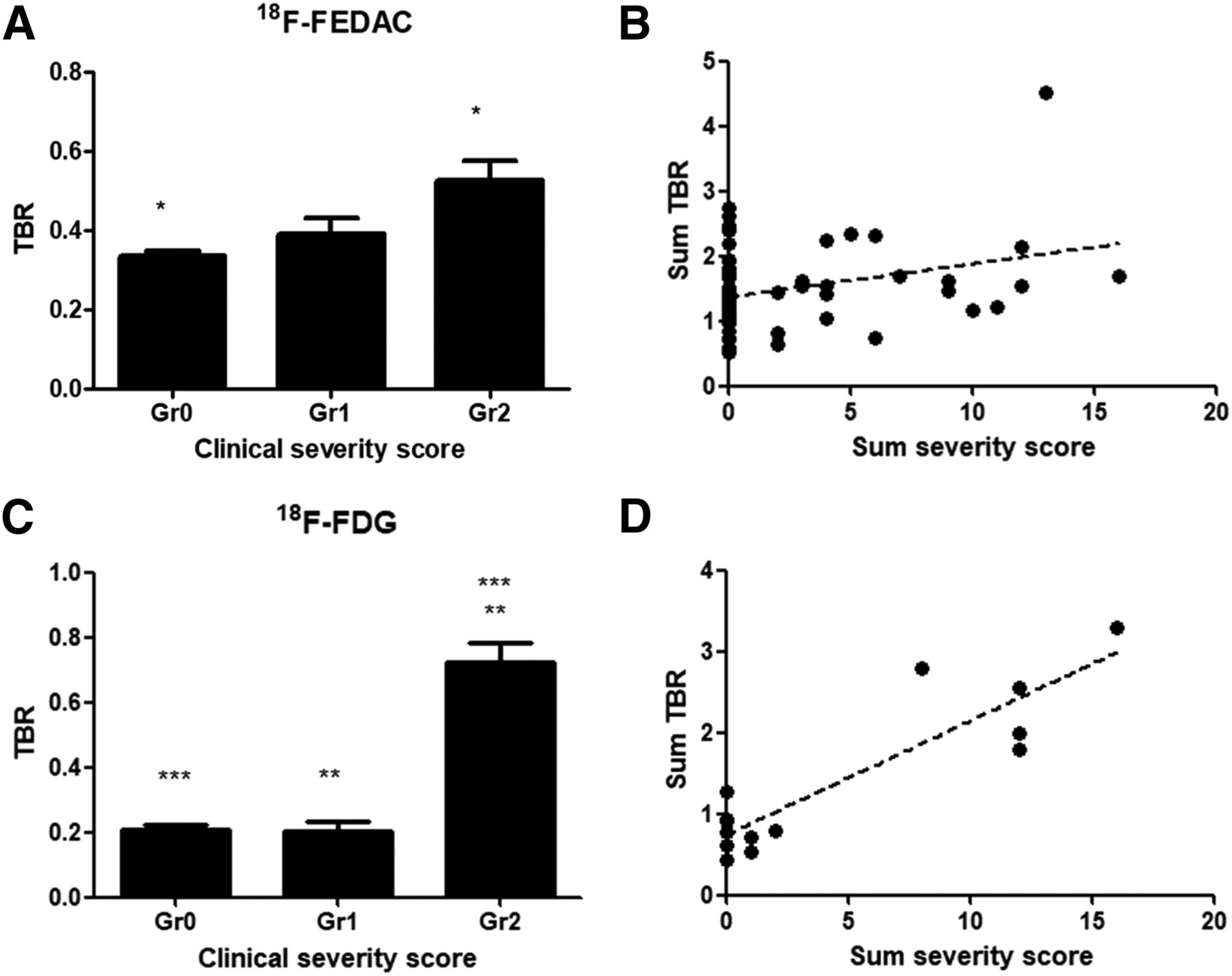
The appearance and severity of arthritis, and the 18F-FEDAC uptake in each paw, differed for each mouse (Supplemental Fig. 6). The 18F-FEDAC TBR of each paw was significantly different according to the associated clinical severity score (P < 0.001, Fig. 4A). The total 18F-FEDAC TBR of all paws from PET imaging correlated weakly with the summed severity score (P = 0.019, r = 0.313, Fig. 4B). The 18F-FDG TBR of each arthritic paw was significantly different according to the clinical severity score (P < 0.001, Fig. 4C). The total 18F-FDG TBR of all paws from PET imaging correlated strongly with the summed severity score (P < 0.001, r = 0.897, Fig. 4D). The correlation between 18F-FDG TBR and the summed severity score was significantly stronger than the correlation between 18F-FEDAC TBR and the summed severity score (P < 0.001).

(A and C) 18F-FEDAC and 18F-FDG uptake according to clinical severity score (grade 0 = score 0, grade 1 = score 1, grade 2 = scores 2–4). (B and D) Scatterplot of summed 18F-FEDAC and 18F-FDG uptake by summed severity score (sum of all scores in mouse).
Predictive Performance of 18F-FEDAC Uptake on Day 23 for Development of Clinical Arthritis
Although arthritis was not detected in CIA mice on day 23, the 18F-FEDAC TBR was significantly higher than that in control mice. Such a result indicates the potential of 18F-FEDAC TBR to reflect subclinical inflammation. Thus, we evaluated the predictive performance of 18F-FEDAC on day 23 for the development of clinical arthritis by receiver-operating-characteristic curve analyses (Fig. 2B). The optimal cutoff was 0.27 (area under the curve, 0.668; P = 0.031), and 18F-FEDAC uptake on day 23 predicted the development of clinical arthritis with a sensitivity of 92.6% and a specificity of 51.7%.
Specific Binding of 18F-FEDAC in CIA Model
We confirmed specific 18F-FEDAC binding on inflammatory joints in CIA mice. After blocking of TSPO with an excess of PK11195, arthritic joint uptake of 18F-FEDAC was significantly decreased compared with uptake in joints with nonblocked TSPO (P = 0.007, Fig. 5).
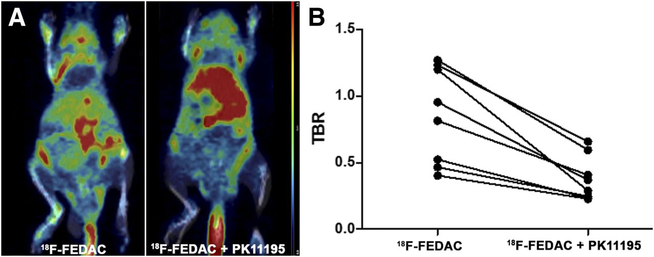
Coronal PET/CT section (A) and graph (B) before and after blocking in same mouse. 18F-FEDAC uptake in arthritic joint was significantly decreased after blockade.
Presence of Activated Macrophages in Arthritic Paws
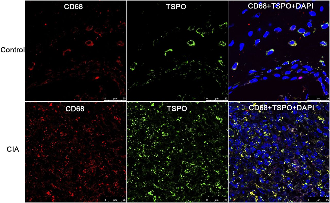
Hematoxylin and eosin staining of the arthritic joints of CIA mice showed a massive infiltration of mainly polymorphonuclear cells in hyperplastic synovium, with pannus formation and bone destruction (Fig. 6). Immunofluorescence staining demonstrated a prominent influx of macrophages characterized by CD68-positive and TSPO-positive cells at the inflammation site (Fig. 7). Staining specificity was verified using a negative control (Supplemental Fig. 7).
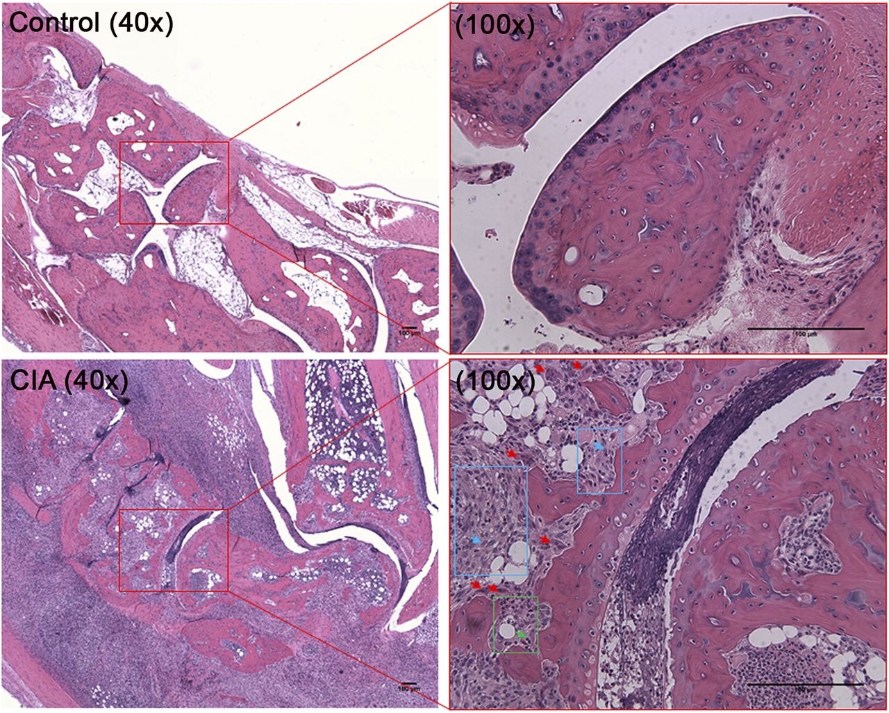
Hematoxylin- and eosin-stained sections from control and CIA joints. In control joint, synovial membrane and cartilage are intact. In CIA joint at ×100 magnification, influx of macrophages (regions in blue boxes) and neutrophils (region in green box) are prominent. Subset of osteoclasts (red arrows) indicates joint destruction. Blue arrows indicate individual macrophages (representative ones) in macrophage influx site (blue box). Green arrows indicate individual neutrophils (representative ones) in neutrophil influx site (green box).

Immunofluorescence staining for CD68 (red) and TSPO (green) in synovium of control and CIA joints. Merged images at far right show increased number and TSPO expression of CD68-positive macrophages, compared with controls.
DISCUSSION
In this study, the increase of TSPO expression corresponded to the increase of 18F-FEDAC uptake in activated macrophages. Real-time PCR and Western blotting revealed higher TSPO messenger RNA and protein expression in activated macrophages than in resting macrophages. Histologic sections of arthritic joints demonstrated an increase in macrophage infiltration and TSPO expression. Upregulation of TSPO in activated macrophages has been reported previously and corresponds with our data (20,24,25). An increase of 18F-FEDAC uptake in lipopolysaccharide-stimulated macrophage cells and arthritic joints was demonstrated, indicating a corresponding overexpression of TSPO. The specific binding of 18F-FEDAC to TSPO was identified by an in vitro and in vivo blocking study.
We investigated time-dependent uptake of 18F-FEDAC compared with 18F-FDG using CIA mice. Among several RA models, the CIA model has been the most widely investigated, and it has been reported that the first signs of arthritis are visually detectable between 21 and 28 d after immunization (23). Here, the first sign of joint swelling was detected on day 26, and swelling gradually increased until day 37. On the basis of previous and current results regarding the clinical course of CIA models, we selected days 23 and 37 as the early and late time points, respectively, for arthritis.
18F-FEDAC uptake increased in the joints of CIA mice on day 23, although no CIA mouse developed signs of clinical arthritis. With a cutoff of 0.27, 18F-FEDAC uptake on day 23 can predict the development of clinical arthritis with a sensitivity of 92.6%. From days 23 to 37, 18F-FEDAC uptake did not increase and remained constant despite the deterioration of clinical arthritis. The serial uptake pattern of 18F-FEDAC, which can be described as an early increase–late plateau, indicated that TSPO expression in activated macrophages at the inflammation site increased during the early stage. However, TSPO levels may be considerably low in the late phase, despite macrophage proliferation and recruitment during RA progression.
Unlike 18F-FEDAC, 18F-FDG uptake did not increase in the joints of CIA mice on day 23. Therefore, 18F-FDG has limited use in early RA detection. On day 37, 18F-FDG uptake increased along with the deterioration of clinical arthritis. The serial uptake pattern of 18F-FDG, which can be described as a time-dependent increase, indicated that diverse inflammatory cells congregate in the inflammation site at later stages of RA, which induces an increase in 18F-FDG uptake. Previous studies also reported no 18F-FDG uptake in subclinical arthritis, whereas increased 18F-FDG uptake in the acute and chronic stages was detected (7,26).
Therefore, we speculate that compared with 18F-FDG, 18F-FEDAC is a more applicable radiotracer for early detection of RA. Although the precise function of TSPO is not known, it may be involved in the initiation of autoimmune arthritis. Temporal TSPO expression during the early and late phases should be analyzed using arthritic postmortem joint tissues. Moreover, validating the potential of 18F-FEDAC as a therapeutic indicator for therapies targeting macrophages or macrophage-related cytokines will be useful.
In addition, we investigated the uptake of 18F-FEDAC and 18F-FDG based on the clinical severity of arthritis. Although 18F-FDG has a limited use in subclinical arthritis, its uptake in arthritic joints has been reported to reflect disease activity (9,11). Similar to previous studies, we also observed a strong correlation between 18F-FDG and disease severity. This correlation was stronger than that observed for 18F-FEDAC. Therefore, 18F-FDG is a valuable tool for evaluation of disease severity and therapeutic responses during late RA stages.
A limitation of this study was that we could not perform a head-to-head comparison between 18F-FEDAC and 18F-FDG PET scans in the same mouse at the same time, because of the long half-life of 18F. Although PET scans were performed for different mice, CIA was induced under the same conditions, and the course and severity of arthritis between the 18F-FEDAC and 18F-FDG groups were similar. Simultaneous use of the 11C-labeled TSPO ligand and 18F-FDG can be considered for head-to-head comparison at the same time during the development of arthritis. Especially, 11C-PBR28 has been proven to have a higher binding affinity for TSPO than PK11195 (27). A head-to-head comparison between 18F-FEDAC and 11C-PBR28 with cold-form PBR28 blocking might also be considered to confirm the specificity of the tracer in the CIA model.
CONCLUSION
18F-FEDAC can be used to visualize active inflammation in arthritic joints in a CIA mouse model by targeting TSPO expression in activated macrophages, which is the major effector of RA pathogenesis. The results of this study suggest the potential usefulness of 18F-FEDAC imaging in early stages of RA.
DISCLOSURE
This study was supported by a grant from the Korea Health Technology R&D Project through the Korea Health Industry Development Institute (KHIDI), funded by the Ministry of Health & Welfare, Republic of Korea to Gi Jeong Cheon (grant HI14C1072), by a grant from the National Research Foundation to Hai-Jeon Yoon (2015R1C1A2A01054113) and to Keon Wook Kang (2015M3D6A1065663), and by a grant from the Global Core Research Center (GCRC) (no. 2011-0030001) to June-Key Chung. No other potential conflict of interest relevant to this article was reported.
Footnotes
↵* Contributed equally to this work.
Published online Jan. 11, 2018.
- © 2018 by the Society of Nuclear Medicine and Molecular Imaging.
REFERENCES
- Received for publication August 11, 2017.
- Accepted for publication December 13, 2017.


Jump to section
Related Articles
Cited By...
- No citing articles found.