


Abstract
18F-FDG PET is a sensitive, promising method for visualizing disease activity in rheumatoid arthritis. This study aimed to assess the association between changes in 18F-FDG joint uptake after 2 wk of infliximab treatment and clinical outcome. Methods: Scans were obtained at the initiation of treatment and at 2 wk. Uptake in metacarpophalangeal and wrist joints was quantified using standardized uptake values. Results: Changes in mean standardized uptake value at 0–2 wk significantly correlated with the disease activity score (DAS) at 14 and 22 wk and contributed significantly to the prediction of DAS at these time points. No significant correlation was found between changes in acute-phase reactants at 0–2 wk and the DAS at later time points. Conclusion: Early changes in 18F-FDG uptake in joints during infliximab treatment of rheumatoid arthritis patients, using PET, may predict clinical outcome.
Rheumatoid arthritis (RA) is a disabling joint disease. Stringent monitoring of disease activity and, if necessary, changing to another, more effective, drug treatment has been shown to improve the outcome of patients (1).
Anti–tumor necrosis factor (TNF) α-blocking agents, such as infliximab, potently bind TNF and block inflammation by inhibiting the downstream effects of this cytokine. Anti-TNF treatment is effective in reducing disease activity and improving outcome, although not in all patients (2).
In general, the response to anti-TNF treatment is assessed between 14 and 22 wk after initiation of the drug. At present, there are no markers that can predict response to anti-TNF treatment at an earlier stage (3–6). Considering the high costs of anti-TNF treatment, that is, $30,500–$38,700 per discounted quality-adjusted life-year gained (7), early prediction of response could reduce the overall costs of treatment.
Several reports have shown that inflammation in synovial tissue in RA patients can be detected using 18F-FDG PET (8–12). Therefore, 18F-FDG PET may be an interesting tool to assess response early during anti-TNF treatment.
This explorative study investigated the potential ability of early 18F-FDG PET changes to predict clinical outcome at a later stage.
MATERIALS AND METHODS
Patients and Study Protocol
All patients (11 women, 5 men) fulfilled the American College of Rheumatology criteria for RA (13). The presence of at least 2 metacarpophalangeal or wrist joints with signs of arthritis was required. Patients were treated with infliximab, and infusions were administered at 0, 2, 6, 14, and 22 wk. At all time points, tenderness and swelling of proximal interphalangeal and metacarpophalangeal joints of hands, elbows, shoulders, and knees (28 joints in total) were determined. Erythrocyte sedimentation rate (ESR) and C-reactive protein (CRP) were measured, and the disease activity scores (DASs) of 28 joints were calculated (14). 18F-FDG PET scans of metacarpophalangeal and wrist joints were acquired at 0 and 2 wk. Informed consent was obtained of all participating patients. The protocol was approved by the local medical ethical committee.
PET
PET was performed using an ECAT EXACT HR+ scanner (CTI/Siemens). Before scanning, the patients fasted for at least 6 h. All plasma glucose levels, measured just before intravenous administration of 18F-FDG, were within normal limits. Patients were injected with 382 ± 44 MBq (mean ± SD) of 18F-FDG. The hands were positioned on top of the chest to include the heart and aorta within the field of view of the scanner for blood pool analysis, allowing quantification of 18F-FDG uptake. After the patients had been positioned, a dynamic emission scan of 60 min was acquired, preceded by a transmission scan (85,000 kilocounts). Venous samples (8 mL) were collected at 35, 45, and 55 min. PET scans were corrected for attenuation, decay, scatter, randoms, and dead time. Summed images (40–60 min) were reconstructed as 128 × 128 matrices using filtered backprojection with a Hanning filter (cutoff, 0.5 cycles/pixel), resulting in a transaxial spatial resolution of about 7 mm in full width at half maximum.
Image Analysis
Coronal, sagittal, and transverse images were used for analysis. Regions of interest were drawn on the ascending and descending aorta, the aortic arch, and the left atrium and left ventricle of the heart to obtain arterial input function. 18F-FDG uptake in hand and wrist joints was measured through manual drawing of regions of interest on PET-positive metacarpophalangeal joints (cluster of the second to fifth metacarpophalangeal joints per hand) and on PET-positive wrists, yielding a maximum of 4 regions per scan. Patlak graphical analysis (15) was applied to the dynamic 18F-FDG scan to quantitate the metabolic rate of glucose (Mrglu) in joints between 10 and 60 min. In addition, standardized uptake value (SUV) was calculated using the summed 18F-FDG images and the same ROIs and was normalized for injected dose and body surface (16–18). The mean SUV and Mrglu of all regions of interest per patient were calculated and used for statistical analysis.
Statistical Analysis
Pearson correlations were used to assess the relationship between changes in mean 18F-FDG joint uptake, ESR, CRP, and DAS from 0 to 2 wk and DAS at 6, 14, and 22 wk. A stepwise regression analysis was performed to investigate which of these parameters was most predictive of DAS at 14 and 22 wk. Because of the small sample size, a forward selection was chosen. Both DAS values at 14 wk and DAS values at 22 wk were used as dependent variables, and since DAS was not normally distributed, its natural logarithm was used. Changes in mean 18F-FDG joint uptake, ESR, CRP, and DAS from 0 to 2 wk were used as independent variables. A separate analysis was performed between SUV and Mrglu, using Pearson correlation coefficients and linear regression analysis. A P value smaller than 0.05 was considered to be significant. Unless stated otherwise, point estimates and distribution data are reported as mean and SD.
RESULTS
Demographic and Clinical Data
Patients were 53 ± 10 y old and had a disease duration of 8 ± 6 y. At inclusion, all patients had active disease. After 14 and 22 wk, DAS decreased to 4.3 (±1.5) and 3.9 (±1.3), respectively. The individual response to infliximab was heterogeneous. At 22 wk, there were 5 good and 8 moderate responders and 3 nonresponders, based on the response criteria of the European League Against Rheumatism (19). The DAS course in relation to response is depicted in Table 1.
Course of DAS in Responders and Nonresponders to Infliximab Therapy
PET Data
Before infliximab treatment, all patients showed enhanced 18F-FDG uptake in at least one metacarpophalangeal region or wrist (Fig. 1)
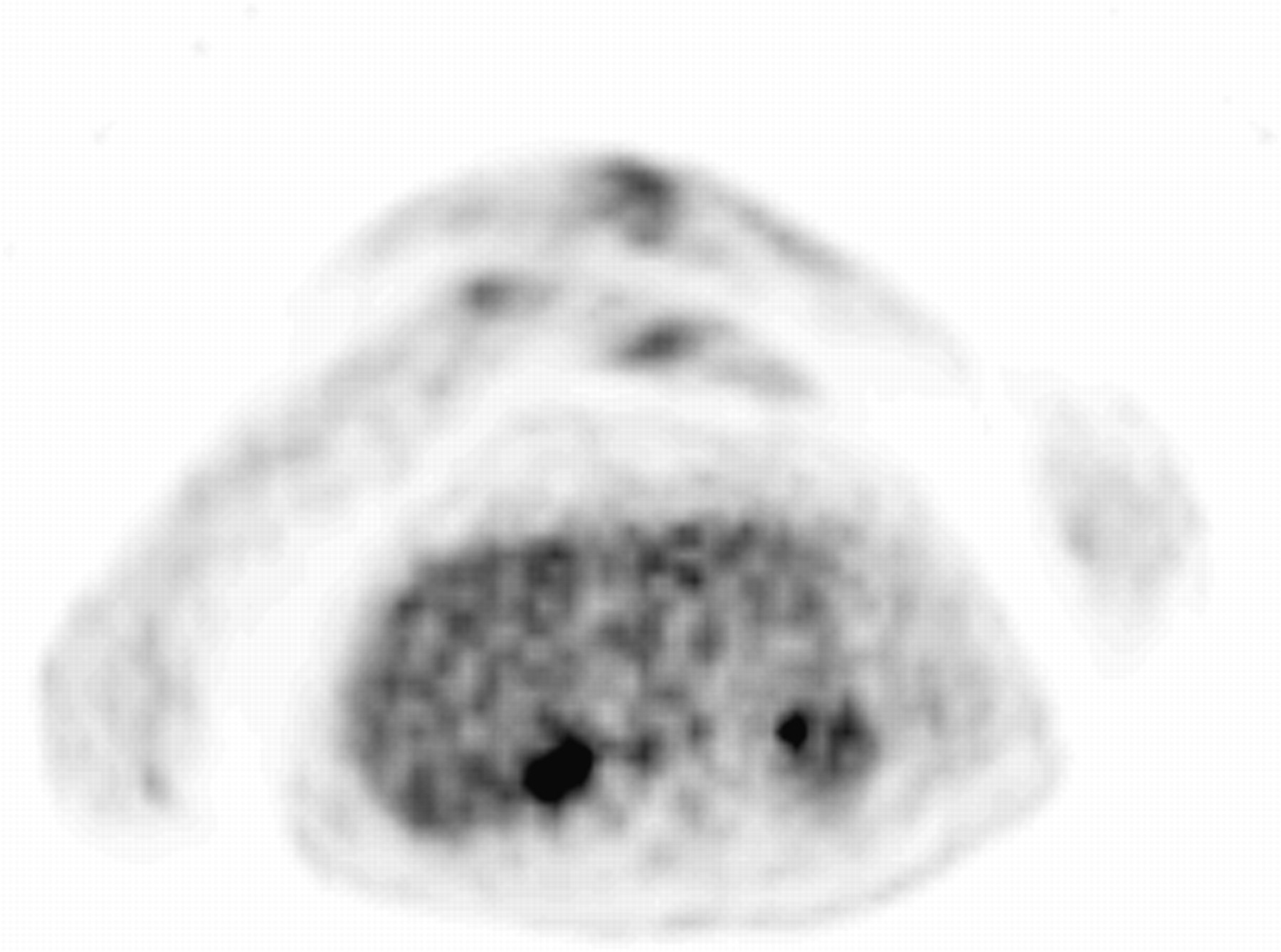
PET image of patient with 18F-FDG uptake in metacarpophalangeal joints of hands and wrists. Arms and hands are positioned on top of thorax.
Both at 0 wk and at 2 wk, Mrglu and SUV were closely correlated (r = 0.93, P < 0.001, and r = 0.88, P < 0.001, respectively) (Fig. 2). A significant correlation was also found between the change in Mrglu and SUV from 0 to 2 wk (r = 0.82, P < 0.001). For analysis of the possible pharmacokinetic effect of infliximab on 18F-FDG metabolism, regression analysis was done at 0 and 2 wk with SUV as dependent and Mrglu as independent variable. No difference between the 2 regression lines was observed. These findings allowed the use of SUV data as a measurement of 18F-FDG joint uptake for further analyses.

Correlation between mean SUV and mean Mrglu at 0 wk (r = 0.93, P < 0.001 [A]) and 2 wk (r = 0.88, P < 0.001 [B]).
The images showed a median of two 18F-FDG–positive clustered metacarpophalangeal and wrist joints (range, 1–4). The change in mean SUV between baseline and 2 wk was heterogeneous. Mean SUV decreased in 11 patients and increased in 5. SUV changes for the different response groups are demonstrated in Table 2.
Course of SUV in Responders and Nonresponders to Infliximab Therapy
Comparison of PET, Laboratory Parameters, and Clinical Data
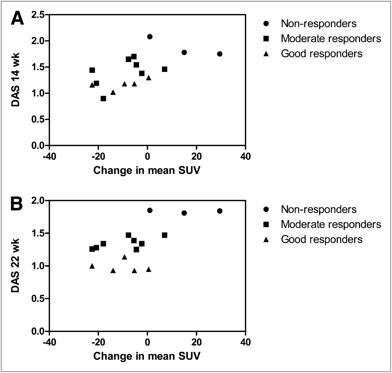
The change in mean SUV after 2 wk of infliximab treatment correlated significantly with DAS at 14 and 22 wk (r = 0.62, P < 0.05, and r = 0.65, P < 0.01, respectively; Figs. 3A and 3B, respectively), but at 6 wk no significant correlation was found (r = 0.26). Changes in ESR, CRP, and DAS after 2 wk did not correlate with DAS at any time point up to 22 wk (r ≤ 0.4, P > 0.05). None of the changes in any components of DAS (ESR, joint pain and swelling, visual analog scale) from 0 to 2 wk correlated with DAS at 14 or 22 wk. In addition, if only those metacarpophalangeal and wrist joints included in the PET analysis were considered, we found no correlation with disease activity at 14 or 22 wk.

Correlation between change in mean SUV and natural logarithm of DAS at 14 wk (r = 0.62, P < 0.05 [A]) and 22 wk (r = 0.65, P < 0.01 [B]).
The forward stepwise regression analysis showed that the change in mean SUV between baseline and 2 wk was the only significant contributor to the prediction of DAS at both 14 and 22 wk (14 wk: β = 0.62, P < 0.05; 22 wk: β = 0.65, P < 0.01). There was no significant predictive value to changes in ESR, CRP, and DAS from baseline to 2 wk (β values < 0.40). None of the changes in any other components of DAS contributed to a better prediction of DAS at 14 and 22 wk. Furthermore, if only those metacarpophalangeal and wrist joints included in the PET analysis were considered, no contribution of changes of swelling or tenderness of these joints to DAS was found at 14 or 22 wk.
DISCUSSION
In a small group of RA patients treated with infliximab, this explorative study showed a significant correlation between early changes in 18F-FDG uptake in hand joints and clinical disease activity after 14 and 22 wk of treatment. At a group level, the results indicate that early 18F-FDG changes are predictive of clinical outcome. In contrast, no predictive value was found for changes in ESR, CRP, and DAS after 2 wk of treatment. 18F-FDG PET may therefore be a valuable technique for predicting the efficacy of infliximab therapy as early as 2 wk after initiation of treatment. To our knowledge, this is the first PET study in RA patients showing the potential of 18F-FDG PET for predicting the response to infliximab therapy early in the course of treatment. Previous 18F-FDG PET studies visualized the treatment effects of infliximab in RA patients but did not investigate the predictive value of 18F-FDG PET for clinical response (4–6,8,9,20). In addition, various other markers have been tested, but—in line with our results—no single marker has consistently been shown to be a predictor of response to infliximab (3–6).
Quantitative determination of the level of inflammation in metacarpophalangeal and wrist joints by 18F-FDG PET could not simply be replaced by clinical scores of arthritis, since no correlation with clinical outcome and no predictive contribution were found for early changes (0–2 wk) in either tender or swollen joint counts of metacarpophalangeal and wrist joints, nor for DAS changes between 0 and 2 wk. Whether PET of inflamed joints outside the hands could also be predictive of clinical outcome in RA patients should be examined in a separate study.
As a consequence of the positioning of the hands in the scanner, individual metacarpophalangeal joints could not be analyzed, making a direct comparison with clinical joint examination at a joint level impossible. Nevertheless, the composite 18F-FDG score of clustered metacarpophalangeal and wrist joints (10 joints per patient) proved to be a sensitive parameter to predict disease activity at 14–22 wk of infliximab treatment. Apparently, early changes in local inflammatory levels in metacarpophalangeal and wrist joints, as measured by PET, were representative of later changes in clinical disease activity with respect to the whole body of the patient. The fact that early 18F-FDG changes correlated with clinical disease activity from 14 wk onward stresses the advantage of the sensitive imaging qualities of 18F-FDG PET. The good correlation between quantified 18F-FDG joint uptake and calculated SUV showed that infliximab had no pharmacokinetic effect on 18F-FDG metabolism. Thus, in future the procedure can be simplified to a static scanning of joints of interest at a rate of a few minutes of 18F-FDG PET imaging per field of view, as a monitoring tool for therapeutic intervention in RA.
CONCLUSION
The promising results for the use of 18F-FDG PET to monitor response to infliximab early in RA patients encourage further investigations in larger groups of patients. In addition, similar studies should be performed in RA patients treated with other drugs to assess whether the predictive value of 18F-FDG PET can be extrapolated to those agents. Moreover, future application of more specific PET tracers may increase the sensitivity of detection of inflammatory changes in RA joints.
Acknowledgments
This work was supported by an educational grant by Schering-Plough, The Netherlands. Schering-Plough did not in any way interfere in the design of the study; the collection, analysis, or interpretation of data; the writing of the manuscript; or the decision to submit the manuscript for publication.
- © 2011 by Society of Nuclear Medicine
REFERENCES
- Received for publication February 26, 2010.
- Accepted for publication September 8, 2010.


Jump to section
Related Articles
Cited By...
- Whole-Body Macrophage Positron Emission Tomography Imaging for Disease Activity Assessment in Early Rheumatoid Arthritis
- Early prediction of treatment response in rheumatoid arthritis by quantitative macrophage PET
- Prospective, simultaneous assessment of joint and vascular inflammation by PET/CT in tofacitinib-treated patients with rheumatoid arthritis: associations with vascular and bone status
- A model of chronic enthesitis and new bone formation characterized by multimodal imaging
- Scintigraphic detection of TNF-driven inflammation by radiolabelled certolizumab pegol in patients with rheumatoid arthritis and spondyloarthritis
- Detection of Subclinical Synovitis with Macrophage Targeting and Positron Emission Tomography in Patients with Rheumatoid Arthritis without Clinical Arthritis
- Evaluation of synovial angiogenesis in patients with rheumatoid arthritis using 68Ga-PRGD2 PET/CT: a prospective proof-of-concept cohort study
- EULAR recommendations for the use of imaging of the joints in the clinical management of rheumatoid arthritis