Main menu

  • Home
  • Content
    • Current
    • Ahead of print
    • Past Issues
    • JNM Supplement
    • SNMMI Annual Meeting Abstracts
    • Continuing Education
    • JNM Podcasts
  • Subscriptions
    • Subscribers
    • Institutional and Non-member
    • Rates
    • Journal Claims
    • Corporate & Special Sales
  • Authors
    • Submit to JNM
    • Information for Authors
    • Assignment of Copyright
    • AQARA requirements
  • Info
    • Reviewers
    • Permissions
    • Advertisers
  • About
    • About Us
    • Editorial Board
    • Contact Information
  • More
    • Alerts
    • Feedback
    • Help
    • SNMMI Journals
  • SNMMI
    • JNM
    • JNMT
    • SNMMI Journals
    • SNMMI

User menu

  • Subscribe
  • My alerts
  • Log in
  • My Cart

Search

  • Advanced search
Journal of Nuclear Medicine
  • SNMMI
    • JNM
    • JNMT
    • SNMMI Journals
    • SNMMI
  • Subscribe
  • My alerts
  • Log in
  • My Cart
Journal of Nuclear Medicine

Advanced Search

  • Home
  • Content
    • Current
    • Ahead of print
    • Past Issues
    • JNM Supplement
    • SNMMI Annual Meeting Abstracts
    • Continuing Education
    • JNM Podcasts
  • Subscriptions
    • Subscribers
    • Institutional and Non-member
    • Rates
    • Journal Claims
    • Corporate & Special Sales
  • Authors
    • Submit to JNM
    • Information for Authors
    • Assignment of Copyright
    • AQARA requirements
  • Info
    • Reviewers
    • Permissions
    • Advertisers
  • About
    • About Us
    • Editorial Board
    • Contact Information
  • More
    • Alerts
    • Feedback
    • Help
    • SNMMI Journals
  • View or Listen to JNM Podcast
  • Visit JNM on Facebook
  • Join JNM on LinkedIn
  • Follow JNM on Twitter
  • Subscribe to our RSS feeds

About the Cover

June 1, 2004; Volume 45,Issue 6

Cover image

Cover image expansion

ON THE COVER

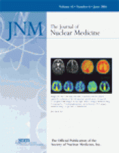
The 2 MR images show no cerebral infarction. The 2 SPECT images, obtained with the patient at rest (left) and after injection of acetazolamide (right), show a mild reduction of cerebral blood flow and normal reactivity to acetazolamide in the left cerebral hemisphere. From left to right, the 5 PET images show a mild reduction of cerebral blood flow, a mild reduction of the cerebral metabolic rate for oxygen, no abnormality of cerebral blood volume, no abnormality of oxygen extraction fraction, and a mild reduction of binding potential for 11C-flumazenil in the left cerebral hemisphere.

Back to top
PreviousNext

In this issue

Journal of Nuclear Medicine: 45 (6)
Journal of Nuclear Medicine
Vol. 45, Issue 6
June 1, 2004
  • Table of Contents
  • About the Cover
  • Index by author
Sign up for alerts

Jump to

  • SNM Newsline
  • Invited Perspectives
  • Clinical Investigations
  • Continuing Education
  • Basic Science Investigations
  • Departments
  • Letters to the Editor
  • Most Cited
  • Most Read
Loading
  • MIRD Pamphlet No. 28, Part 1: MIRDcalc—A Software Tool for Medical Internal Radiation Dosimetry
  • Absorbed Dose–Response Relationship in Patients with Gastroenteropancreatic Neuroendocrine Tumors Treated with [177Lu]Lu-DOTATATE: One Step Closer to Personalized Medicine
  • Prediction of 177Lu-DOTATATE PRRT Outcome Using Multimodality Imaging in Patients with Gastroenteropancreatic Neuroendocrine Tumors: Results from a Prospective Phase II LUMEN Study
  • Renal and Multiorgan Safety of 177Lu-PSMA-617 in Patients with Metastatic Castration-Resistant Prostate Cancer in the VISION Dosimetry Substudy
  • High-Temporal-Resolution Lung Kinetic Modeling Using Total-Body Dynamic PET with Time-Delay and Dispersion Corrections
More...
SNMMI

© 2025 SNMMI

Powered by HighWire