
Main menu

  • Home
  • Content
    • Current
    • Ahead of print
    • Past Issues
    • JNM Supplement
    • SNMMI Annual Meeting Abstracts
    • Continuing Education
    • JNM Podcasts
  • Subscriptions
    • Subscribers
    • Institutional and Non-member
    • Rates
    • Journal Claims
    • Corporate & Special Sales
  • Authors
    • Submit to JNM
    • Information for Authors
    • Assignment of Copyright
    • AQARA requirements
  • Info
    • Reviewers
    • Permissions
    • Advertisers
  • About
    • About Us
    • Editorial Board
    • Contact Information
  • More
    • Alerts
    • Feedback
    • Help
    • SNMMI Journals
  • SNMMI
    • JNM
    • JNMT
    • SNMMI Journals
    • SNMMI

User menu

  • Subscribe
  • My alerts
  • Log in
  • My Cart

Search

  • Advanced search
Journal of Nuclear Medicine
  • SNMMI
    • JNM
    • JNMT
    • SNMMI Journals
    • SNMMI
  • Subscribe
  • My alerts
  • Log in
  • My Cart
Journal of Nuclear Medicine

Advanced Search

  • Home
  • Content
    • Current
    • Ahead of print
    • Past Issues
    • JNM Supplement
    • SNMMI Annual Meeting Abstracts
    • Continuing Education
    • JNM Podcasts
  • Subscriptions
    • Subscribers
    • Institutional and Non-member
    • Rates
    • Journal Claims
    • Corporate & Special Sales
  • Authors
    • Submit to JNM
    • Information for Authors
    • Assignment of Copyright
    • AQARA requirements
  • Info
    • Reviewers
    • Permissions
    • Advertisers
  • About
    • About Us
    • Editorial Board
    • Contact Information
  • More
    • Alerts
    • Feedback
    • Help
    • SNMMI Journals
  • View or Listen to JNM Podcast
  • Visit JNM on Facebook
  • Join JNM on LinkedIn
  • Follow JNM on Twitter
  • Subscribe to our RSS feeds
OtherBasic Science Investigations

Evaluation of 3′-Deoxy-3′-18F-Fluorothymidine for Monitoring Tumor Response to Radiotherapy and Photodynamic Therapy in Mice

Masahiro Sugiyama, Harumi Sakahara, Kengo Sato, Norihiro Harada, Dai Fukumoto, Takeharu Kakiuchi, Toru Hirano, Eiji Kohno and Hideo Tsukada
Journal of Nuclear Medicine October 2004, 45 (10) 1754-1758;
Masahiro Sugiyama
  • Find this author on Google Scholar
  • Find this author on PubMed
  • Search for this author on this site
Harumi Sakahara
  • Find this author on Google Scholar
  • Find this author on PubMed
  • Search for this author on this site
Kengo Sato
  • Find this author on Google Scholar
  • Find this author on PubMed
  • Search for this author on this site
Norihiro Harada
  • Find this author on Google Scholar
  • Find this author on PubMed
  • Search for this author on this site
Dai Fukumoto
  • Find this author on Google Scholar
  • Find this author on PubMed
  • Search for this author on this site
Takeharu Kakiuchi
  • Find this author on Google Scholar
  • Find this author on PubMed
  • Search for this author on this site
Toru Hirano
  • Find this author on Google Scholar
  • Find this author on PubMed
  • Search for this author on this site
Eiji Kohno
  • Find this author on Google Scholar
  • Find this author on PubMed
  • Search for this author on this site
Hideo Tsukada
  • Find this author on Google Scholar
  • Find this author on PubMed
  • Search for this author on this site
  • Article
  • Figures & Data
  • Info & Metrics
  • PDF
Loading

Article Figures & Data

Figures

  • Tables
  • FIGURE 1.
    • Download figure
    • Open in new tab
    • Download powerpoint
    FIGURE 1.

    Biodistribution of 18F-FLT in C3H/He mice transplanted with SCCVII and in BALB/c nu/nu mice transplanted with HeLa at 1 h after injection. Data are expressed as mean ± SD.

  • FIGURE 2.
    • Download figure
    • Open in new tab
    • Download powerpoint
    FIGURE 2.

    Changes in relative tumor volume in control mice and x-ray–irradiated mice. Mice bearing SCCVII received 20 Gy at day 0. Data are expressed as mean ± SD. Radiotherapy resulted in tumor shrinkage and no tumor regrowth was found until 14 d after radiation. Tumors grew again after 14 d.
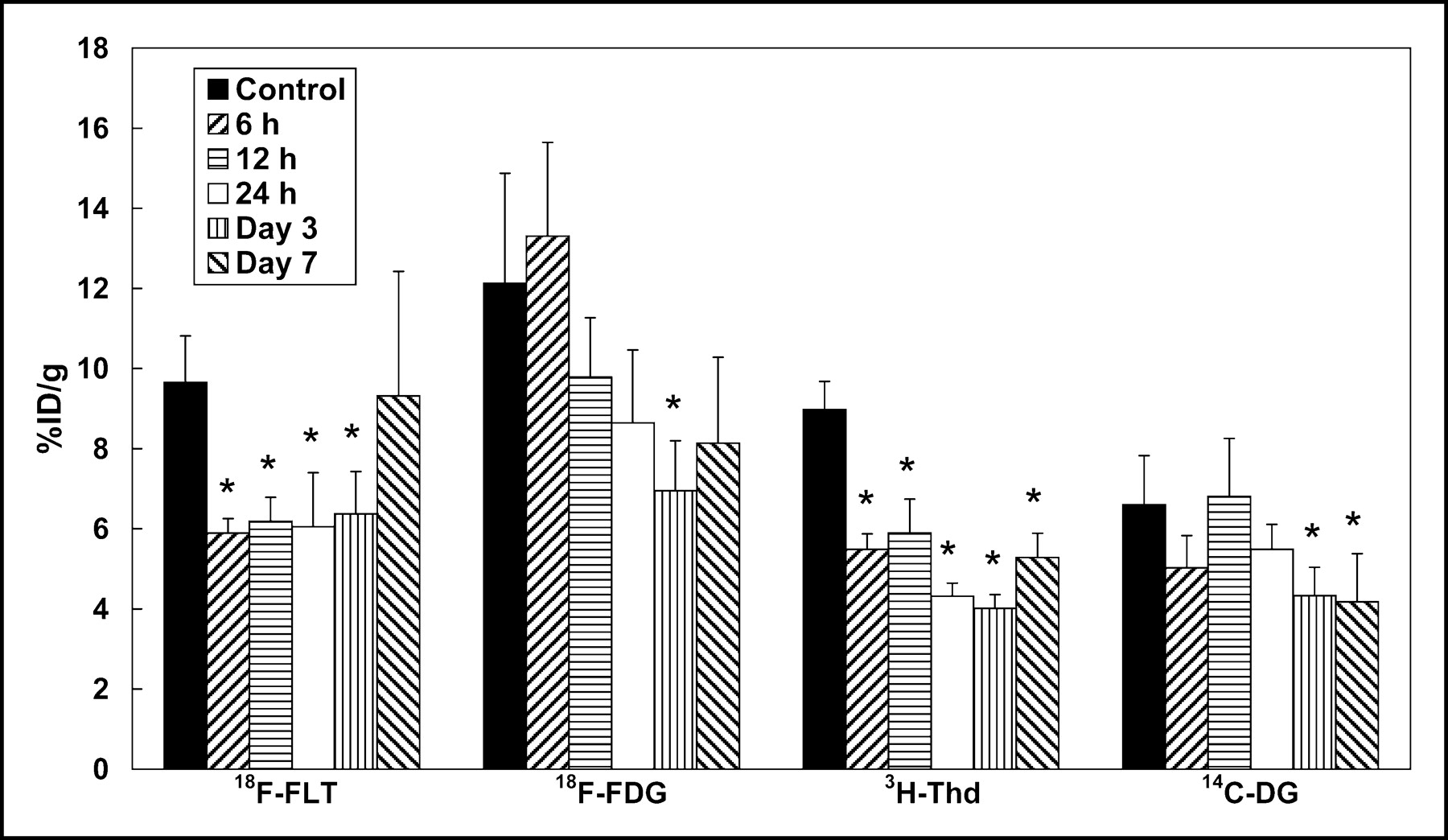
  • FIGURE 3.
    • Download figure
    • Open in new tab
    • Download powerpoint
    FIGURE 3.

    Tumor uptake of 4 tracers after radiotherapy. Data are expressed as mean ± SD. Asterisks indicate statistically significant differences compared with untreated controls (P < 0.05). Tumor uptake of 18F-FLT decreased significantly at 6 h, 12 h, 24 h, and 3 d after radiotherapy compared with untreated controls. There was a significant decrease in 3H-Thd uptake at 6 h, 12 h, 24 h, 3 d, and 7 d compared with untreated controls. Tumor uptake of 18F-FDG and 14C-DG did not show a statistically significant decrease at 6, 12, and 24 h after radiotherapy.
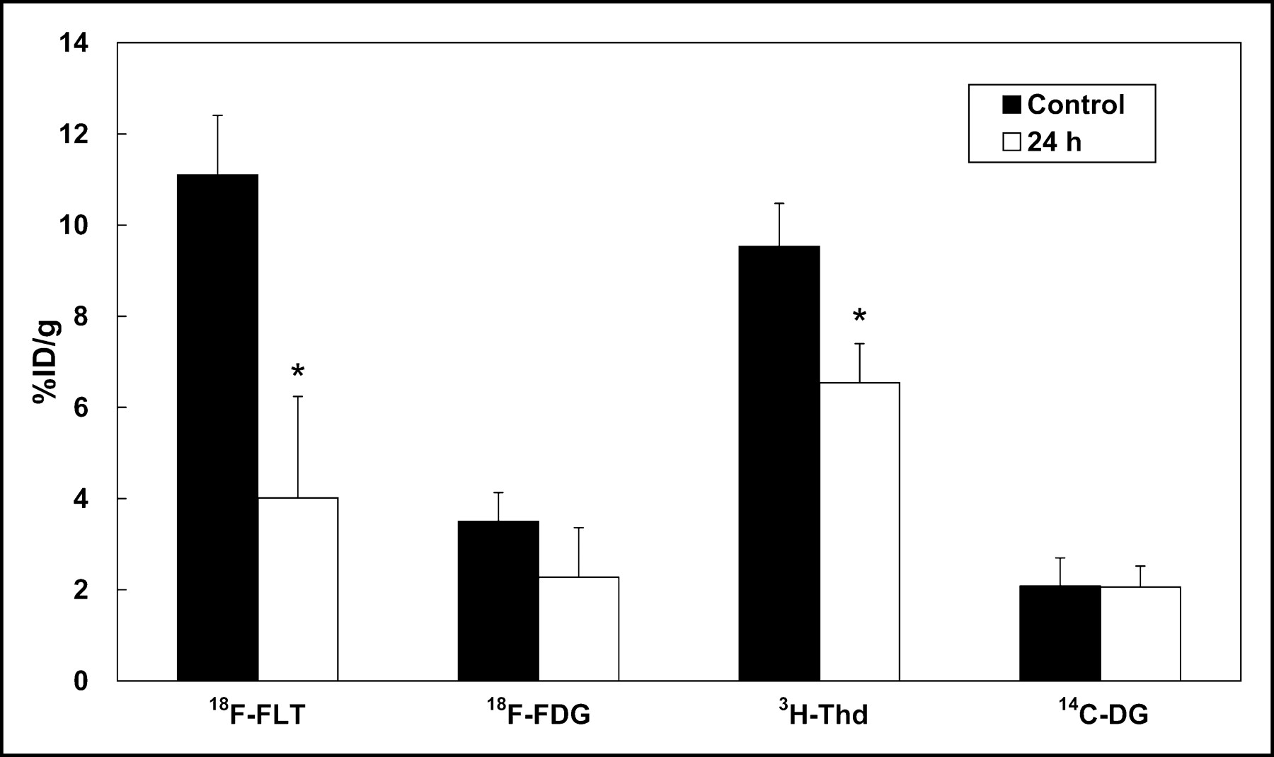
  • FIGURE 4.
    • Download figure
    • Open in new tab
    • Download powerpoint
    FIGURE 4.

    Tumor uptake of 4 tracers at 24 h after PDT. Data are expressed as mean ± SD. Asterisks indicate statistically significant differences compared with untreated controls (P < 0.05). A significant decrease of tumor uptake of 18F-FLT and 3H-Thd was observed in PDT-treated tumors. There was no significant difference between untreated controls and PDT-treated tumors in the uptake of 18F-FDG and 14C-DG.

Tables

  • Figures
    • View popup
    TABLE 1

    PCNA Labeling Index in Tumors After Radiotherapy and PDT

    GroupSCCVII tumor treated with 20-Gy irradiationHeLa tumor treated with PDT
    Control53.2 ± 8.782.0 ± 8.6
    6 h38.5 ± 5.3*ND
    24 h36.8 ± 5.3*13.5 ± 12.7*
    • ↵* P < 0.05 vs. untreated control group.

    • ND = not done.

    • Data are expressed as mean ± SD.

PreviousNext
Back to top

In this issue

Journal of Nuclear Medicine: 45 (10)
Journal of Nuclear Medicine
Vol. 45, Issue 10
October 1, 2004
  • Table of Contents
  • About the Cover
  • Index by author
Print
Download PDF
Article Alerts
Sign In to Email Alerts with your Email Address
Email Article

Thank you for your interest in spreading the word on Journal of Nuclear Medicine.

NOTE: We only request your email address so that the person you are recommending the page to knows that you wanted them to see it, and that it is not junk mail. We do not capture any email address.

Enter multiple addresses on separate lines or separate them with commas.
Evaluation of 3′-Deoxy-3′-18F-Fluorothymidine for Monitoring Tumor Response to Radiotherapy and Photodynamic Therapy in Mice
(Your Name) has sent you a message from Journal of Nuclear Medicine
(Your Name) thought you would like to see the Journal of Nuclear Medicine web site.
Citation Tools
Evaluation of 3′-Deoxy-3′-18F-Fluorothymidine for Monitoring Tumor Response to Radiotherapy and Photodynamic Therapy in Mice
Masahiro Sugiyama, Harumi Sakahara, Kengo Sato, Norihiro Harada, Dai Fukumoto, Takeharu Kakiuchi, Toru Hirano, Eiji Kohno, Hideo Tsukada
Journal of Nuclear Medicine Oct 2004, 45 (10) 1754-1758;

Citation Manager Formats

  • BibTeX
  • Bookends
  • EasyBib
  • EndNote (tagged)
  • EndNote 8 (xml)
  • Medlars
  • Mendeley
  • Papers
  • RefWorks Tagged
  • Ref Manager
  • RIS
  • Zotero
Share
Evaluation of 3′-Deoxy-3′-18F-Fluorothymidine for Monitoring Tumor Response to Radiotherapy and Photodynamic Therapy in Mice
Masahiro Sugiyama, Harumi Sakahara, Kengo Sato, Norihiro Harada, Dai Fukumoto, Takeharu Kakiuchi, Toru Hirano, Eiji Kohno, Hideo Tsukada
Journal of Nuclear Medicine Oct 2004, 45 (10) 1754-1758;
Twitter logo Facebook logo LinkedIn logo Mendeley logo
  • Tweet Widget
  • Facebook Like
  • Google Plus One
Bookmark this article

Jump to section

  • Article
    • Abstract
    • MATERIALS AND METHODS
    • RESULTS
    • DISCUSSION
    • CONCLUSION
    • Acknowledgments
    • Footnotes
    • REFERENCES
  • Figures & Data
  • Info & Metrics
  • PDF

Related Articles

  • THIS MONTH IN JNM
  • PubMed
  • Google Scholar

Cited By...

  • Molecular Imaging to Plan Radiotherapy and Evaluate Its Efficacy
  • Predictive Value of Early-Stage Uptake of 3'-Deoxy-3'-18F-Fluorothymidine in Cancer Cells Treated with Charged Particle Irradiation
  • Early Response Monitoring with 18F-FDG PET and Cetuximab-F(ab')2-SPECT After Radiotherapy of Human Head and Neck Squamous Cell Carcinomas in a Mouse Model
  • Acute Cytotoxic Effects of Photoimmunotherapy Assessed by 18F-FDG PET
  • Usefulness of 3'-Deoxy-3'-18F-Fluorothymidine PET for Predicting Early Response to Chemoradiotherapy in Head and Neck Cancer
  • 18F-FDG PET/CT for Image-Guided and Intensity-Modulated Radiotherapy
  • Radiopharmaceuticals in Preclinical and Clinical Development for Monitoring of Therapy with PET
  • Evaluation of D-18F-FMT, 18F-FDG, L-11C-MET, and 18F-FLT for Monitoring the Response of Tumors to Radiotherapy in Mice
  • Kinetic Modeling of 3'-Deoxy-3'-18F-Fluorothymidine for Quantitative Cell Proliferation Imaging in Subcutaneous Tumor Models in Mice
  • [18F]Fluorothymidine Positron Emission Tomography before and 7 Days after Gefitinib Treatment Predicts Response in Patients with Advanced Adenocarcinoma of the Lung
  • Imaging of Cell Proliferation: Status and Prospects
  • Dynamic Small-Animal PET Imaging of Tumor Proliferation with 3'-Deoxy-3'-18F-Fluorothymidine in a Genetically Engineered Mouse Model of High-Grade Gliomas
  • Preclinical Efficacy of the c-Met Inhibitor CE-355621 in a U87 MG Mouse Xenograft Model Evaluated by 18F-FDG Small-Animal PET
  • Early Detection of Chemoradioresponse in Esophageal Carcinoma by 3'-Deoxy-3'-3H-Fluorothymidine Using Preclinical Tumor Models
  • Positron Emission Tomography As an Imaging Biomarker
  • Dynamic Imaging of Transient Metabolic Processes by Small-Animal PET for the Evaluation of Photosensitizers in Photodynamic Therapy of Cancer
  • Reproducibility of 3'-Deoxy-3'-18F-Fluorothymidine MicroPET Studies in Tumor Xenografts in Mice
  • Google Scholar

More in this TOC Section

  • 11C-Methionine PET of Myocardial Inflammation in a Rat Model of Experimental Autoimmune Myocarditis
  • Counting Rate Characteristics and Image Distortion in Preclinical PET Imaging During Radiopharmaceutical Therapy
  • Design and Fabrication of Kidney Phantoms for Internal Radiation Dosimetry Using 3D Printing Technology
Show more Basic Science Investigations

Similar Articles

SNMMI

© 2025 SNMMI

Powered by HighWire