


Abstract
The aim of this study was to investigate whether 3′-deoxy-3′-18F-fluorothymidine (18F-FLT) can monitor the early response of tumor cell proliferation to charged particle irradiation in vitro and in vivo. Methods: In vitro, after 0.1, 0.5, 1, 5, and 10 Gy of proton or carbon ion irradiation, 18F-FLT cell uptake was examined at 24 h and cell proliferation ability was measured from days 1 to 4. In vivo, after 0.5, 1, and 5 Gy of proton or carbon ion irradiation, 18F-FLT PET imaging was performed on tumor-bearing BALB/c nu/nu mice at 24 h and tumor growth was measured from days 1 to 7. Tumor-to-background ratios of standardized uptake values were calculated to assess the 18F-FLT accumulation in tumors. Both cells and mice also received x-irradiation as a control. Results: In vitro, 18F-FLT cell uptake was significantly lower after 1 Gy of proton irradiation (P < 0.05) and carbon ion irradiation (P < 0.05) and after 5 Gy of x-irradiation (P < 0.01), but cell proliferation ability at these doses did not show significant differences until day 3. In vivo, 18F-FLT tumor uptake was significantly lower after 1 Gy of proton (P < 0.001) and carbon ion irradiation (P < 0.01) and after 5 Gy of x-irradiation (P < 0.001), but tumor growth did not significantly differ at these doses until day 4 after proton irradiation, day 3 after carbon ion irradiation, and day 5 after x-irradiation. Conclusion: The reduction in 18F-FLT uptake after charged particle irradiation was more rapid than the change in tumor growth in vivo or the change in cell proliferation ability in vitro. Therefore, 18F-FLT is a promising tracer for monitoring the early response of cancer to charged particle irradiation.
- 3′-deoxy-3′-18F-fluorothymidine (18F-FLT)
- positron emission tomography (PET)
- charged particle irradiation
- tumor volume
- cell proliferation ability
Charged particle therapy, the newest technique in radiotherapy, has physical and radiobiologic advantages over conventional x-ray therapy. Charged particles such as protons and carbon ions deposit most of their energy at the end of their path (called the Bragg peak) and little energy along the way, producing favorable dose distributions in the tumor and little radiation damage to the surrounding normal tissues (1–3). Although maintaining the same irradiation to the tumor, the integral dose by protons has been found to decrease by 50% or more compared with x-rays (4,5). A lower integral dose may also reduce the risk of secondary malignancies, which is a serious concern for those treated with intensity-modulated radiotherapy. However, the treatment cost for charged particle therapy is much higher than that for x-ray therapy because of the high investment cost required to build accelerators, beam transport systems, and gantries (6). Therefore, we need to know the likelihood of an early response to radiotherapy in order to evaluate whether the medical benefit justifies the high cost and to develop a better treatment plan.
The efficacy of radiotherapy is often identified by morphologic imaging techniques, which depict responses such as changes in tumor size and composition. However, these changes frequently take time, leading to difficulty in evaluating the effect of treatment early (7). By being able to show the metabolic and physiologic changes that precede a change in size, PET solved this problem and has been widely used for diagnosing cancer, monitoring therapy response, and planning radiotherapy (8,9). 18F-FDG is the most commonly used PET tracer for routine clinical use (10). However, during or immediately after a course of radiation therapy, the overall glucose utilization and signal seen on 18F-FDG PET are unclear. Also, the inflammatory response to radiotherapy limits the ability of 18F-FDG PET to detect the early response of a tumor to radiation (11,12). Therefore, tracers that can distinguish tumors from inflammatory tissues are required.
3′-deoxy-3′-18F-fluorothymidine (18F-FLT) PET imaging was first introduced in 1998 as a noninvasive tool for visualizing tumor cell proliferation (13). The correlation between 18F-FLT uptake and proliferation has been validated for various tumors (14–17). Several studies have suggested that 18F-FLT is more cancer-specific (18–20) and sensitive (21) than 18F-FDG. The potential detectability of 18F-FLT during the early response to irradiation has also been reported (7,22–24). However, because of the high cost and long treatment-planning time of charged particle therapy, few studies have evaluated the use of 18F-FLT for monitoring the efficacy of proton and, especially, carbon ion radiotherapy. Recently, a study by Inubushi et al. on head and neck mucosal malignant melanoma showed for the first time that 18F-FLT PET/CT imaging is useful for predicting the therapeutic outcome of carbon ion radiotherapy (25).
The purpose of this study was to investigate whether 18F-FLT can be used to monitor the early response of cell proliferation to charged particle irradiation in vitro and in vivo, thereby facilitating early modification of the treatment plan and providing a reference for the cost effectiveness of charged particle therapy.
MATERIALS AND METHODS
Cell Culture and Animal Models
For the in vitro study, colon 26, a mouse colon carcinoma cell line, was obtained from the Cell Resource Center for Biomedical Research, Tohoku University, Sendai, Japan; maintained in Dulbecco modified Eagle medium, nutrient mixture F-12, supplemented with 10% fetal bovine serum and 1% penicillin/streptomycin (Gibco); and incubated at 37°C in 5% CO2.
For the in vivo study, 5- to 6-wk-old male BALB/c nu/nu mice (Japan SLC) were used. Colon 26 cells were inoculated subcutaneously into the left shoulder at cell densities from 6 × 10⁶ to 1 × 10⁷/200 μL together with Matrigel matrix (BD Biosciences). The tumor size was determined by caliper measurements using the formula [length × (width)2]/2. Irradiation was applied when the xenografts reached approximately 600 mm3. All animal experiments were fully accredited by the Laboratory Animal Care Committee of the University of Fukui.
Irradiation
Both cells and tumor-bearing mice were treated with charged particle irradiation and x-irradiation. The accelerator complex has been described elsewhere (26). The details are described in the supplemental data (available at http://jnm.snmjournals.org).
18F-FLT Synthesis
18F-FLT was radiosynthesized in a TRACERlab MX-FDG (GE Healthcare) using an FLT kit from ABX GmbH. The details are described in the supplemental data.
Cell Counting and 18F-FLT Cell Uptake Experiments
From days 1 to 4 after irradiation, the cells were trypsinized to detach them from the flasks and assayed using the trypan blue dye-exclusion method for cell-counting experiments (5 flasks per dose, each day). The 18F-FLT cell uptake experiment was assessed at 24 h after irradiation; 0.37 MBq of 18F-FLT were added to each flask, and the cells were further incubated for 1 h. After incubation, the cells were rinsed 3 times with ice-cold phosphate-buffered saline and then immediately lysed using 0.1N NaOH (Nacalai Tesque, Inc.). Radioactivity in the cells was measured in a γ counter (Wallac Wizard3 1480; Perkin-Elmer), and protein levels were quantified using a bicinchoninic acid assay kit (Thermo Scientific Pierce) according to the manufacturer’s instructions. Cell uptake levels are expressed as percentage of input radioactivity normalized to milligram of protein.
Small-Animal PET Imaging Experiment and Follow-up Study
Tumor-bearing mice were imaged using a small-animal PET scanner (SHR-41000; Hamamatsu Photonics) (27) at 24 h after irradiation treatment. The details are described in the supplemental data.
Statistical Analysis
Statistics were analyzed using Prism, version 5 (GraphPad Software). All data are expressed as mean ± SD. Differences between untreated controls and irradiated groups were analyzed using 1-way ANOVA with adjustment by the Bonferroni method to compare all pairs of columns. A P value of less than 0.05 was considered statistically significant.
RESULTS
Cell Proliferation Ability After Irradiation
After x-irradiation, a significant difference in cell number in the group receiving 10 Gy (compared with the control group) was observed on day 1 (Fig. 1A, P < 0.05) and persisted until day 4 (P < 0.001). X-irradiation at 5 Gy did not result in a significant difference until day 3 (P < 0.001). No significant difference was observed at any time from days 1 to 4 after x-irradiation at 0.1, 0.5, or 1 Gy. After both proton irradiation and carbon ion irradiation, significant differences in cell number in the groups receiving 5 and 10 Gy were observed on day 1 (Figs. 1B and 1C, P < 0.05) and continued to day 4 (P < 0.001). Furthermore, the group treated with 1 Gy also showed significant differences from day 3 (P < 0.01 and P < 0.001, respectively) to day 4 (P < 0.001). The groups receiving 0.1 and 0.5 Gy showed no significant differences at any time.

Cell proliferation ability curves of colon 26 cells after various doses of x-irradiation (A), proton irradiation (B), and carbon ion irradiation (C). Cells were irradiated at doses of 0.1, 0.5, 1, 5, and 10 Gy. *P < 0.05, **P < 0.01, and ***P < 0.001 for irradiated group vs. control group.
In Vitro Uptake of 18F-FLT After Irradiation
After x-irradiation, significantly lower 18F-FLT uptake (compared with the control group) was seen in the groups receiving 5 Gy (Fig. 2A, P < 0.01) and 10 Gy (P < 0.001). After charged particle irradiation, significantly lower 18F-FLT uptake was seen in the groups receiving 5 Gy, 10 Gy (Figs. 2B and 2C, P < 0.001), and 1 Gy (Figs. 2B and 2C, P < 0.05). Relative uptake was significantly lower for carbon ion irradiation than for x-irradiation at 5 Gy and 10 Gy (Fig. 2D, P < 0.001) and significantly lower for proton irradiation than for x-irradiation at 10 Gy (P < 0.001). Furthermore, significant differences between proton irradiation and carbon ion irradiation were observed at both 5 Gy and 10 Gy (P < 0.001).
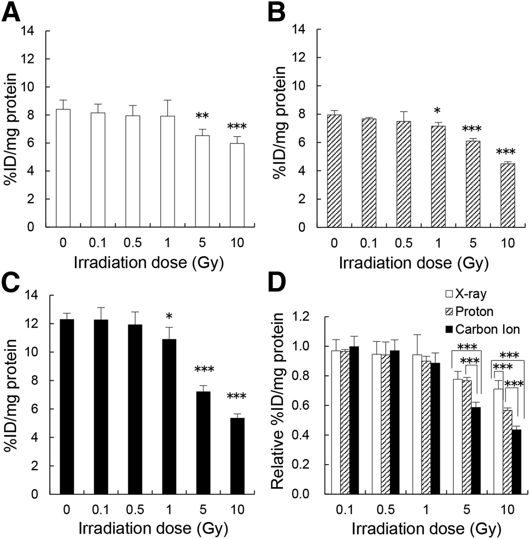
(A–C) 18F-FLT uptake in colon 26 cells after x-irradiation (A), proton irradiation (B), and carbon ion irradiation (C). Cells were irradiated at same doses as for cell counting: 0.1, 0.5, 1, 5, and 10 Gy. *P < 0.05, **P < 0.01, and ***P < 0.001 for irradiated group vs. control group. (D) Relative 18F-FLT uptake values in colon 26 cells among the 3 irradiations were compared.
In Vivo Validation of Predictive Value of 18F-FLT PET for Irradiation Monitoring
The results for 18F-FLT PET imaging after irradiation are given in Figures 3–5.

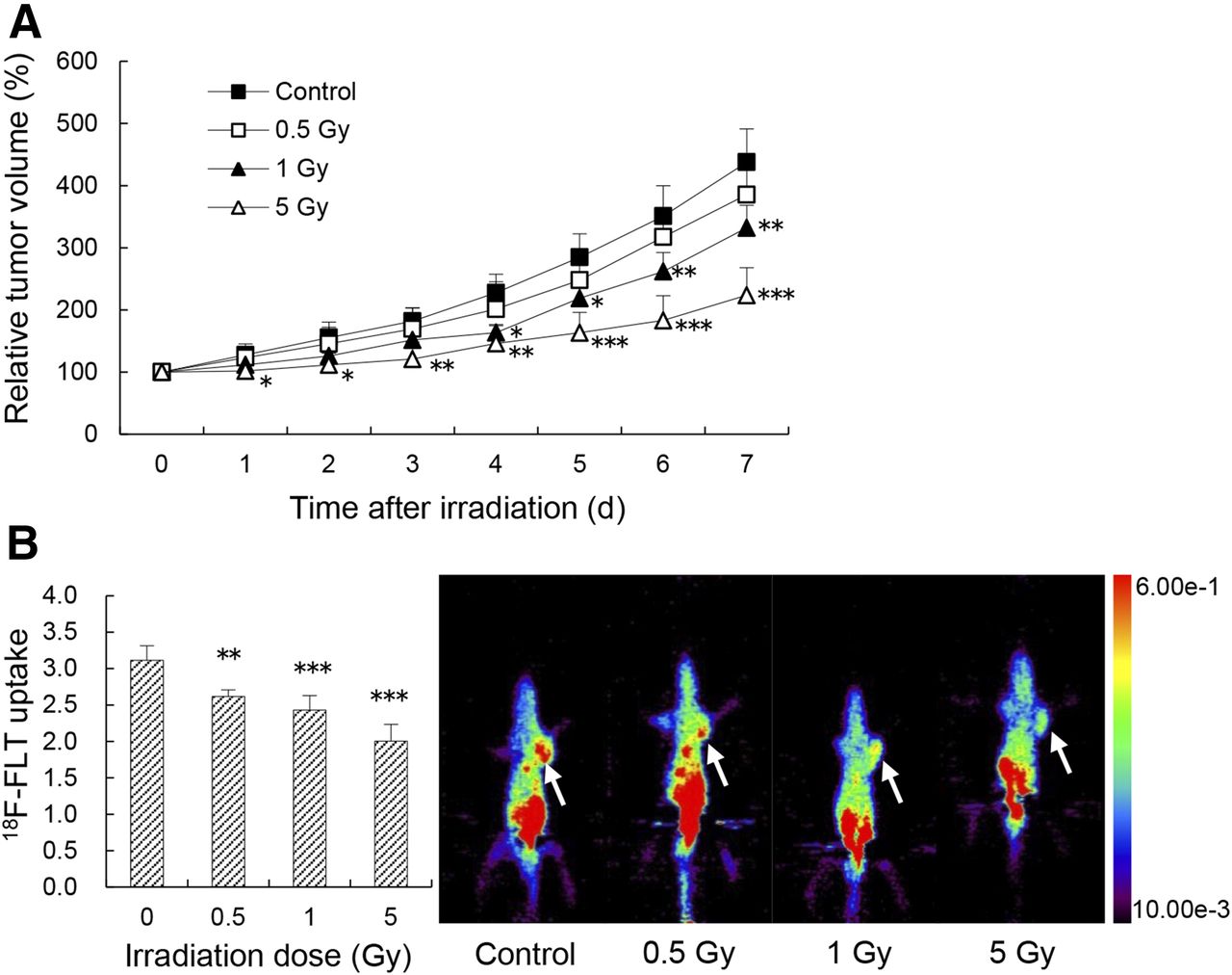
Relative growth curves of colon 26 tumors (A), uptake of 18F-FLT by tumors (B, left), and representative images of mice (B, right) after x-irradiation. Tumors on left shoulder of mice (arrows) were irradiated at doses of 0.5, 1, and 5 Gy on day 0. *P < 0.05, **P < 0.01, and ***P < 0.001 for irradiated group vs. control group.
Relative growth curves of colon 26 tumors (A), uptake of 18F-FLT by tumors (B, left), and representative images of mice (B, right) after proton irradiation. Tumors on left shoulder of mice (arrows) were irradiated at doses of 0.5, 1, and 5 Gy on day 0. *P < 0.05, **P < 0.01, and ***P < 0.001 for irradiated group vs. control group.

Relative growth curves of colon 26 tumors (A), uptake of 18F-FLT by tumors (B, left), and representative images of mice (B, right) after carbon ion irradiation. Tumors on left shoulder of mice (arrows) were irradiated at doses of 0.5, 1, and 5 Gy on day 0. *P < 0.05, **P < 0.01, and ***P < 0.001 for irradiated group vs. control group.
X-Irradiation
A significant difference in relative tumor volume was observed on day 5 between the group receiving 5 Gy of x-irradiation and the control group (a volume increase of 218% and 309%, respectively) (Fig. 3A, P < 0.05). This significance persisted until day 7, when the increase was 301% and 413%, respectively (Fig. 3A, P < 0.01). However, compared with the control group, the 0.5- and 1-Gy groups showed no significant differences during the 7 d after receiving the x-irradiation. On 18F-FLT PET imaging, a remarkable decrease in tumor uptake was observed on day 1 for the group receiving 5 Gy of x-irradiation, compared with the control group (Fig. 3B, left, P < 0.001). For the groups receiving 0.5 or 1 Gy, however, no significant reduction was seen. Figure 3B (right) shows representative images of each group.
Proton Irradiation
A significant difference in relative tumor volume was observed on day 1 between the group receiving 5 Gy of proton irradiation and the control group (a volume increase of 102% and 128%, respectively) (Fig. 4A, P < 0.05) and on day 4 between the group receiving 1 Gy of proton irradiation and the control group (a volume increase of 164% and 228%, respectively, P < 0.05). Tumor growth in both the 5-Gy and the 1-Gy groups was significantly lower (224% and 332% [P < 0.001 and P < 0.01], respectively) than in the control group (438%) until day 7. The 0.5-Gy group had a slower growth rate than the control group, but the difference was not statistically significant (a 386% increase on day 7). Uptake of 18F-FLT in tumor was dramatically less on day 1 in each of the groups receiving proton irradiation (5 Gy [Fig. 4B, left] [P < 0.001], 1 Gy [P < 0.001], and 0.5 Gy [P < 0.01]) than in the control group. Representative images of each group are shown in Figure 4B (right).
Carbon Ion Irradiation
A significant difference in relative tumor volume was observed on day 1 between the group receiving 5 Gy of carbon ion irradiation and the control group (a volume increase of 105% and 124%, respectively) (Fig. 5A, P < 0.05). There was a significant difference on day 3 between the 1-Gy group and the control group (a volume increase of 150% and 199%, respectively, P < 0.05). Both treated groups remained significantly lower until day 7, with 209% and 318% increases, respectively, compared with the control group, which had a 426% increase (P < 0.001 and P < 0.05, respectively). The relative tumor volume was 357% in the 0.5-Gy group on day 7, but the difference from the control group was not statistically significant. Uptake of 18F-FLT in tumor was significantly less on day 1 in each of the groups receiving carbon ion irradiation (5 Gy [Fig. 5B, left] [P < 0.001], 1 Gy [P < 0.01], and 0.5 Gy [P < 0.05]) than in the control group. Representative images of each group are shown in Figure 5B (right).
DISCUSSION
The present study showed that 18F-FLT uptake in tumors receiving charged particle irradiation was significantly lower than that in nonirradiated tumors at as early as 24 h—much earlier than significant changes in tumor size were seen in vivo or significant changes in cell proliferation ability were seen in vitro. This finding is consistent with previous findings for x-irradiation (7,22,23), although only a few studies using carbon ion radiotherapy have been reported (25,28,29). Charged particle irradiation has been associated with better treatment outcomes and less damage to normal tissue than x-irradiation. The 18F-FLT cell uptake values and tumor-to-background standardized uptake value ratios reflect proliferation of the tumor after irradiation. Our findings of 18F-FLT uptake after irradiation suggest that 18F-FLT is able to predict the early response of cell proliferation to irradiation. Early response may be an important prognostic marker for the outcome of charged particle irradiation and should facilitate modification of treatment plans at an early stage. The results of our study provide more evidence of the predictive value of 18F-FLT for charged particle irradiation.
PET with various tracers that allow monitoring of proliferation, glucose metabolism, amino acid metabolism, hypoxia, and lipid metabolism can be used to plan radiation treatment. A cellular proliferation decrease and cell death usually happen immediately after irradiation and, over the long term, lead to an overall reduction in metabolism, including reduced glucose consumption, protein synthesis/amino acid transport, and DNA synthesis. 18F-FDG PET, although not appropriate for detecting early tumor response to radiation, can be performed several months after completion of radiotherapy (30,31). On the other hand, DNA analogs or amino acids and their analogs, which are more specific to tumor characteristics than 18F-FDG, may be more suitable for monitoring early radiotherapy response (10). For example, 11C-methionine is currently one of the best PET tracers available for delineating brain tumor contour (32). 11C- and 18F-labeled choline derivatives are promising tracers for prostate cancer (33). 18F-fluoromisonidazole, 18F-fluoroazomycin arabinoside, and 64Cu-diacetyl-bis(N4-methylthiosemicarbazone) are common PET tracers for imaging hypoxia, because tumor hypoxia is considered an important factor for resistance to radiotherapy and appears to be an independent risk factor for tumor progression (34). 18F-FLT, which allows monitoring of cell proliferation, is a promising tracer as a marker of early response of cell proliferation to radiotherapy (7,22–24).
In our animal study, 18F-FLT uptake at 24 h was significantly low in tumors receiving 5 Gy of charged particle irradiation; this finding is in accordance with the significant difference in tumor growth at 24 h. 18F-FLT uptake at 24 h was also significantly different in tumors receiving 1 Gy of charged particle irradiation and 5 Gy of x-irradiation, but relative growing tumor volume did not show a significant difference until day 4 after proton irradiation, day 3 after carbon ion irradiation, and day 5 after x-irradiation. The remarkable reductions in tumor uptake of 18F-FLT preceded the changes in tumor volume, suggesting the prognostic potential of 18F-FLT PET for monitoring early response after irradiation. These findings are consistent with previous studies using x-irradiation in murine SCCVII tumors (7,22), as well as with clinical trials (23), and a recent report demonstrated the usefulness of 18F-FLT PET/CT imaging for predicting the therapeutic outcome of carbon ion radiotherapy in head and neck mucosal malignant melanoma (25). Furthermore, after 0.5 Gy of charged particle irradiation, tumor uptake of 18F-FLT was significantly different from that in controls whereas tumor growth was not, indicating that the 0.5-Gy dose may not be sufficient to induce an anticancer effect in vivo. Murayama et al. (24) also reported a significant difference in tumor uptake of 18F-FLT on day 1 in a 2-Gy group, although no significant differences in tumor growth were found 14 d after irradiation. Our results support their interpretation that the reduction in tumor uptake of 18F-FLT on day 1 after a low irradiation dose may reflect sublethal damage, since DNA damage by a low dose might be repairable, as demonstrated by the tumor growth curve. Our study also showed slight differences in 18F-FLT uptake between tumors receiving proton irradiation and tumors receiving carbon ion irradiation at 5 Gy (decreased to 64.3% vs. 70.6%), 1 Gy (78.1% vs. 78.3%), and 0.5 Gy (84.2% vs. 84.5%). Wilkens and Oelfke found that the model used in their study did not predict a biologic advantage of carbon ions (35). This finding may be related to uncertainty in the relative biological effectiveness of carbon ions, which can cause large variations in the actual delivered dose (35). Also, carbon ions are heavier than protons and cannot travel very far through tissue. But our results did prove the advantage of charged particle irradiation over x-irradiation in vivo (1).
In vitro, cell uptake of 18F-FLT was significantly lower at 24 h after 5 Gy and 10 Gy of charged particle irradiation and after 10 Gy of x-irradiation. These changes were also reflected by the decrease in cell proliferation ability at 24 h after these doses. Our findings are consistent with a previous report of significantly fewer surviving murine SCCVII tumor cells at 24 h after 10 Gy of x-irradiation (22). At 24 h, cell uptake of 18F-FLT was also significantly lower after 1 Gy of proton or carbon ion irradiation (10% and 11% reduction, respectively) than after 5 Gy of x-irradiation (∼22% reduction). However, the cell proliferation ability after these doses was not significantly different until day 3. These results indicate that 18F-FLT reflects the early response of tumor cell proliferation to irradiation, as reported previously (22,24,36). Wang et al. showed that reduced uptake of 18F-FLT by 2 other human colorectal cancer cell lines at 24 h after x-irradiation preceded a significant reduction in cell number in the S phase at 72 h after irradiation (36). In addition, the relative 18F-FLT uptake seemed to reflect the higher efficacy of charged particle irradiation than of x-irradiation in vitro, as agrees well with our in vivo study. Furthermore, carbon ion irradiation seemed to be more effective than proton irradiation at 5 Gy (41% vs. 23% reduction, respectively) and 10 Gy (56% vs. 43% reduction, respectively), probably because, in vitro, without traveling through the tissue, carbon ions have a higher relative biological effectiveness in the target and cause greater DNA damage, leading to a therapeutic advantage over protons (35).
According to the mechanism of charged particle irradiation and x-irradiation, the irradiation directly or indirectly damages the DNA, leading to decreased DNA synthesis or cell proliferation, as was confirmed in our study. Because 18F-FLT is taken up by proliferating cells and phosphorylated by TK1 during the S phase, when cells primarily rely on the salvage pathway as a dominant source for DNA synthesis, we suppose that decreased DNA synthesis or cell proliferation resulted in the decreased 18F-FLT uptake.
One limitation of the current study is that it was conducted on only one model of cancer: colorectal carcinoma in mice. However, in other cancer types (with other molecular–genetic alterations), the temporal dynamics and magnitude of radiation dose–response in tumor proliferation as assessed by 18F-FLT PET may be different. Besides, although the tumors grew more slowly in the irradiated groups than in the control group, the tumors did not shrink but kept growing after irradiation. Further studies with more cancer types and higher doses will be needed to provide more reliable data for both preclinical and clinical studies.
CONCLUSION
The reduction in 18F-FLT uptake at 24 h after charged particle irradiation preceded the change in tumor growth in vivo and the change in cell proliferation ability in vitro. Although further investigations using various cancer types are needed, 18F-FLT is a promising tracer for monitoring the early response of tumor cell proliferation to charged particle irradiation.
DISCLOSURE
The costs of publication of this article were defrayed in part by the payment of page charges. Therefore, and solely to indicate this fact, this article is hereby marked “advertisement” in accordance with 18 USC section 1734. This study was partially supported by research funds from the Wakasa Wan Energy Research Center and by grant 24249065 from JSPS KAKENHI. No other potential conflict of interest relevant to this article was reported.
Acknowledgments
We thank Takashi Hasegawa and the staff of the Research and Development Division, Wakasa Wan Energy Research Center, and we thank Akira Ito of CMI (Century Medical, Inc.) for operating the cyclotron.
Footnotes
Published online Mar. 12, 2015.
- © 2015 by the Society of Nuclear Medicine and Molecular Imaging, Inc.
REFERENCES
- Received for publication December 16, 2014.
- Accepted for publication February 3, 2015.


