


Abstract
O-18F-fluoromethyl-d-tyrosine (d-18F-FMT) is a promising novel agent for tumor imaging by PET. The aim of this study was to evaluate the potential of d-18F-FMT and the other conventional ligands used for tumor imaging, namely, 18F-FDG, l-11C-methionine (l-11C-MET), and 3′-deoxy-3′-18F-fluorothymidine (18F-FLT), as a PET ligand for monitoring early responses to radiotherapy in tumor-bearing mice. Methods: C3H/HeN mice inoculated with murine squamous cell carcinomas were treated with a single dose of x-ray irradiation at 2, 6, 20, or 60 Gy. Tumor uptake of each ligand was examined 1, 3, and 7 d after the irradiation. Results: Tumor uptake of d-18F-FMT was decreased on day 1 after irradiation at 6, 20, or 60 Gy, and the decrease persisted until day 7. Tumor uptake of 18F-FDG was elevated on days 1 and 3 after irradiation at 2, 6, or 20 Gy, followed by a decrease in uptake on day 7 in mice irradiated at 20 or 60 Gy. Decreased tumor uptake of l-11C-MET was observed only on day 3 after the irradiation. Decreased tumor uptake of 18F-FLT was detected on day 1 after irradiation at 2, 6, 20, or 60 Gy; thereafter, the dose-dependent decrease in uptake was no longer seen. Only for d-18F-FMT were significant positive correlations found between ligand uptake at all the time points examined and tumor volume on day 14 after various doses of irradiation. Conclusion: The findings suggest that d-18F-FMT is a promising PET ligand for early-phase detection and prediction of the effects of radiation therapy.
- O-18F-fluoromethyl-d-tyrosine
- d-18F-FMT
- 18F-fluoro-2-deoxy-d-glucose
- 18F-FDG
- l-11C-methionine
- l-11C-MET
- 3′-deoxy-3′-18F-fluorothymidine
- 18F-FLT
- radiation therapy
Although 18F-FDG, a glucose analog, is the most commonly used PET ligand for tumor imaging and monitoring of treatment efficacy, several limitations have been reported, such as nonspecific uptake by foci of infection or inflammation (1). Considerable effort has been made to create more suitable PET ligands, the imaging results of which would correspond more directly to tumor aggressiveness or its response to therapy than do those of 18F-FDG. An increased cellular proliferation rate is potentially more tumor-specific than high glucose use and may correspond more directly to tumor aggressiveness and tumor responsiveness to therapy. Significant correlations of the uptake of 3′-deoxy-3′-18F-fluorothymidine (18F-FLT) (2), a thymidine analog, with other measures of cellular proliferation, including the Ki-67 score, have been demonstrated for a variety of human cancers (3,4). Increased amino acid metabolism is also a well-known characteristic of tumors, in which amino acid transport or protein synthesis rates are enhanced. Hence, natural and unnatural labeled amino acid analogs have been proposed as PET ligands for tumor detection. Their higher uptake by tumor cells than by normal cells reflects the facilitated amino acid transport or protein synthesis in tumor cells and is related to cellular proliferation (5). l-11C-methionine (l-11C-MET), a carbon-labeled natural amino acid, has been widely used because of its simple labeling procedure and high uptake by tumors (6,7). In addition, fluorine-labeled artificial amino acid analogs, such as O-18F-fluoromethyl-l-tyrosine (l-18F-FMT) (8), O-18F-fluoroethyl-l-tyrosine (l-18F-FET) (9), and O-18F-fluoropropyl-l-tyrosine (l-18F-FPT) (10) have also been developed as candidate PET ligands for tumor imaging. Different from natural amino acid–based ligands such as l-11C-MET, they are unnatural amino acid–based ligands that reflect amino acid transporter activity; that is, these amino acids are transported into the cells but not actually incorporated into the protein synthetic pathway. Recently, we demonstrated that the d-isomers of 18F-fluoroalkyl tyrosine analogs were more suitable than their l-isomers as ligands for PET of tumors (11,12). Among the l- and d-isomers of 18F-FMT, 18F-FET, and 18F-FPT, O-18F-fluoromethyl-d-tyrosine (d-18F-FMT) was found to show the highest tumor-to-blood and tumor-to-liver ratios (11,12).
PET is currently relied on as a noninvasive technique for the detection of early-phase responses of tumor cells to therapy (e.g., chemotherapy and radiotherapy) (4,13–17). l-11C-MET and 18F-FLT imaging has been reported to show more sensitivity and faster response to irradiation than does 18F-FDG imaging (4,13–15). The aim of this study was to evaluate the potential usefulness of d-18F-FMT, 18F-FDG, l-11C-MET, and 18F-FLT as a PET ligand for monitoring tumor responses to radiotherapy in C3H/HeN mice inoculated with murine squamous cell carcinoma SCCVII.
MATERIALS AND METHODS
Chemicals
Acetonitrile, dimethylsulfoxide, N,N-dimethylformamide, tetrahydrofuran, lithium aluminum hydride (1.0 M in tetrahydrofuran), l-homocysteine thiolactone, and d-tyrosine were purchased from Sigma-Aldrich. Hydroiodic acid was purchased from Nacalai Tesque. Mannose triflate and 5′-O-(4,4′-dimethoxytrityl)-2,3′-anhydrothymidine were purchased from ABX.
Syntheses of Labeled Compounds
11C and 18F were produced by 14N(p,α)11C and 18O(p,n)18F nuclear reactions, respectively, using a cyclotron (HM-18; Sumitomo Heavy Industry) at the Hamamatsu Photonics PET Center. 11C-methyl iodide was prepared from 11C-CO2. l-11C-MET was labeled by S-methylation of l-homocysteine thiolactone (7). d-18F-FMT was synthesized by reactions of 18F-fluoromethyl bromide with d-tyrosine by a previously described method (11). 18F-FLT was synthesized by nucleophilic fluorination of 5′-O-(4,4′-dimethoxytriphenylmethyl)-2,3′-anhydrothymidine (18). 18F-FDG was produced by nucleophilic fluorination of mannose triflate after basic hydrolysis of 2-18F-fluoro-1,3,4,6-tetra-O-acetyl-d-glucose (19).
Animal Model
Female C3H/HeN Slc mice (Japan SLC, Inc.) inoculated with SCCVII tumors were used for the assessments. SCCVII, a murine squamous cell carcinoma cell line, was inoculated subcutaneously into the right thigh of 8-wk-old mice at a cell density of 1 × 10⁵ in 0.05 mL of saline. The tumor size was determined by caliper measurements using the following formula (20): tumor volume (Tvol) = πlwh/6 (where l is the length, w the width, and h the height of the tumor). Treatment was started when the tumors grew to 0.4–1.0 cm3. All experiments were approved by the Institutional Review Board of Tokai University School of Medicine and conformed to the guidelines of the Central Research Laboratory, Hamamatsu Photonics KK.
Radiation Treatment
Irradiation was performed with 4 MV of x-rays by a linear accelerator (Clinac 600C; Varian Medical Systems, Inc.), at room temperature. The mice were restrained in acrylic holders without anesthesia, and local irradiation of the tumors was executed at the dose rate of 1.2 Gy/min. The mice received 4 doses of radiation, namely, 2, 6, 20, and 60 Gy, which represent, respectively, a commonly used daily fractionated dose in clinical therapy, the daily fractionated dose in SCCVII tumors, a dose commonly used in previous PET studies, and a dose that yields local control of 50% of the SCCVII tumors treated.
Experimental Design
The tumor-bearing mice (80 for each ligand) were randomized into 4 groups; group 1 was used for the tumor growth study (6 mice for each group), and groups 2–4 (3 or 4 mice for each time point of the control and irradiated groups) were used for the ligand uptake study. The tumors were locally exposed to a single radiation dose of 2, 6, 20, or 60 Gy on day 0, and nonirradiated mice served as controls. The tumor size was measured at various time points in group 1, and growth curves were prepared. The Tvol at 14 d of irradiation was determined. PET ligand uptake studies were performed on the animals of groups 2, 3, and 4 on days 1, 3, and 7 of irradiation.
Ligand Uptake Study
The uptake of d-18F-FMT, 18F-FDG, l-11C-MET, and 18F-FLT by the SCCVII tumors was measured on days 1, 3, and 7. Untreated mice served as the controls. The mice were injected with 1.5–3.0 MBq of d-18F-FMT, 18F-FDG, or 18F-FLT, or with 5.0–10 MBq of l-11C-MET, via the tail vein and were sacrificed by decapitation under halothane anesthesia at 60 min after the ligand injection. Samples of blood and tumor were then rapidly collected and weighed. Radioactivity in the samples was measured with a γ-counter (ARC-2000; Aloka). Tissue radioactivity was expressed as a dimensionless standardized uptake value (dpm [disintegrations per minute] measured per gram of tissue/dpm injected per gram of body weight) (11).
Statistical Analysis
Statistical analysis was performed with SPSS software for Windows, version 15.0 (SPSS Inc.). All data were expressed as mean ± SD. The differences between untreated controls and the irradiated groups with regard to the Tvol on day 14 (group 1; tumor growth study) and the uptake of each PET ligand (groups 2, 3, and 4; ligand uptake study) were analyzed by the nonparametric Kruskal–Wallis H test and the Mann–Whitney U test. Correlations between PET ligand uptake by the tumor and Tvol on day 14 were tested using linear regression analysis. A probability value of less than 0.05 was considered to denote statistical significance.
RESULTS
The dose–response curves for tumor growth are shown in Figure 1. After irradiation with 2 and 6 Gy, although no shrinkage of the tumors was noted, growth rate was less than in nonirradiated controls. Irradiation of the tumors at 20 and 60 Gy resulted in similar responses of tumor swelling until day 2, and tumor shrinkage was observed on day 8 at both doses. Regrowth was seen later than day 10 after irradiation at 20 Gy. No tumor regrowth was observed after irradiation at 60 Gy, with the tumors showing progressive shrinkage until they disappeared completely. Significant differences in the Tvol on day 14, compared with controls, were observed in each of the groups after irradiation at 6 Gy (P < 0.05), 20 Gy (P < 0.001) and 60 Gy (P < 0.001).
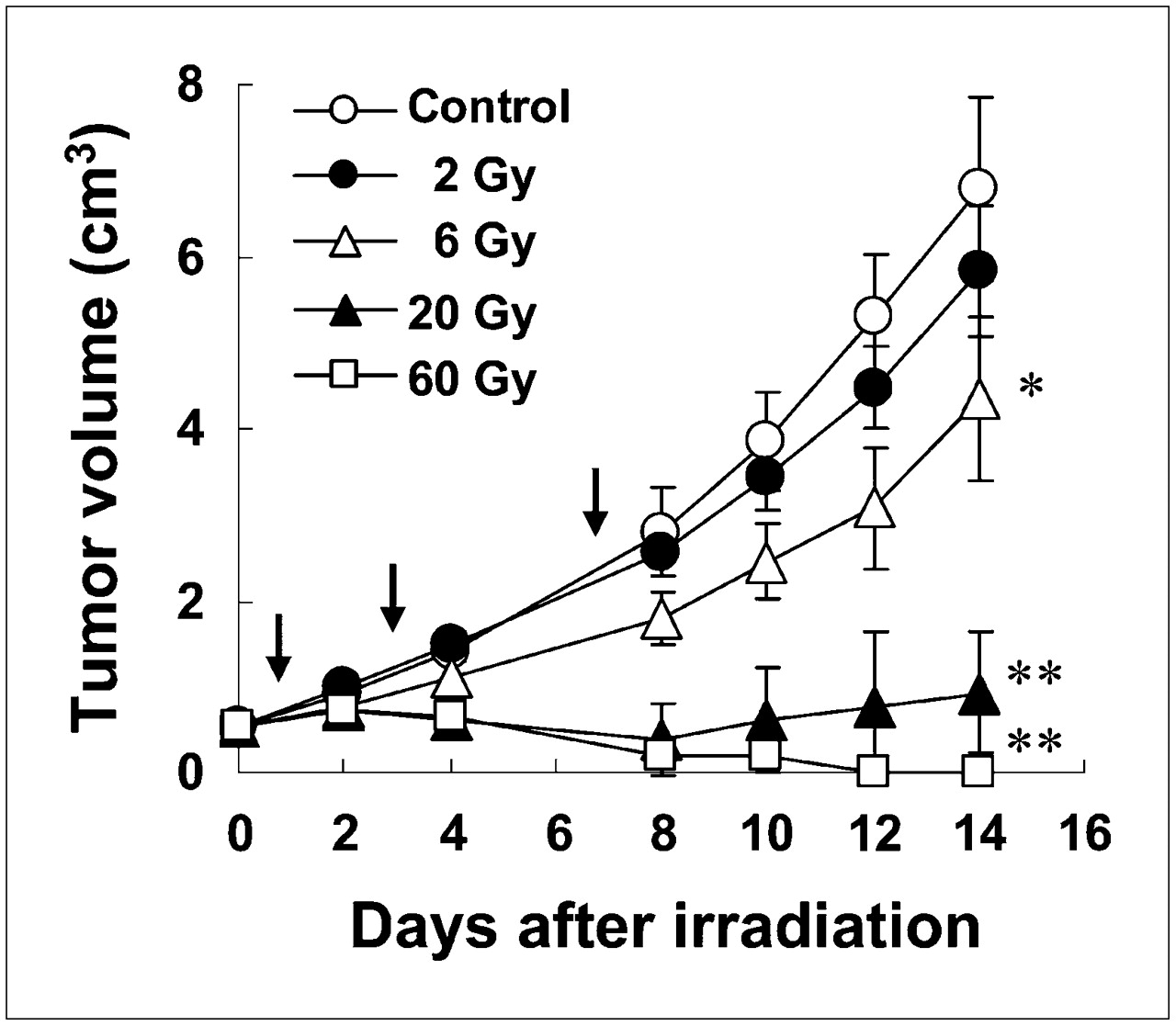
Growth curves of SCCVII tumors after various doses of irradiation. Tumors on right thighs of mice were irradiated on day 0. Each point represents mean ± SD of Tvol in 6 mice. Ligand uptake studies were performed on days 1, 3, and 7 after irradiation, as denoted by arrows. Asterisks indicate that Tvol on day 14 was significantly different from control (*P < 0.05; **P < 0.001).
Tumor uptake of PET ligands was compared after irradiation at 2, 6, 20, and 60 Gy (Fig. 2). In the controls on day 0 (Tvol = ∼0.7 cm3), the uptake values of d-18F-FMT, 18F-FDG, l-11C-MET, and 18F-FLT were 0.8, 1.2, 1.6, and 2.0 (as standardized uptake value), respectively, based on our pooled data (11,12). In the nonirradiated controls, the relationship between tumor uptake and tumor size varied among the PET ligands. The uptake of 3 of the 4 ligands, except 18F-FLT, by the control tumors was relatively constant until day 3 (Tvol < 2 cm3). On day 7, a slight increase in the uptake of d-18F-FMT and 18F-FDG and an apparent decrease in the uptake of l-11C-MET were observed, compared with the values on day 0; the respective uptake values were 111.8%, 109.8%, and 65.9%. However, in the case of 18F-FLT, a marked decrease in uptake by the control tumors was observed on day 3 (Tvol < 2 cm3). The uptake values of 18F-FLT by the control tumors on days 1, 3, and 7 were 84.1%, 56.5%, and 59.8%, respectively, compared with those on day 0.
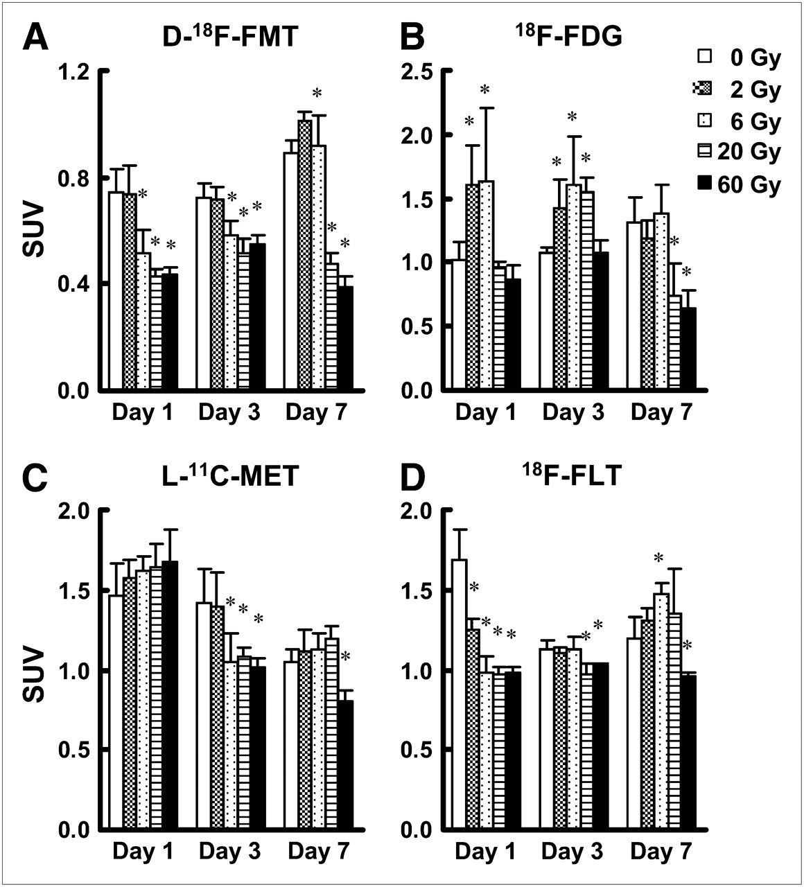
Changes in ligand uptake of d-18F-FMT (A), 18F-FDG (B), l-11C-MET (C), and 18F-FLT (D) by tumors after various doses of irradiation. Tumors were irradiated at doses of 2, 6, 20, and 60 Gy on day 0, and ligand uptake studies were performed on days 1, 3, and 7. Tissue radioactivity was expressed as standardized uptake value (SUV). Each column represents mean ± SD. Asterisks indicate statistically significant difference from control in each group (P < 0.05).
After irradiation at 6, 20, and 60 Gy, the uptake of d-18F-FMT was decreased on day 1 to 69.3%, 57.3%, and 58.8%, respectively, of that in the control tumor (P < 0.05). This significant decrease of d-18F-FMT uptake persisted until day 7 after irradiation at 20 and 60 Gy; however, in the group irradiated with 6 Gy, the significant reduction in ligand uptake noted on day 1 was no longer seen on day 7 (Fig. 2A). In regard to 18F-FDG, tumor uptake of this ligand remained unchanged or became significantly higher than in the control by day 3 for all radiation doses examined. A significant decrease in tumor uptake of 18F-FDG was observed only on day 7 after irradiation at 20 and 60 Gy; those uptake values were 56.4% and 48.7%, respectively, of the values in the control tumors (P < 0.05) (Fig. 2B). On day 1, no significant difference in the uptake of l-11C-MET was observed between irradiated tumors and nonirradiated control tumors (Fig. 2C). On day 3, a significant decrease in tumor uptake of l-11C-MET was observed after irradiation at 6, 20, and 60 Gy (74.2%, 76.0%, and 71.4%, respectively), compared with uptake in the control tumor (P < 0.05). On day 7, only the tumor irradiated at a dose of 60 Gy showed a significant reduction in the uptake of l-11C-MET, compared with the control tumor (P < 0.05) (Fig. 2C). After the irradiation at 2, 6, 20, and 60 Gy, 18F-FLT uptake was decreased on day 1 to 74.3%, 58.6%, 57.7%, and 58.2%, respectively, of the corresponding values in the control tumor (P < 0.05), and the ratios of the standardized uptake value of the tumor to that of blood were 1.3, 1.0, 0.94, and 0.94, respectively. On day 3, only the tumors irradiated at doses of 20 and 60 Gy showed a significant reduction of 18F-FLT uptake, compared with the control tumor (P < 0.05). This trend persisted until day 7; thus, only tumors irradiated at a dose of 60 Gy showed a significant reduction of 18F-FLT uptake, compared with the control tumor (P < 0.05) (Fig. 2D).
Figure 3 compares the uptake of each PET ligand into the tumor in relation to the Tvol on day 14 after various doses of radiation. Significant positive correlations were found in uptake of d-18F-FMT on day 1 (P < 0.001), day 3 (P < 0.001), and day 7 (P < 0.001). In contrast, a correlation was found only on day 7 for 18F-FDG (P < 0.001), only on day 3 for l-11C-MET (P = 0.002), and only on days 1 and 3 for 18F-FLT (P = 0.002, P = 0.005, respectively).

Correlation between ligand uptake by tumor and Tvol on day 14 after various doses of irradiation. Plots of standardized uptake value (SUV), measured with mice in groups 2–4, vs. Tvol, measured with mice in group 1, are shown for each ligand.
DISCUSSION
The present study evaluated the potential usefulness of d-18F-FMT, 18F-FDG, l-11C-MET, and 18F-FLT as PET ligands to detect the responses of tumors to radiation therapy, especially in the early phase of treatment. Minn et al. (21) found that clinical responders to radiation therapy showed a more prominent decrease of the 18F-FDG uptake than did nonresponders. In most of these clinical 18F-FDG PET studies, the evaluation was scheduled late in the course of radiation therapy or after treatment completion, when the patients had already received 30 Gy of radiation (21). On the other hand, an increase in 18F-FDG uptake was also reported during radiation therapy both in irradiated animals bearing xenografts of human tumors and in cancer patients (22,23). Recent studies have shown that a decrease of the cellular proliferation rate is one of the earliest events in response to successful tumor treatment. This finding spurred interest in the imaging of cellular proliferation by PET, so that direct detection of the responses of tumors to treatment even in the early phase could potentially serve as a useful guideline for selecting individualized treatments for patients. For this purpose, the potential detectability of the early responses of tumors to radiation therapy has been studied using l-11C-MET and 18F-FLT (4,13–15). We expected d-18F-FMT to be a promising ligand not only for tumor imaging (11,12) but also for predicting tumor responses to therapy. This present trial was, to our knowledge, the first to evaluate d-18F-FMT, an artificial and unnatural d-amino acid–based PET ligand, for monitoring tumor responses to radiation therapy.
In this study, the relationship between ligand uptake and tumor size in nonirradiated control tumors differed among the ligands examined. The most significant changes in tumor uptake were for 18F-FLT, with a negative correlation noted between uptake of 18F-FLT and tumor size in the range of 0.1–1.5 cm3. It is well known that the presence of hypoxic cells in solid tumors, thought to be in that state because of oxygen and nutrient deprivation but still to be clonogenic, is an important cause of failure of radiation therapy and some chemotherapy regimens. It appears that prolonged hypoxia could decrease the DNA synthesis rate, the growth rate of cells, and the proportion of cells synthesizing DNA, consequently increasing the fraction of cells in the G1 phase. In SCCVII tumors, an increase in the hypoxic fraction has been reported along with tumor growth, with the hypoxic fraction in tumors 5, 10, and 22 mm in diameter being, on average, 0.86%, 10%, and 23%, respectively (24,25). Because tumors of 10 and 22 mm almost corresponded to the tumor size on days 0 and 7 in this study, the hypoxic fraction of tumor on day 7 was estimated to be twice as high as that on day 0.
As for the effects of ionizing radiation, several PET studies have demonstrated decreased 18F-FDG uptake into tumors treated with 20 Gy relatively early after irradiation (15,26). However, in the present study the 18F-FDG uptake after this dose of radiation did not change in the early phase and decreased only by day 7. These results were consistent with the results of previous studies using the same tumor cell line (13,14) showing that tumor uptake of 18F-FDG does not decrease within 24 h after irradiation. Moreover, in the present study, irradiation with small doses of 2 and 6 Gy significantly elevated the tumor uptake of 18F-FDG, compared with uptake by nonirradiated tumors, until day 3, uptake thereafter decreased to the same level as in control tumors on day 7. The present results were consistent with previously reported data (22,23). Thus, radiosensitive human xenografts exhibited a 2.3-fold higher 18F-FDG uptake at 2 h after 10 Gy of irradiation than did nonirradiated tumors (22). Clinical 18F-FDG PET scans showed an initial increase in uptake into metastases after 6 Gy of irradiation (daily fractions of 2 Gy for 3 d), with uptake returning to the initial level after a total dose of 30 Gy (23). The mechanism of the increased tumor uptake of 18F-FDG soon after the start of irradiation has not been clarified. However, inflammation induced by radiation might be one possibility (23,27), and another might be the radiation-induced elevation of glucose metabolism (22). The increase in tumor uptake of 18F-FDG until day 3 in the present study was consistent with the conclusion of Hautzel et al. (23) that 18F-FDG PET during and immediately after completion of radiation therapy does not accurately determine the treatment response.
All radiation doses significantly reduced tumor uptake of 18F-FLT in the early phase of irradiation. Dose-dependent differences in tumor uptake of 18F-FLT on day 1 were observed between 2 and 6 Gy but not among 6, 20, and 60 Gy. Yang et al. (14) reported that 18F-FLT-PET could not detect differences between radiation doses. The doses of radiation used in both studies may have been too high to show a dose dependency of radiation on tumor uptake of 18F-FLT, since its uptake was reduced almost to the background level by 10 Gy in the previous study. Moreover, in the present study, the tumor-to-blood ratio of 18F-FLT in tumor at doses of 2, 6, 20, and 60 Gy decreased to the background level on day 1, except at 2 Gy. The 26% reduction of tumor 18F-FLT uptake at 2 Gy of irradiation on day 1 might reflect sublethal damage, since damaged DNA at 2 Gy might be repaired as demonstrated by the tumor growth curve. Yang et al. (14) also demonstrated that the surviving fraction of SCCVII cells determined by clonogenic assay was 80% after 10 Gy of irradiation, despite a marked reduction in the 18F-FLT uptake to the background level at 24 h after a single irradiation session. Our results also supported their interpretation that the reduction of 18F-FLT uptake did not represent the ratio of killed tumor cells but rather represented cells showing biologic impairment of proliferation. Therefore, this early-phase response to irradiation of decreased tumor uptake of 18F-FLT might reflect a reduction of cells in the S phase and cell growth arrest, as reported previously (14). Recovery of the tumor uptake values of 18F-FLT to the control level was observed by day 3 in tumors irradiated at 2 and 6 Gy. This recovery might be attributable to both sublethal DNA damage repair and subsequent repopulation. Significantly decreased uptake of 18F-FLT in the early phase in tumors irradiated at 20 Gy was also completely restored to the control level on day 7, as confirmed previously (13,14). Tumor uptake of 18F-FLT on day 7 after 20 Gy of irradiation was almost the same as uptake in the control cells, irrespective of tumor size. The reason may be differences in the proliferative fraction and growth rate among tumors of different sizes, as was supported by the results of the present study showing that tumor uptake of 18F-FLT was higher at recurrence. Tumor uptake of 18F-FLT on day 7 may not provide quantitative information on tumor growth but provides qualitative information on clonogenic activity after irradiation, namely, the proliferative activity of the cells.
In contrast to 18F-FDG, a lower uptake of l-11C-MET has been expected in inflammatory tissues, suggesting that amino acid–based PET ligands may be potentially more suitable than 18F-FDG to discriminate between tumors and inflammatory tissues. However, recent rodent and clinical studies of l-11C-MET-PET have demonstrated a definite increase in the tissue accumulation of l-11C-MET in various inflammatory processes (28–30). The uptake of 18F-FDG, l-11C-MET, and l-18F-FET, an amino acid transport ligand, in tumor-infiltrated and immunologically stimulated lymph nodes revealed increased uptake of 18F-FDG and l-11C-MET in both tissues (31). In contrast, l-18F-FET proved to be a specific PET ligand for discrimination between tumor-infiltrated and inflammatory lymph nodes in the murine models studied. l-14C-MET and 3H-thymidine were reported to show a similar pattern of dose-dependent decrease after irradiation (15). In the present study, the uptake patterns of l-11C-MET and 18F-FLT by tumors irradiated at each dose were similar on day 7. Similar to 18F-FLT, the tumor uptake pattern of l-11C-MET on day 7 might provide only qualitative information about the proliferative fraction of tumor cells after irradiation.
The tumor uptake of d-18F-FMT revealed a significant response to radiation therapy on day 1 after irradiation. The uptake patterns on day 1 were completely different between d-18F-FMT and l-11C-MET, reflecting the previous finding that d-18F-FMT was not taken up by inflammatory tissues (11). Although the reason for the differential uptake of a natural amino acid–based ligand, l-11C-MET, and an unnatural amino acid-based transporter ligand, d-18F-FMT, by inflammatory tissues remains unclear, differential expression of transport subtypes may be a valid explanation (31). l-11C-MET is taken up by both the leucine-preferring and the alanine-preferring transport system (32). In contrast, d-18F-FMT is taken up mostly by the l-transport system, similar to l-leucine and l-tyrosine (Takeo Urakami et al., unpublished data, July 2005). After transport into the free amino acid pool, although l-11C-MET was incorporated into proteins via conversion into amino-acyl-tRNA (6,33), d-18F-FMT was not taken up into the acid-precipitable protein fraction (11). The most absolute difference in tumor uptake between l-11C-MET and d-18F-FMT might be related to whether the ligand is used for protein synthesis. In contrast to tumor uptake of 18F-FLT and l-11C-MET on day 7, tumor uptake of d-18F-FMT on day 7 was not sufficiently sensitive to provide accurate qualitative information on the clonogenicity of tumor cells but provided quantitative information on tumor growth after irradiation. According to the results of the present study, d-18F-FMT was superior to 18F-FDG and l-11C-MET for obtaining quantitative information on tumor growth after irradiation, without inflammatory artifacts.
In the present study, a significant correlation was found between d-18F-FMT uptake on days 1, 3, and 7 and Tvol on day 14 after irradiation. In contrast, no such correlation between ligand uptake at all the 3 time points and Tvol on day 14 was found for the other 3 tracers; a correlation was found only on day 7 for 18F-FDG, only on day 3 for l-11C-MET, and on days 1 and 3 for 18F-FLT. A significant correlation was noted between the uptake of both 18F-FDG and l-11C-MET and the Tvol on day 14, even after the inflammatory effects of irradiation had disappeared. These results demonstrated that prognostic information after radiation therapy was provided by only d-18F-FMT among the 4 ligands tested at all time points during the first week after irradiation. On the other hand, the correlation was found only at limited time points for the other 3 tracers. It would be of great benefit to patients on radiation therapy if the PET time points for monitoring and predicting efficacy after irradiation were not severely restricted.
CONCLUSION
Tumor uptake of d-18F-FMT and 18F-FLT, but not of 18F-FDG and l-11C-MET, was effectively decreased on day 1 after irradiation. Prognostic information after radiation therapy was obtained with d-18F-FMT at all time points the first week after irradiation but only at limited time points for the 3 conventional ligands, 18F-FDG, l-11C-MET, and 18F-FLT. The results suggest that d-18F-FMT is a promising ligand for tumor imaging by PET, not only for tumor detection but also for monitoring early-phase responses to radiation therapy.
Acknowledgments
We gratefully acknowledge the technical assistance provided by Kengo Sato. This study was in part supported by Research and Development of Technology for Measuring Vital Function Merged with Optical Technology, Research and Development Project Aimed at Economic Revitalization.
Footnotes
-
COPYRIGHT © 2009 by the Society of Nuclear Medicine, Inc.
References
- Received for publication August 20, 2008.
- Accepted for publication October 9, 2008.


Jump to section
Related Articles
Cited By...
- Harnessing Preclinical Molecular Imaging to Inform Advances in Personalized Cancer Medicine
- Predictive Value of Early-Stage Uptake of 3'-Deoxy-3'-18F-Fluorothymidine in Cancer Cells Treated with Charged Particle Irradiation
- First Clinical Results of (D)-18F-Fluoromethyltyrosine (BAY 86-9596) PET/CT in Patients with Non-Small Cell Lung Cancer and Head and Neck Squamous Cell Carcinoma
- D-18F-Fluoromethyl Tyrosine Imaging of Bone Metastases in a Mouse Model