


Article Figures & Data
Figures
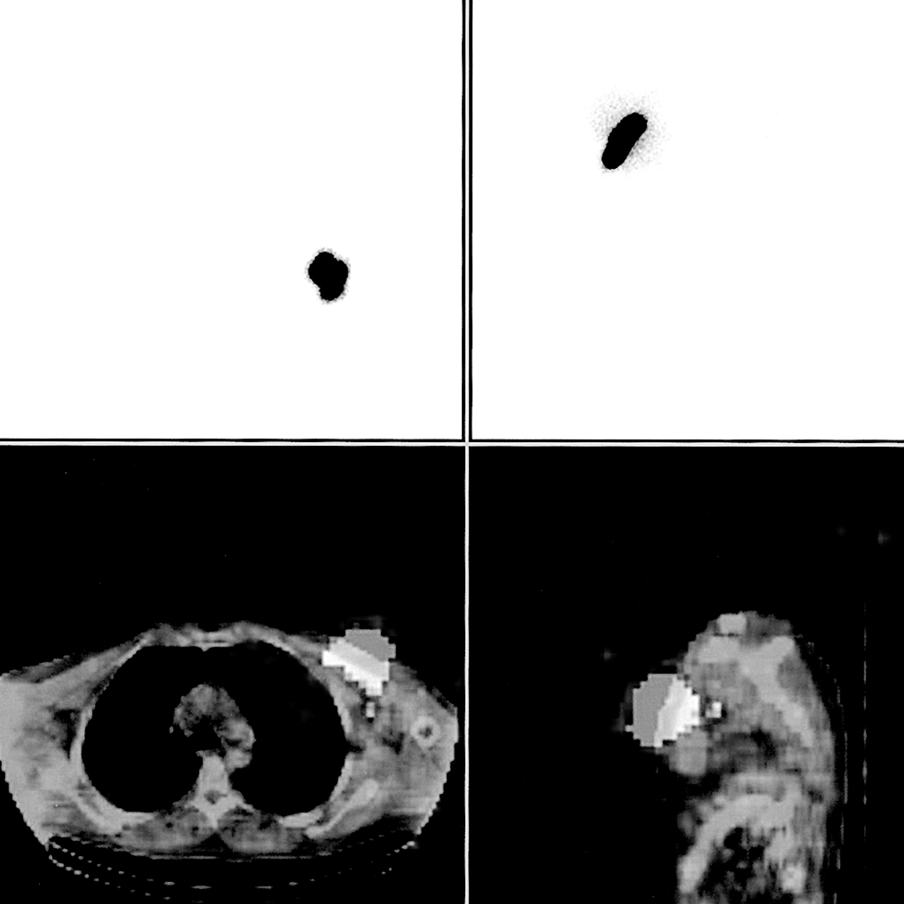
- FIGURE 1.
Patient with cutaneous malignant melanoma in chest wall. SN hidden by scattered radiation of injection site. Multiprojection planar lymphoscintigraphy failed to identify SN (top images: selected planar images in anterior and lateral projections). On fused SPECT/CT images, hot node was identified in axilla (bottom images: transaxial and sagittal slices).
- FIGURE 2.
Patient with cutaneous malignant melanoma in right upper back. SNs in multiple basins. Multiprojection planar images identified hot node in axilla (bottom right image: anterior projection). Fused SPECT/CT images identified additional supraclavicular and low jugular nodes (top: transaxial slices; bottom left: coronal slices).
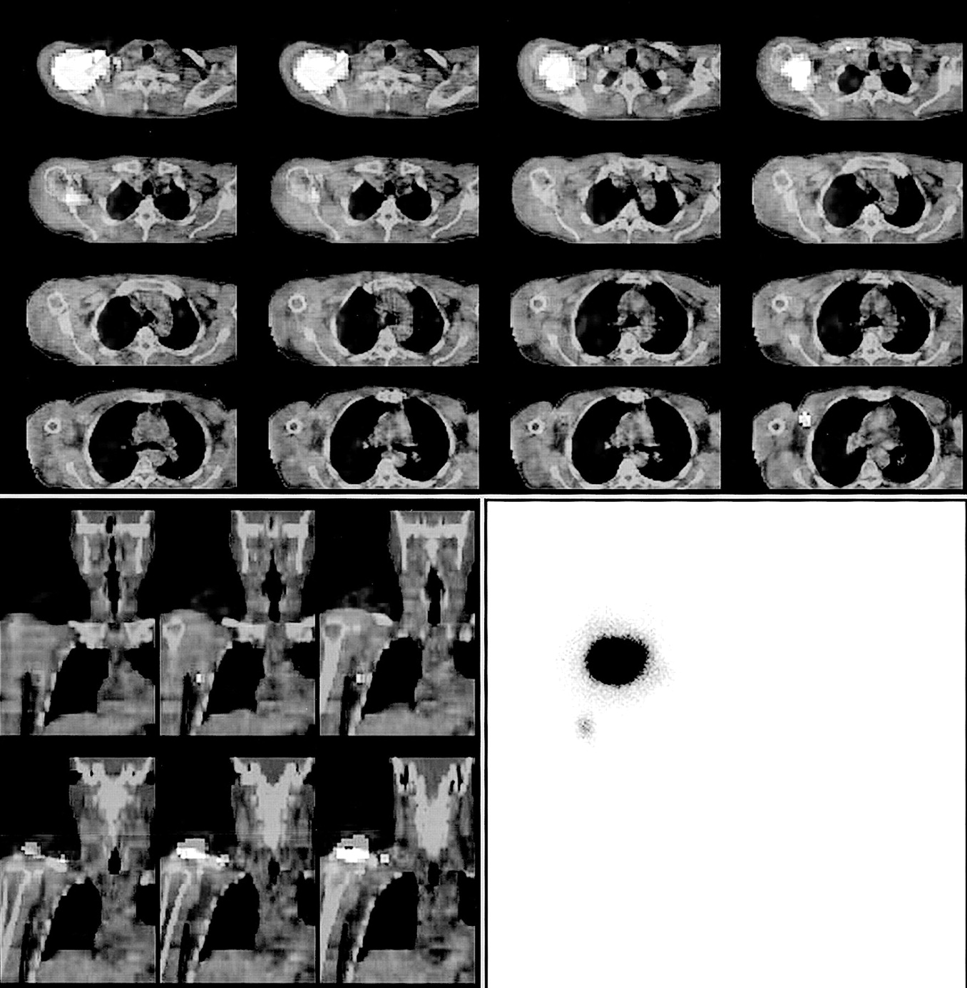
- FIGURE 3.
Patient with melanoma in back. Anterior (bottom left) and lateral (bottom right) planar images detect multiple nodes. Transaxial SPECT/CT images (top) localize nodes in anterior axilla and in aberrant superficial location.


Tables
- TABLE 1
Scintigraphic and Histopathologic Findings in Patients with Tumor Localized in Head and Neck Region
Patient no. Age (y) Sex Tumor type and location Planar imaging* Localization of nodes seen on planar imaging SPECT/CT imaging Surgical and histopathologic results First nodes detected Additional nodes Single vs. multiple basins Additional nodes† Single vs. multiple basins Clinically relevant added value‡ 1 70 M SCC, tongue Midjugular Supraclavicular Unclear Mid- and lower jugular chain + jugulo-digastric (hidden) Multiple + Positive SN (cervical) 2 50 F SCC, tongue High jugular 4 along jugular chain Single Jugular chain + submental (hidden) Single + Negative 3 43 M SCC, tongue High jugular 1 midjugular Single Jugular chain − Single − Positive SN (cervical) 4 72 M SCC, tongue Supraclavicular, suspected submandibular − Unclear Supraclavicular Second node was ruled out (injection site) Single + Positive SN (supraclavicular) 5 27 M SCC, tongue High jugular 3 along jugular chain Single Jugular chain + posterior cervical triangle Multiple + Multiple involved nodes 6 54 M SCC, oral mucosa High jugular 4 along jugular chain Single Jugular chain − Single − Negative 7 81 M MM, head 2 preauricular 2 submandibular Unclear 2 preauricular, 1 submandibular, 1 jugulodigastric − Unclear − Negative 8 67 M MM, scalp Faint occipital − Single Posterior cervical triangle − Single − Multiple involved nodes 9 55 M MM, scalp High jugular, contralateral occipital 1 cervical Multiple 2 jugular, 2 contralateral occipital − Multiple − Negative ↵* Including images obtained with 57Co flood source.
↵† Additional nodes detected on SPECT/CT only. Hidden = hidden by scatter radiation of injection site.
↵‡ SPECT/CT added data were considered clinically relevant if they guided surgeon to SNs that were undetected on planar images or to SNs in another basin.
MM = malignant melanoma.
- TABLE 2
Scintigraphic and Histopathologic Findings in Patients with Malignant Melanoma of Trunk
Patient no. Age (y) Sex Tumor type and location Planar imaging* Localization of nodes seen on planar imaging SPECT/CT imaging Surgical and histopathologic results First nodes detected Additional nodes Single vs. multiple basins Additional nodes† Single vs. multiple basins Clinically relevant added value‡ 1 52 M L shoulder L axilla − Single L axilla, central − Single − Negative 2 24 M R shoulder R axilla − Single Apical, R axilla + (IT) Single + Negative 3 73 M Midupper back L axilla 2 L axilla, 1 R axilla Multiple In L axilla, 1 subscapular node and 1 central node + pectoral node Multiple + Negative 4 61 M L upper back L axilla 2 L axilla Unclear 2 central, 1 pectoral axillary node + prescapular (IT) Multiple + Negative 5 71 M R upper back R axilla 2 R axilla Single Central axillary nodes − Single − Negative 6 67 M Midupper back L jugular chain, 2 nodes L axilla, L occipital Multiple L jugular, axillary, and occipital nodes − Multiple − Negative 7 59 F Midupper back L axilla L axilla Single Central axillary nodes − Single − 1 positive SN node (axillary) 8 52 F Midupper back R axilla, 2 nodes L axilla, 2 nodes Multiple Bilateral central axillary nodes − Multiple − Negative 9 60 M R upper back R axilla − Single 1 axillary central node + supraclavicular node, low jugular node Multiple + Negative 10 55 M Midlower back L axilla L groin Multiple L central axilla node, L groin node − Multiple − 1 positive node (groin) 11 62 M Chest wall − − − − + 1 central axillary node (hidden) Single + Negative 12 26 F Abdominal wall L groin, aberrant uptake − Unclear L groin + (?) parailiac nodes, aberrant uptake Unclear − Negative (parailiac nodes and aberrant uptake not examined) ↵* Including images obtained with 57Co flood source.
↵† Additional nodes detected on SPECT/CT only. Hidden = hidden by scatter radiation of injection site; IT = in-transit.
↵‡ SPECT/CT added data were considered clinically relevant if they guided surgeon to SNs that were undetected on planar images or to SNs in another basin.
In this issue


Jump to section
Related Articles
Cited By...
- Radioguided Surgery
- 89Zr-Nanocolloidal Albumin-Based PET/CT Lymphoscintigraphy for Sentinel Node Detection in Head and Neck Cancer: Preclinical Results
- Sentinel lymph node biopsy using dynamic lymphoscintigraphy combined with ultrasound-guided fine needle aspiration in penile carcinoma
- SPECT/CT
- Multimodality Molecular Imaging of Tumor Angiogenesis
- Optimal 57Co Flood Source Activity and Acquisition Time for Lymphoscintigraphy Localization Images
- The Additional Value of SPECT/CT in Lymphatic Mapping in Breast Cancer and Melanoma
- Evaluation and Localization of Lymphatic Drainage and Sentinel Lymph Nodes in Patients with Head and Neck Melanomas by Hybrid SPECT/CT Lymphoscintigraphic Imaging
- Improved Sentinel Node Identification by SPECT/CT in Overweight Patients with Breast Cancer
- Procedure Guideline for SPECT/CT Imaging 1.0
- Role of Nuclear Medicine in the Management of Cutaneous Malignant Melanoma
- Fusion of SPECT and Multidetector CT Images for Accurate Localization of Pelvic Sentinel Lymph Nodes in Prostate Cancer Patients