


Article Figures & Data
Figures
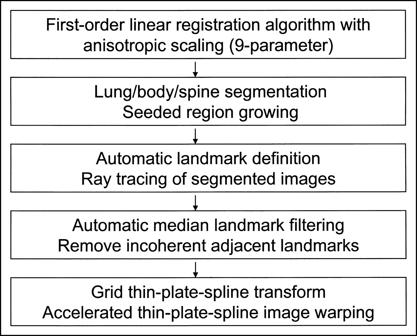
- FIGURE 1.
Flow chart with steps performed during automatic image registration.
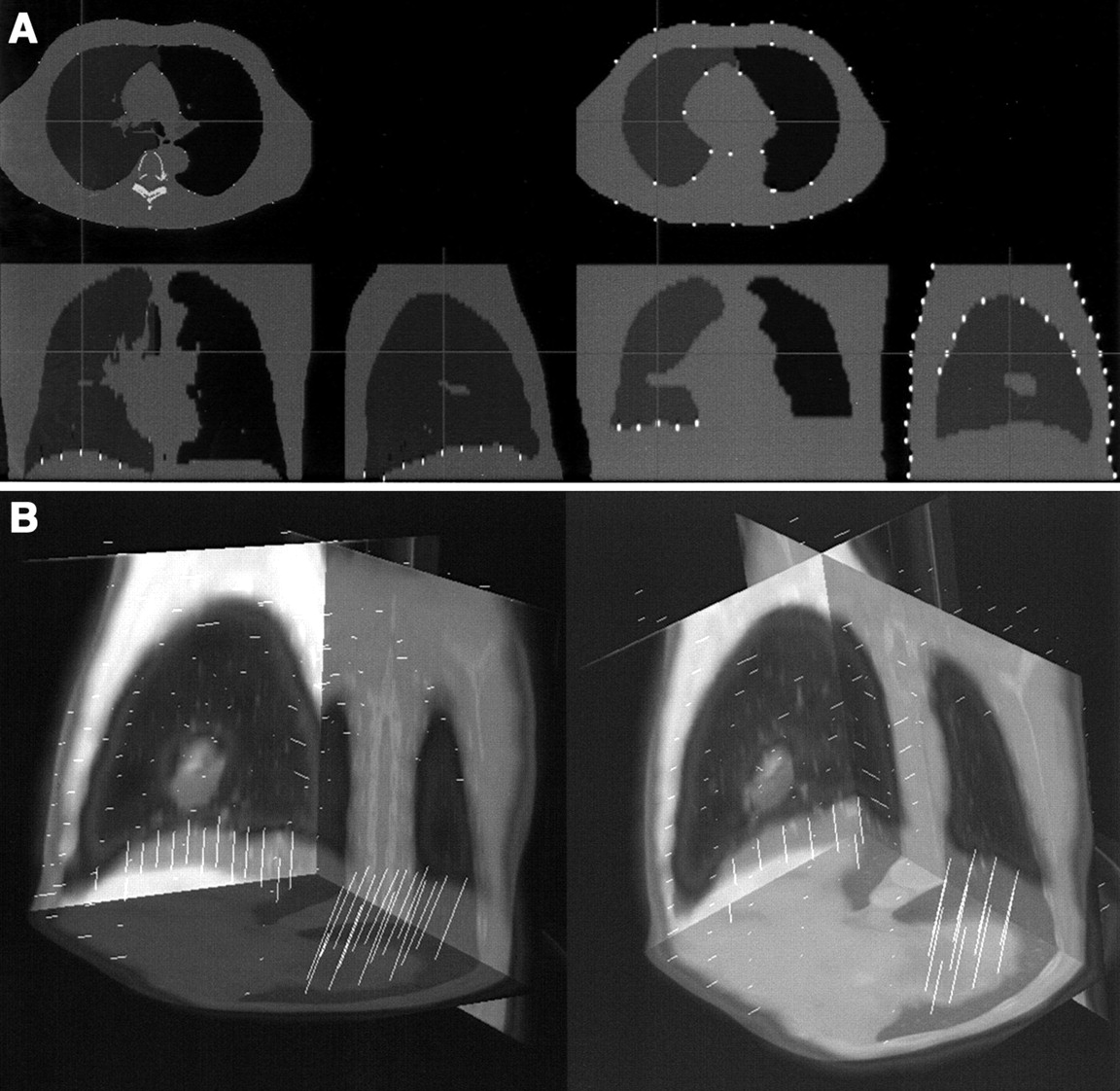
- FIGURE 2.
Automatically identified landmarks on segmented CT (left) and segmented transmission map (right) identified by algorithm for nonlinear step (A) and resulting displacement vectors created by joining corresponding CT and transmission map landmarks (B).
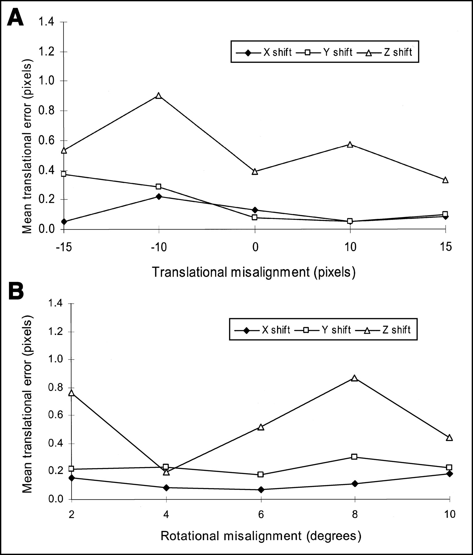
- FIGURE 3.
Errors in translational parameters after image registration as function of initial translational (A) and rotational (B) image misalignment.
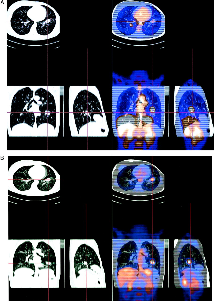
- FIGURE 4.
Results of registration in case of deep-inspiration CT acquisition registered with normal-breathing PET (A) and normal breathing during CT and PET acquisition (B). In both cases, only linear registration module was used without warping correction.
- FIGURE 5.
Example of warping artifact due to opposite displacement vectors in close proximity (left) and result of median filtering (right). Arrows indicate location of artifact.
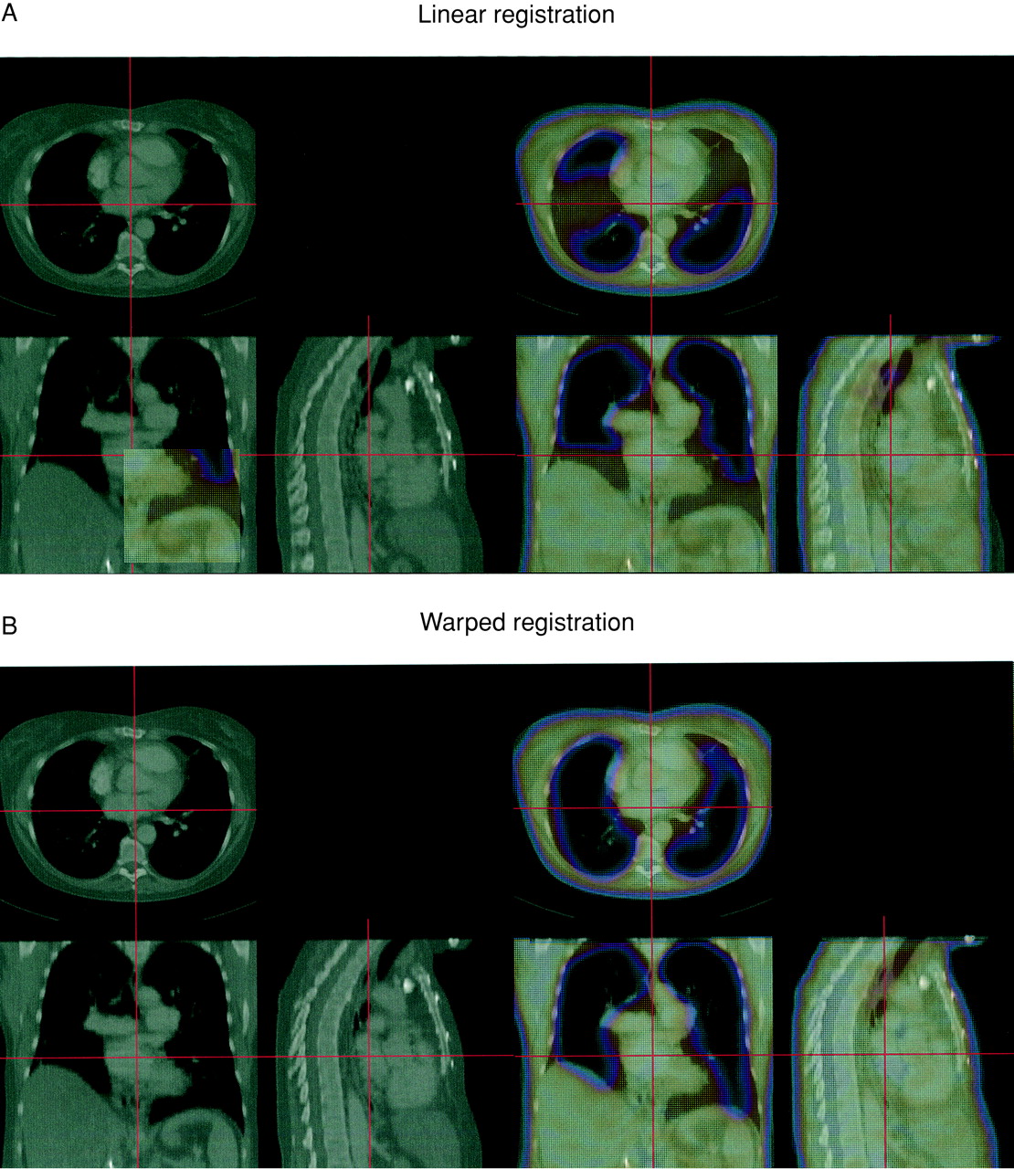
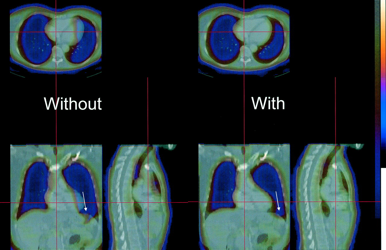
- FIGURE 6.
Final registration results in case of large diaphragmatic displacement shown on transmission–CT fusion images before warping (A) and after warping has been applied (B).
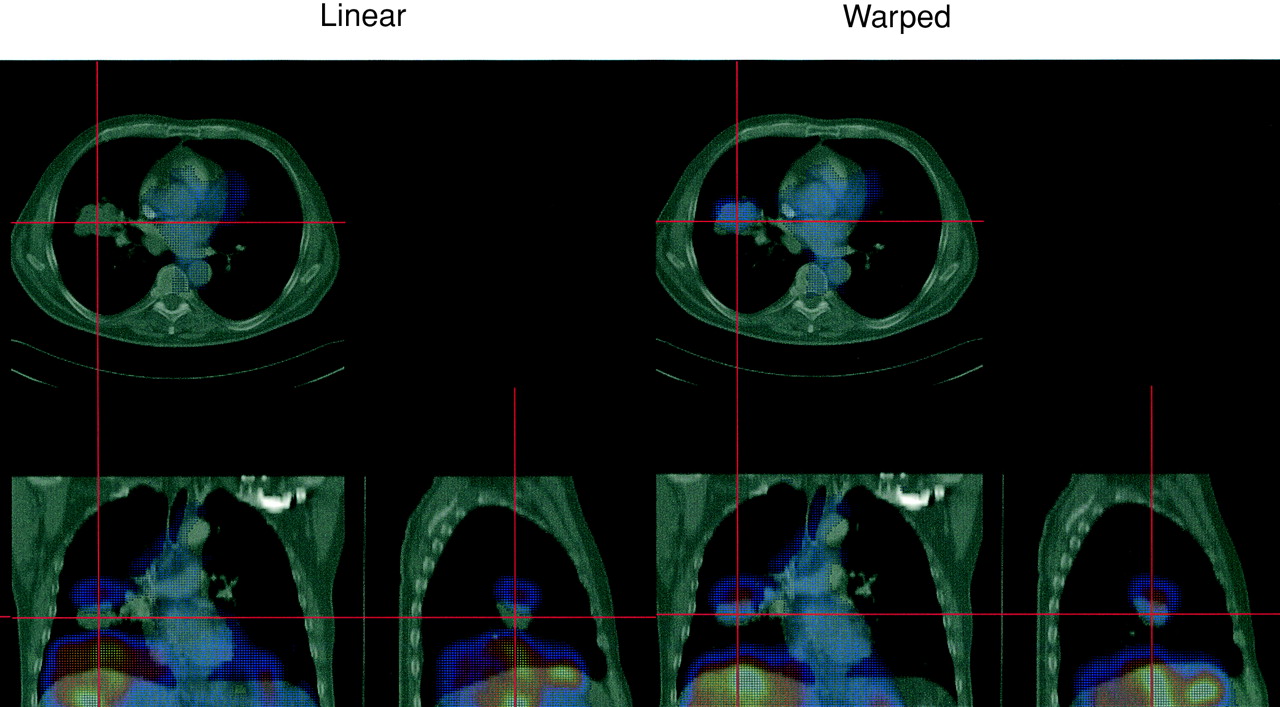
- FIGURE 7.
Final registration results in case shown on emission PET/CT fusion images before warping (left) and after warping applied (right).


Tables
Data Translation Scaling Rotation TR+EM 0.3 ± 0.5 1.1 ± 1.6 0.3 ± 0.2 TR 0.3 ± 0.4 1.1 ± 1.5 0.2 ± 0.5 EM 0.3 ± 0.4 1.8 ± 2.2 0.4 ± 0.4 Units of reproducibility are pixels for translations, % for scaling, and degrees for rotation.
Criteria Translation Scaling Rotation MI 0.3 ± 0.5 1.1 ± 1.6 0.3 ± 0.2 NMI 0.4 ± 0.6 1.5 ± 1.7 0.3 ± 0.4 Units of reproducibility are pixels for translations, % for scaling, and degrees for rotation.
Parameter Multi-res1 Multi-res2 Multi-res4 Time (s) 363 ± 187 290 ± 89 189 ± 106 Iterations 204 ± 85 351 ± 77 598 ± 104 - TABLE 4
Lung Volumes Measured on Segmented CT Scans, Original Transmission Scans, and Warped Transmission Scans
Scan Lung volume (L) Right Left CT 2.2 ± 1.0 1.8 ± 0.5 Transmission 1.7 ± 0.4 1.5 ± 0.4 Warped transmission 2.2 ± 0.7 1.9 ± 0.5
In this issue


Jump to section
Related Articles
Cited By...
- Automated Motion Correction for Myocardial Blood Flow Measurements and Diagnostic Performance of 82Rb PET Myocardial Perfusion Imaging
- Nonrigid Versus Rigid Registration of Thoracic 18F-FDG PET and CT in Patients with Lung Cancer: An Intraindividual Comparison of Different Breathing Maneuvers
- Quantitative Analysis of Myocardial Perfusion SPECT Anatomically Guided by Coregistered 64-Slice Coronary CT Angiography
- Motion-Frozen Myocardial Perfusion SPECT Improves Detection of Coronary Artery Disease in Obese Patients
- Dual-Modality Imaging: Combining Anatomy and Function
- Optimized Contrast-Enhanced CT Protocols for Diagnostic Whole-Body 18F-FDG PET/CT: Technical Aspects of Single-Phase Versus Multiphase CT Imaging
- Automated 3-Dimensional Elastic Registration of Whole-Body PET and CT from Separate or Combined Scanners
- Comparison Between 18F-FDG PET, In-Line PET/CT, and Software Fusion for Restaging of Recurrent Colorectal Cancer
- "Motion-Frozen" Display and Quantification of Myocardial Perfusion
- PET/CT: Panacea, Redundancy, or Something in Between?
- Software Approach to Merging Molecular with Anatomic Information