


Article Figures & Data
Figures
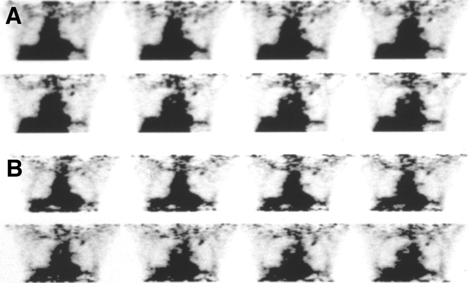
- FIGURE 1.
A 72-y-old woman presented with right lower lobe nodule measuring 1.8 × 1.0 cm. SUV increased from 1.45 on scan 1 at 65 min after injection (A) to 1.72 on scan 2 at 123 min after injection (B). Biopsy of lesion revealed moderately differentiated adenocarcinoma.
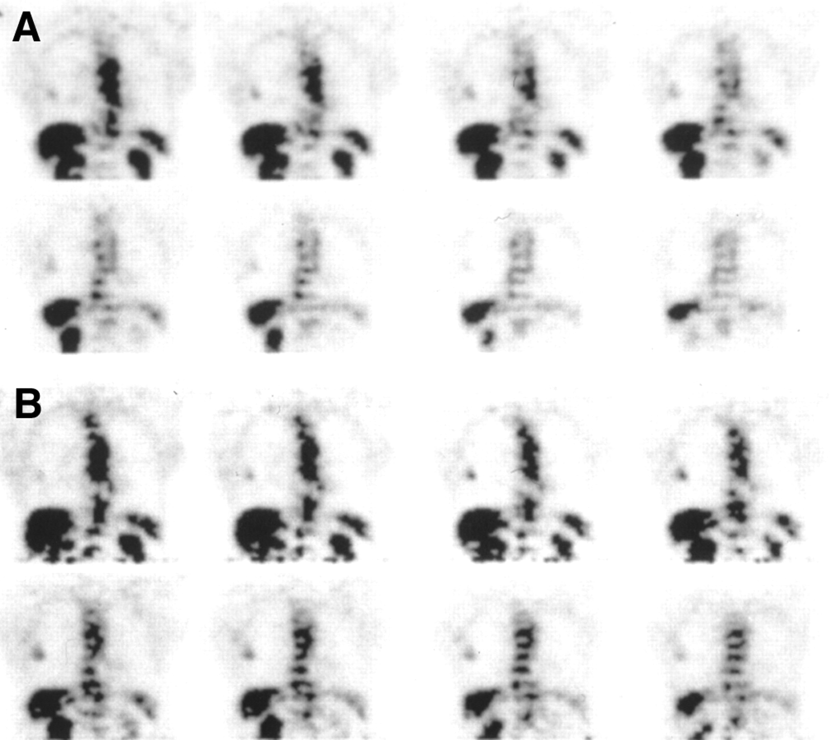
- FIGURE 2.
A 69-y-old woman presented with 0.8-cm density in left upper lobe. SUV was not significantly different between scan 1 at 54 min after injection (A), where SUV was 1.54, and scan 2 at 100 min after injection (B), where SUV was 1.50. Excised lesion was granuloma.

Tables
Lesion no. Patient’s age (y) Patient’s sex Lesion size (cm)/location Follow-up/pathology SUV1* SUV2† % Change‡ 1 78 F 2.5/RLL Adenocarcinoma (Bx) 3.10 4.00 29.0 2 71 M 2.0/LUL Adenocarcinoma (Res) 1.12 1.33 18.8 3 79 M 4.2/RUL Adenocarcinoma (Res) 4.64 5.56 19.1 4 75 M 3.0/RUL Adenocarcinoma (Bx) 4.72 5.38 14.0 5 73 F 2.4 × 2.1/LLL§ SCC (Bx) 8.34 10.53 26.3 6 1.8/LLL| SCC (Bx) 4.58 5.97 30.3 7 69 F 1.5/RUL Adenocarcinoma (Bx) 1.69 2.18 29.0 8 75 M 2.0/LUL Adenocarcinoma (Res) 1.22 1.36 11.5 9 63 F 1.8 × 1.0/RLL Adenocarcinoma (Bx) 1.45 1.72 18.6 10 88 F 3.5 × 5.0/LLL Adenocarcinoma (Bx) 7.06 8.24 16.7 11 83 M 3.4 × 2.7/RUL Adenocarcinoma (Res) 3.09 4.27 38.2 12 54 F 0.6/LUL Probable lung cancer: no Bx, but response to XRT 2.80 3.21 14.6 13 73 F 1.7 × 1.4/RUL SCC (Res) 2.60 2.87 10.4 14 70 F 6.0/RUL SCC (Res) 5.23 5.93 13.4 15 71 M 3.5 × 2.5/LUL Metastasis from prostate cancer (Bx) 2.57 2.90 12.8 16 67 M 2.5/RML Adenocarcinoma (Bx) 2.54 3.08 21.3 17 68 M 4.0 × 3.0/LUL Adenocarcinoma (Bx) 3.35 4.36 30.1 18 71 F 1.9 × 1.2/RUL SCC (Bx) 3.63 4.08 12.4 19 83 F 2.8 × 2.7/LUL Adenocarcinoma (Bx) 3.12 3.56 14.1 20 67 F 3.0 × 3.0/RUL Adenocarcinoma (Res) 6.25 8.04 28.6 ↵* Mean ± SD of SUV1 = 3.66 ± 1.95.
↵† Mean ± SD of SUV2 = 4.43 ± 2.43.
↵‡ Mean = 20.5%.
↵§ Lesion 1.
↵| Lesion 2.
RLL = right lower lobe; Bx = biopsy; LUL = left upper lobe; Res = surgical resection; RUL = right upper lobe; LLL = left lower lobe; SCC = squamous cell carcinoma; XRT = x-ray therapy; RML = right middle lobe.
Lesion no. Patient’s age (y) Patient’s sex Lesion size (cm)/location Follow-up: period of stability on CXR/CT or result of resection SUV1* SUV2† % Change‡ 21 69 M 1.8 × 1.2/RUL 24 mo 1.12 0.73 −39.0 22 44 F 1.3/RML Resection: inflammatory cells 0.43 0.41 −4.7 23 51 F 1.5/LUL 30 mo 1.58 1.43 −9.5 24 52 M 0.8/RUL 24 mo 0.83 0.46 −44.6 25 56 M 0.9/LUL 28 mo 0.99 0.95 −4.0 26 57 F 1.7 × 1.2/RUL 20 mo 0.68 0.73 7.4 27 51 F 1.0/LLL 23 mo 0.43 0.47 9.3 28 64 M 0.9/LUL 25 mo 0.63 0.71 12.7 29 69 F 0.8/LUL Resection: granuloma 1.54 1.50 −2.6 30 36 F 0.5/RUL 28 mo 1.33 1.41 6.0 31 48 F 1.1/RLL 24 mo 1.47 1.41 −4.1 32 62 M 1.2/RLL 22 mo 0.53 0.49 −7.5 33 73 F 2.0/LUL 23 mo 1.05 1.00 −4.8 34 51 F 0.9/RUL 22 mo 0.79 0.63 −20.3 35 72 F 0.9/RUL 23 mo 0.75 0.97 29.3 36 47 M 1.0/RUL Resolved 5 mo after PET scan 1.32 1.11 −15.9 37 54 M 1.0/RML§ 22 mo 2.48 2.64 6.5 38 0.5/RLL| 22 mo 2.65 2.85 7.5
In this issue


Jump to section
Related Articles
Cited By...
- PET/CT in nononcological lung diseases: current applications and future perspectives
- British Thoracic Society guidelines for the investigation and management of pulmonary nodules: accredited by NICE
- Qualitative and Quantitative Comparison of PET/CT and PET/MR Imaging in Clinical Practice
- The value of dual-time-point 18F-FDG PET/CT for identifying axillary lymph node metastasis in breast cancer patients
- Voxel-Based Analysis of Dual-Time-Point 18F-FDG PET Images for Brain Tumor Identification and Delineation
- Time and Again, Children Resemble Their Parents
- Evaluation of Dual-Time-Point 18F-FDG PET for Staging in Patients with Lung Cancer
- Dual Time Point 18F-FDG PET Imaging Detects Breast Cancer with High Sensitivity and Correlates Well with Histologic Subtypes
- 18F-FDG PET After Radiofrequency Ablation: Is Timing Everything?
- Dual-Time-Point 18F-FDG PET for the Evaluation of Gallbladder Carcinoma
- PET Evaluation of Lung Cancer
- Potential of Dual-Time-Point Imaging to Improve Breast Cancer Diagnosis with 18F-FDG PET
- Comparison of Different Methods for Delineation of 18F-FDG PET-Positive Tissue for Target Volume Definition in Radiotherapy of Patients with Non-Small Cell Lung Cancer
- Detection of Klatskin's Tumor in Extrahepatic Bile Duct Strictures Using Delayed 18F-FDG PET/CT: Preliminary Results for 22 Patient Studies
- 18F-FDG PET of Gliomas at Delayed Intervals: Improved Distinction Between Tumor and Normal Gray Matter
- Metabolic significance of the pattern, intensity and kinetics of 18F-FDG uptake in malignant pleural mesothelioma
- Shortened PET Data Acquisition Protocol for the Quantification of 18F-FDG Kinetics
- Delayed 18F-FDG PET for Detection of Paraaortic Lymph Node Metastases in Cervical Cancer Patients
- Value of Dual-Phase 2-Fluoro-2-Deoxy-D-Glucose Positron Emission Tomography in Cervical Cancer
- SUV Varies with Time After Injection in 18F-FDG PET of Breast Cancer: Characterization and Method to Adjust for Time Differences
- 18F-FDG Accumulation with PET for Differentiation Between Benign and Malignant Lesions in the Thorax