


Article Figures & Data
Figures
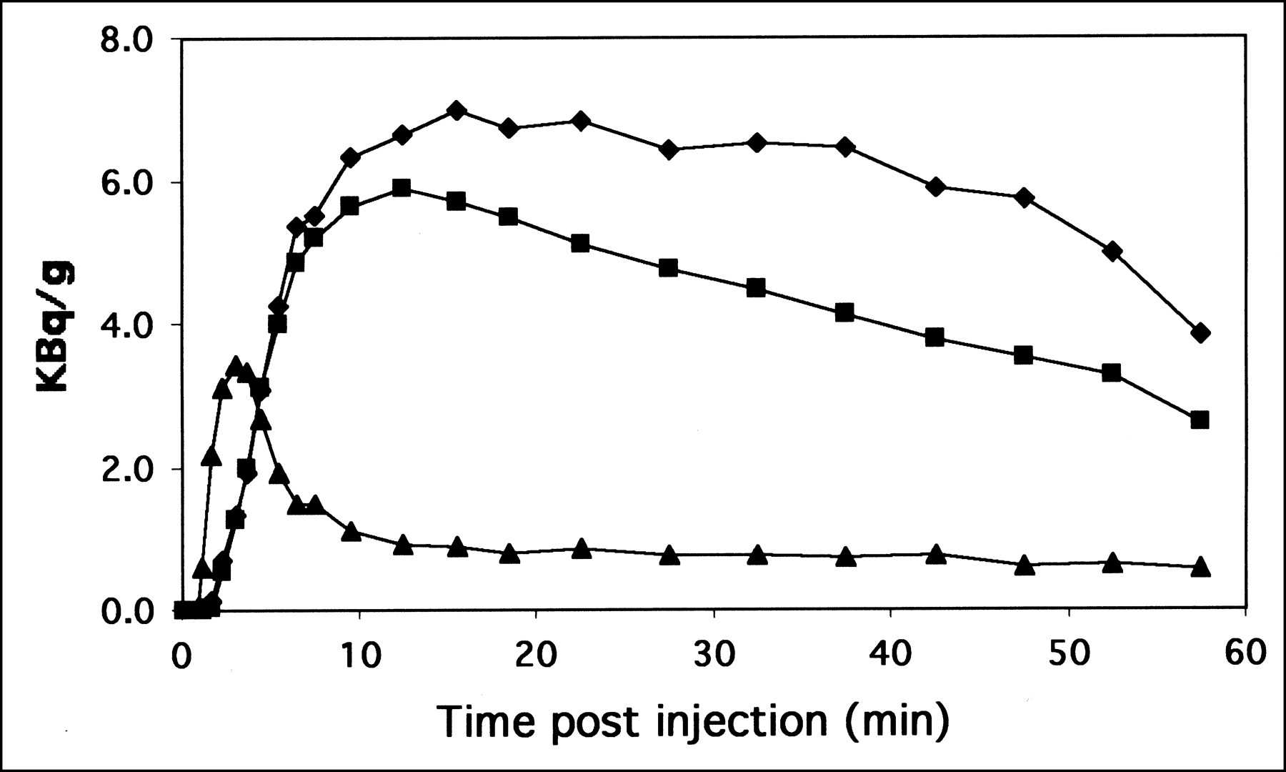
- FIGURE 1.
Example of TACs for FES in gallbladder (⧫), liver (▪), and blood (▴). Data are normalized to 37 MBq injected per 56 kg body weight.
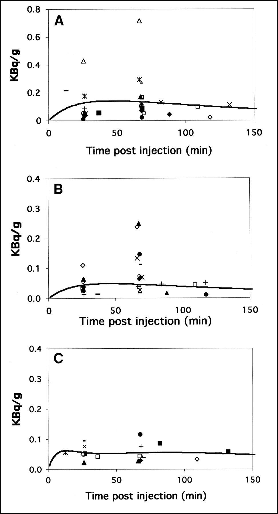
- FIGURE 2.
FES activity in bladder (A), gastrointestinal tract (B), and uterus (C). Different symbols are used for different studies. Data are normalized to 37 MBq injected per 56 kg body weight. Fits are shown as solid lines. Gastrointestinal tract and bladder data were fit with rising exponential (Eq. 2), and uterus data were fit with function described in Equation 3. y-axis for bladder is twice as large as for uterus and intestinal tract.

Tables
Organ No. of studies C̃ Mean (SD) (kBq-h/g) Organ mass (g) Ã Mean (SD) (MBq-h) Breast 47 0.7 (0.3) 361 0.2 (0.1) Gall Bladder 15 31.7 (16.8) 49 1.6 (0.8) Intestines* 1 4 5.8 (7.0) 2 14 (28) 9.8 3 4 plus pooled 6.6 (6.4) 176 (ULI) + 322 (SmI) 3.3 (3.2) Blood 48 2.0 (0.5) 347 (heart contents) 0.7 (0.2) Heart Wall 48 1.8 (0.6) 241 0.4 (0.1) Kidneys 3 4.2 (0.7) 248 1.0 (0.2) Liver 49 18.9 (4.8) 1400 26.4 (6.7) Lungs 48 1.3 (0.5) 651 0.8 (0.3) Red Marrow 47 1.2 (0.6) 1050 1.2 (0.6) Spleen 18 1.1 (0.7) 123 0.1 (0.1) Bladder* 1 2 14.4 (10.1) 2 16 (28) 18.3 3 2 plus pooled 15.7 (7.5) 160 2.5 (1.2) Uterus* 1 3 4.8 (2.7) 2 10 (18) 7.6 3 3 plus pooled 5.5 (2.6) 79 0.4 (0.2) Remainder 50,793† 58.9 (7.6) ↵* These organs also had additional points from more sparsely sampled TACs. 1 = fully sampled curves; 2 = pooled additional data (parentheses indicate number of points used in curve); 3 = mean of fully sampled curves plus additional value from pooled data. These numbers are used for Table 2.
↵† Remainder of body for 56-kg woman.
ULI = large intestine; SmI = small intestine.
Organ Mean* (mGy/MBq) SD (mGy/MBq) 25%† (mGy/MBq) 75%† (mGy/MBq) Adrenals 0.023 (85) 0.003 0.021 0.025 Brain 0.010 (36) 0.001 0.009 0.010 Breasts 0.009 (32) 0.002 0.008 0.010 GB wall 0.102 (379) 0.041 0.075 0.134 LLI 0.012 (45) 0.001 0.011 0.013 Small intestine 0.027 (99) 0.015 0.017 0.038 Stomach 0.014 (50) 0.001 0.013 0.014 ULI 0.030 (110) 0.016 0.019 0.042 Heart wall 0.026 (96) 0.004 0.024 0.029 Kidney 0.035 (128) 0.004 0.032 0.038 Liver 0.126 (466) 0.030 0.105 0.149 Lungs 0.017 (61) 0.002 0.015 0.018 Muscle 0.021 (79) 0.001 0.021 0.022 Ovaries 0.018 (66) 0.002 0.016 0.019 Pancreas 0.023 (84) 0.002 0.021 0.024 Red marrow 0.013 (48) 0.002 0.012 0.014 Bone surface 0.014 (53) 0.001 0.014 0.015 Skin 0.005 (18) 0.000 0.005 0.005 Spleen 0.015 (54) 0.003 0.012 0.017 Testes 0.012 (44) 0.001 0.011 0.012 Thymus 0.014 (50) 0.001 0.013 0.014 Thyroid 0.012 (45) 0.001 0.012 0.013 UB wall 0.050 (186) 0.020 0.036 0.066 Uterus 0.039 (145) 0.013 0.031 0.049 Lens 0.009 (33) 0.000 0.009 0.009
In this issue


Jump to section
Related Articles
Cited By...
- Kinetic Analysis and Metabolism of Poly(Adenosine Diphosphate-Ribose) Polymerase-1-Targeted 18F-Fluorthanatrace PET in Breast Cancer
- Biodistribution of 18F-FES in Patients with Metastatic ER+ Breast Cancer Undergoing Treatment with Rintodestrant (G1T48), a Novel Selective ER Degrader
- Breast Cancer: Evaluating Tumor Estrogen Receptor Status with Molecular Imaging to Increase Response to Therapy and Improve Patient Outcomes
- 18F-Fluoroestradiol PET: Current Status and Potential Future Clinical Applications
- Imaging Diagnostic and Therapeutic Targets: Steroid Receptors in Breast Cancer
- Assessment of Estrogen Receptor Expression in Epithelial Ovarian Cancer Patients Using 16{alpha}-18F-Fluoro-17{beta}-Estradiol PET/CT
- Assessment of Progesterone Receptors in Breast Carcinoma by PET with 21-18F-Fluoro-16{alpha},17{alpha}-[(R)-(1'-{alpha}-furylmethylidene)Dioxy]-19-Norpregn-4-Ene-3,20-Dione
- PET Imaging of Estrogen Receptors as a Diagnostic Tool for Breast Cancer Patients Presenting with a Clinical Dilemma
- Antiretroviral Tissue Kinetics: In Vivo Imaging Using Positron Emission Tomography
- Quantitative Metrics of Net Proliferation and Invasion Link Biological Aggressiveness Assessed by MRI with Hypoxia Assessed by FMISO-PET in Newly Diagnosed Glioblastomas
- Assessment of Human Biodistribution and Dosimetry of 4-Fluoro-11{beta}-Methoxy-16{alpha}-18F-Fluoroestradiol Using Serial Whole-Body PET/CT
- Tumor Receptor Imaging
- Whole-Body Biodistribution and Radiation Dosimetry of the Human Cannabinoid Type-1 Receptor Ligand 18F-MK-9470 in Healthy Subjects
- Tumor-Specific Positron Emission Tomography Imaging in Patients: [18F] Fluorodeoxyglucose and Beyond
- Quantitative Fluoroestradiol Positron Emission Tomography Imaging Predicts Response to Endocrine Treatment in Breast Cancer
- Correlation of Hypoxic Cell Fraction and Angiogenesis with Glucose Metabolic Rate in Gliomas Using 18F-Fluoromisonidazole, 18F-FDG PET, and Immunohistochemical Studies
- Seeing into cells: The promise of in vivo molecular imaging in oncology
- 18F-Fluorothymidine Radiation Dosimetry in Human PET Imaging Studies