


Article Figures & Data
Figures
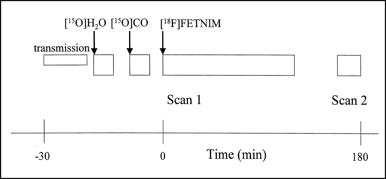
- FIGURE 1.
Diagram of imaging protocol for [15O]H2O, [15O]CO2, and [18F]FETNIM PET shows 2-phase acquisition of [18F]FETNIM data, with scan 1 obtained at 0–120 min and scan 2 obtained at 160–180 min. For clarification, time scale is different on both sides of time 0.
- FIGURE 2.
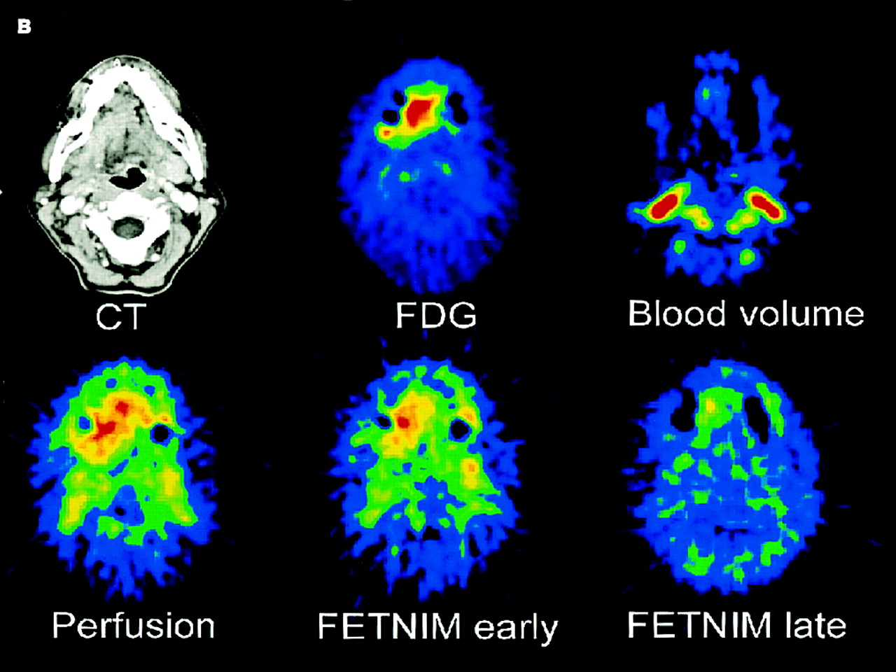
Multiple tomographic PET images of 2 untreated patients with head and neck cancer. Corresponding axial CT scans are depicted in upper left of both image sets. (A) Supraglottic laryngeal cancer (T1 N0) shows high uptake of [18F]FDG (top row, middle) and increased blood flow (bottom row, left). Early distribution pattern of [18F]FETNIM 5–8 min from injection allows easy delineation of tumor (bottom row, middle), whereas in later phase, 120 min from injection (bottom row, right), [18F]FETNIM is distributed more evenly between tumor and ambient tissues in neck and base of mouth. PET images also show transfer of ROI (red line circling tumor) from [18F]FDG image to other PET studies. (B) Right lingual cancer (T4 N1) likewise shows high uptake of [18F]FDG (top row, middle) and high blood flow (bottom row, left). Early [18F]FETNIM image obtained 5–8 min from injection (bottom row, middle) closely resembles that of corresponding perfusion image, whereas later phase [18F]FETNIM image at 120 min (bottom row, right) shows focal uptake, especially in apex of tumor. Tumor is hardly visible in [15O]CO blood volume images (top rows, right) of both (A) and (B).
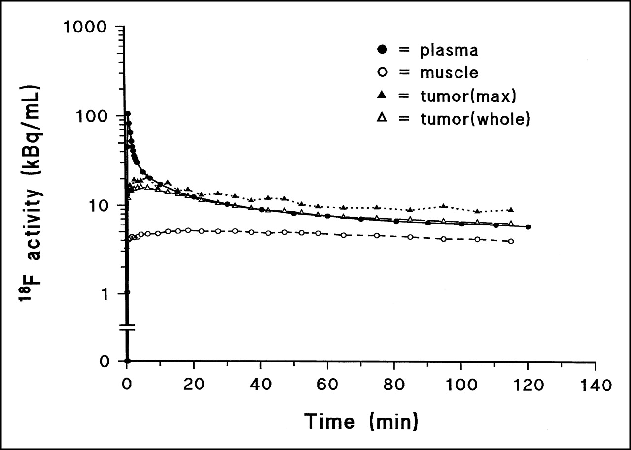
- FIGURE 3.
Time course of uptake of [18F]FETNIM in patient with hypopharyngeal carcinoma. Whole and maximum (max) tumor activity are shown separately.
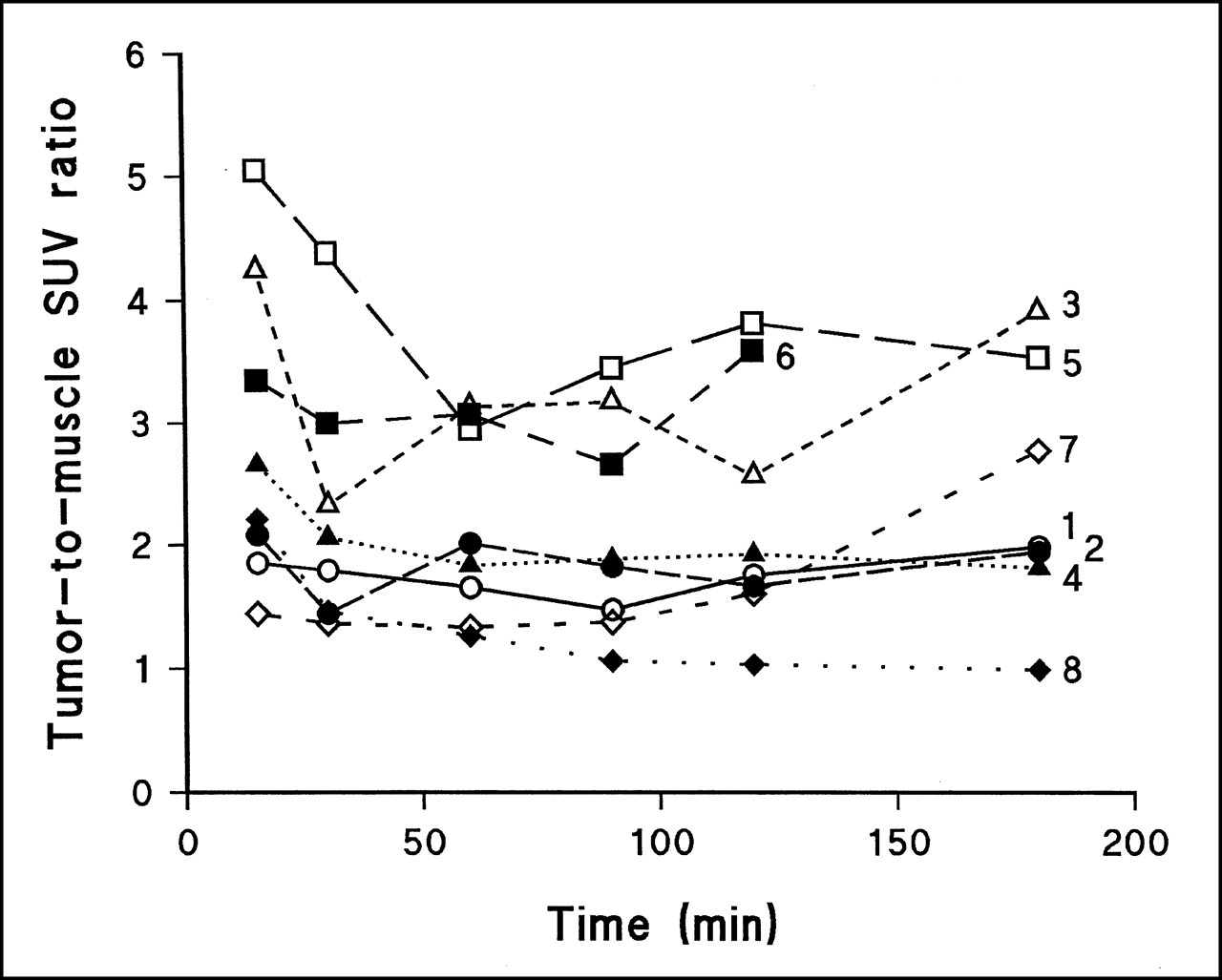
- FIGURE 4.
Time course of [18F]FETNIM uptake expressed as tumor-to-muscle SUV ratio. Numbers within frame refer to patients as numbered in Table 1.
- FIGURE 5.
Relationship between DV of [18F]FETNIM and maximum (max) SUV in tumor at different times during dynamic study.
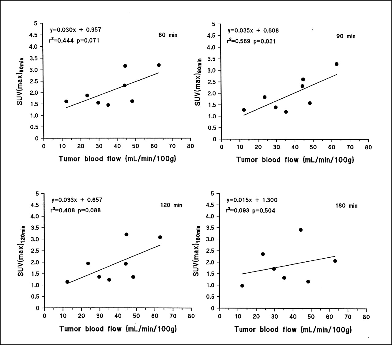
- FIGURE 6.
Relationship between uptake of [18F]FETNIM expressed as maximum (max) SUV and blood flow at different times during dynamic study.


Tables
Patient no. Age (y) Sex Region TNM UICC stage Grade 1 65 M Supraglottic, larynx T1 N0 M0 I 1 2 72 M Glottic, larynx T2 N0 M0 II 1 3 56 M Oral, tongue T3 N2b M0 IV a 3 4 49 M Mandibular, gingiva T4 N2 M0 IV a 2 5 50 F Oral, tongue T4 N1 M0 IV a 2 6 66 M Piriform sinus T1 N3 M0 IV b 3 7 55 M Mandibular, gingiva T4 N2b M0 IV 2 8 62 M Glottic, larynx T2 N0 M0 II 2 UICC = International Union Against Cancer.
Patient no. [18F]FDG Blood flow (mL/100 g/ min) Blood volume (mL/100 g tissue) [18F]FETNIM Early uptake Late uptake SUV Volume* (mL) Twhole/p Tmax/p DV Twhole/p Tmax/p DV 1 18.5 8.3 35.3 6.4 0.74 0.91 0.85 0.75 0.89 0.86 2 14.9 3.4 29.7 4.1 0.70 0.72 0.72 0.82 1.01 0.76 3 19.0 34.1 44.4 7.5 1.00 1.41 1.20 1.04 1.74 1.41 4 13.9 20.1 48.3 6.2 0.79 0.94 0.91 0.75 1.02 0.91 5 8.4 9.9 63.1 4.6 1.10 1.98 1.64 0.81 1.28 1.54 6 17.3 401.6 44.8 ND 1.06 1.49 1.22 ND ND ND 7 11.7 53.6 23.7 5.7 0.95 1.24 1.08 0.98 1.54 1.10 8 7.6 1.7 12.4 5.5 0.83 0.95 0.82 0.80 1.00 0.83 Median 14.4 15.0 39.8 5.7 0.89 1.09 0.99 0.81 1.02 0.91 ↵* Metabolically active tumor volume as determined from [18F]FDG PET.
Twhole/p = whole tumor-to-plasma radioactivity ratio at 90–120 min or 160–180 min; Tmax/p = maximum tumor-to-plasma radioactivity ratio at 90–120 min or 160–180 min; DV = distribution volume at 20–120 min or 160–180 min; ND = not detected.
- TABLE 3
Pixel-by-Pixel Comparison of Blood Flow and [18F]FETNIM Uptake in Head and Neck Tumors 90 Minutes After Tracer Injection
Patient no. Observations (n) r2 P 1 290 0.02 0.02 2 125 0.06 0.005 3 528 0.01 0.1 4 403 0.17 0.0001 5 331 0.43 0.0001 6 1,986 0.00 0.002 7 1,009 0.00 0.2 8 34 0.01 0.5
In this issue


Jump to section
Related Articles
Cited By...
- Multiparametric Imaging of Tumor Hypoxia and Perfusion with 18F-Fluoromisonidazole Dynamic PET in Head and Neck Cancer
- 2-18F-Fluoroethanol Is a PET Reporter of Solid Tumor Perfusion
- Multiparametric Analysis of the Relationship Between Tumor Hypoxia and Perfusion with 18F-Fluoroazomycin Arabinoside and 15O-H2O PET
- 18F-Alfatide II and 18F-FDG Dual-Tracer Dynamic PET for Parametric, Early Prediction of Tumor Response to Therapy
- PET Imaging of Chemokine Receptors in Vascular Injury-Accelerated Atherosclerosis
- PET of Hypoxia: Current and Future Perspectives
- Quantitative Assessment of Hypoxia Kinetic Models by a Cross-Study of Dynamic 18F-FAZA and 15O-H2O in Patients with Head and Neck Tumors
- Innovations in Radiotherapy Planning of Head and Neck Cancers: Role of PET
- 18F-EF5: A New PET Tracer for Imaging Hypoxia in Head and Neck Cancer
- Reproducibility of Tumor Perfusion Measurements Using 15O-Labeled Water and PET
- Reproducibility of Tumor Blood Flow Quantification with 15O-Water PET
- Application of PET/CT in the Development of Novel Anticancer Drugs
- Prognostic Impact of Hypoxia Imaging with 18F-Misonidazole PET in Non-Small Cell Lung Cancer and Head and Neck Cancer Before Radiotherapy
- Hypoxia-Specific Tumor Imaging with 18F-Fluoroazomycin Arabinoside
- Assessment of Inter- and Intrapatient Variability in C15O2 Positron Emission Tomography Measurements of Blood Flow in Patients with Intra-abdominal Cancers
- Positron emission tomographic imaging of angiogenesis and vascular function
- Nuclear medicine in imaging head and neck malignancies
- 18F-Fluoroerythronitroimidazole Radiation Dosimetry in Cancer Studies
- On Measuring Hypoxia in Individual Tumors with Radiolabeled Agents