


Abstract
Fibroblast activation protein (FAP) is overexpressed in cancer-associated fibroblasts and is involved in a variety of tumor-promoting activities such as matrix remodeling, angiogenesis, chemotherapy resistance, and immunosuppression. Because FAP shows low expression in most normal organs, it presents an interesting target for imaging and endoradiotherapy. In this investigation, FAP inhibitors (FAPIs) were modified and optimized for use as theranostic tracers. Methods: FAPIs based on a quinoline structure were synthesized and characterized with respect to binding, internalization, and efflux in cells expressing human and murine FAP as well as CD26. Preclinical pharmacokinetics were determined in tumor-bearing animals with biodistribution experiments and small-animal PET. Finally, a proof-of-concept approach toward imaging and therapy was chosen for 2 patients with metastasized breast cancer. Results: Of 15 synthesized FAPIs, FAPI-04 was identified as the most promising tracer for clinical application. Compared with the previously published ligand, FAPI-02, FAPI-04 showed excellent stability in human serum, higher affinity for FAP as opposed to CD26, and slower excretion in vitro. In vivo, a higher SUV was reached in tumor-bearing animals, leading to larger areas under the curve as calculated from biodistribution experiments. Finally, PET/CT scans with 68Ga-FAPI-04 in 2 patients with metastasized breast cancer revealed high tracer uptake in metastases and a reduction in pain symptoms after therapy with a considerably low dose of 90Y-FAPI-04. Conclusion: FAPI-04 represents a promising tracer for both diagnostic imaging and, possibly, targeted therapy of malignant tumors with a high content of activated fibroblasts, such as breast cancer.
See an invited perspective on this article on page 1412.
Cancer-associated fibroblasts modulate the microenvironment of malignant tumors by secreting factors that regulate both malignant cells and nonmalignant cells such as immune and endothelial cells (1). Although heterogeneous in their origin, cancer-associated fibroblasts have common properties distinct from normal fibroblasts and show expression of proteins not found in their normal counterparts. One of these proteins is the fibroblast activation protein (FAP), a type II transmembrane serine protease with both dipeptidyl peptidase activity and endopeptidase activity (2–4).
FAP expression is associated with a poor prognosis in a variety of tumors, such as colon (5), pancreatic (6), ovarian (7), and hepatocellular carcinoma (8). It plays multiple biologic roles in cancer. First, via its peptidase activity, FAP leads to matrix digestion and remodeling of the tumor microenvironment, enabling invasion and migration of tumor cells (9). Because neuropeptide Y is a natural substrate of FAP and the cleavage product has been shown to be proangiogenic, FAP is considered to be involved in tumor angiogenesis, as has been substantiated by studies showing a correlation between FAP expression and microvessel density in tumors (10,11).
Furthermore, besides its enzyme function, FAP activates cell signaling by forming complexes with other proteins—for example, β1 integrins such as α3β1 (12,13).
Because cancer-associated fibroblasts are the primary source of collagen I, which contributes to decreased chemotherapeutic drug uptake and thereby plays a role in regulating the sensitivity of tumors toward chemotherapy, elimination of these fibroblasts via targeting of FAP may lead to increased uptake of chemotherapeutic drugs and greater therapeutic efficiency (14). An immunosuppressive role has also been described (15).
In addition, FAP has other favorable properties qualifying it as a promising target for diagnosis and therapeutic intervention: besides expression in some nonmalignant conditions such as wound healing, rheumatoid arthritis, atherosclerotic plaques, and diseases leading to fibrosis, FAP is expressed in more than 90% of human epithelial cancers, is absent from normal tissues in adult humans, and has a large extracellular domain, with the catalytic site also located extracellularly. These properties ensure a low background activity with high image contrast and a low frequency of side effects for FAP-targeting molecules, the possibility of application in many different tumor entities, and the design of combination therapies targeting both tumor cells and the stromal components of a tumor. Such targeting of the stromal cells has been done using a variety of molecules, including antibodies (16–18), chimeric antigen receptor T cells (19,20), immunoconjugates (21), peptide–drug complexes bearing the consensus sequence for the enzymatic activity (22,23), vaccines (14,24), and small molecules for the inhibition of FAP enzyme activity (25). Another approach is the use of these small molecules as carriers of radioactivity into the tumor for both imaging and therapy. In a previous paper, we showed that a FAP inhibitor (FAPI), FAPI-02, rapidly accumulates in FAP-expressing cells, tumor xenografts, and patients (26). Although high image contrast was obtained, the tumor retention time was relatively short, a quality that presents no problem for diagnosis but may need improvement for therapy. In this investigation, we designed variants of FAPI-02 with the goal of increasing tumor retention time and thereby developing a theranostic FAP-targeting agent.
MATERIALS AND METHODS
Synthesis and Radiolabeling
As an example of the synthetic pathway toward the FAPI derivatives, the pathway for FAPI-04 is given in Supplemental Figure 1 (supplemental materials are available at http://jnm.snmjournals.org). A detailed compilation of the precursors, the intermediates, and their syntheses can be found in the supplemental material. 177Lu and 68Ga were chelated after pH adjustment with sodium acetate. The reaction mixture was heated to 95°C for 10 min, and the completeness of the reaction was checked by radio–liquid chromatography. The 68Ga compounds were processed by solid-phase extraction before PET. Stability in human serum was determined by precipitation of samples and radiochromatographic analysis of the supernatant. The stability of the compounds was checked for FAPI-04, as an example, by incubation in human serum at 37°C (Supplemental Fig. 2).
Cell Culture
HT-1080 cells transfected with the human or murine FAP gene, as well as CD26-transfected human embryonic kidney cells (obtained from Stefan Bauer, NCT, Heidelberg, Germany (27)), were cultivated in Dulbecco modified Eagle medium containing 10% fetal calf serum at 37°C in 5% carbon dioxide.
For radioligand binding studies, cells were seeded in 6-well plates and cultivated for 48 h to a final confluence of approximately 80%–90% (1.2–2 × 10⁶ cells per well). The medium was replaced by 1 mL of fresh medium without fetal calf serum. The radiolabeled compound (177Lu-labeled FAPIs; specific activity, 200 nmol/GBq) was added to the cell culture and incubated for different intervals ranging from 10 min to 24 h. Competition experiments were performed by simultaneous exposure to unlabeled (10−5 to 10−9 M) and radiolabeled compound for 60 min. In all experiments, the cells were washed twice with 1 mL of phosphate-buffered saline, pH 7.4, and subsequently lysed with 1.4 mL of lysis buffer (0.3 M NaOH, 0.2% sodium dodecyl sulfate). Radioactivity was determined in a γ-counter (Cobra II; Packard), normalized to 106 cells, and calculated as percentage injected dose (%ID). Cell efflux was determined after incubation of the cells with the tracer for 60 min. Afterward, the radioactive medium was removed, and the cells were washed and incubated in nonradioactive medium for 1, 2, 4, or 24 h. After 2 washings, the cells were lysed and the radioactivity counted. Each experiment was performed 3 times, and 3 repetitions per independent experiment were acquired.
For internalization experiments, the cells were incubated with the radiolabeled compound for 60 min at 37°C and 4°C. Cellular uptake was terminated by removing medium from the cells and washing twice with 1 mL of phosphate-buffered saline. Subsequently, the cells were incubated with 1 mL of glycine-HCl (1 M, pH 2.2) for 10 min at room temperature to remove the surface-bound activity. The cells were washed with 2 mL of ice-cold phosphate-buffered saline and lysed with 1.4 mL of lysis buffer to determine the internalized fraction. For the cells incubated at 4°C, all washing and elution steps were performed using ice-cold buffers. The radioactivity was measured using a γ-counter, normalized to 1 million cells, and calculated as %ID.
Animal Studies
For in vivo experiments, 8-wk-old BALB/c nu/nu mice (Charles River) were subcutaneously inoculated into the right trunk with 5 × 10⁶ HT-1080 FAP cells. When the tumor had grown to approximately 1 cm3, the radiolabeled compound (177Lu-labeled FAPIs; specific activity, 200 nmol/GBq) was injected via the tail vein. For organ distribution, the animals (n = 3 for each time point) were sacrificed after the indicated time points (from 30 min to 24 h). The distributed radioactivity was measured in all dissected organs and in blood using a γ-counter (Cobra Autogamma; Packard). The values are expressed as %ID per gram of tissue (%ID/g). PET imaging was performed using a small-animal PET scanner (Inveon; Siemens). The mice were injected with 68Ga-FAPI-02 (80 nmol/GBq). Within the first 60 min, a dynamic scan was performed, followed by a static scan from 120 to 140 min after injection. Images were reconstructed iteratively using the 3-dimensional maximum a priori ordered-subset expectation maximization (Siemens) and were converted to SUV images. Quantification was done using a region-of-interest technique and expressed as SUV. Imaging was also done in animals bearing SK-LMS-1 tumors, a model in which the tumor cells are FAP-negative and only activated mouse fibroblasts are present. A blocking experiment was performed by adding 30 nmol of unlabeled precursor to the solution of 68Ga-FAPI-04 before injection. All animal experiments were conducted in compliance with the German animal protection laws.
Clinical PET/CT Imaging
Diagnostic imaging of 2 patients with 68Ga-FAPI-04 PET/CT for medical reasons was performed under the conditions of the updated declaration of Helsinki (section 37, unproven interventions in clinical practice) and in accordance with the German Pharmaceuticals Law (section 13, 2b). The tracer was injected intravenously (80 nmol/GBq), and images were obtained 10 min, 1 h, and 3 h later. The images were obtained on a Biograph mCT Flow PET/CT scanner (Siemens Medical Solutions) using the following parameters: a 5-mm slice thickness, an increment of 3–4 mm, a soft-tissue reconstruction kernel, and CARE Dose4D (Siemens Medical Solutions). Immediately after the CT component had been acquired, whole-body PET was performed in 3 dimensions (matrix, 200 × 200) in FlowMotion (Siemens Medical Solutions) at a rate of 0.7 cm/min. The emission data were corrected for random, scatter, and decay events. Images were reconstructed using ordered-subset expectation maximization with 2 iterations and 21 subsets and were Gauss-filtered to a transaxial resolution of 5 mm in full width at half maximum. Attenuation was corrected using the low-dose nonenhanced CT data. SUVs were quantitatively assessed using a region-of-interest technique. All patients gave written informed consent. The evaluation was approved by our institutional ethical review board (approval S-016/2018).
Therapeutic Application
One patient with metastasized breast cancer was treated with 2.9 GBq of 90Y-FAPI-04 (24 nmol/GBq). FAPI-04 was labeled with a 90Y-chloride solution (Yttriga; Eckert and Ziegler Radiopharma GmbH) and injected intravenously via a low-protein-binding sterile filter system (Filtropur S 0.2; Sarstedt). γ-camera images of the bremsstrahlung were acquired 3 h and 1 d after tracer administration using a Hawkeye SPECT/CT system (GE Healthcare).
RESULTS
Synthesis and Radiolabeling
Following the synthetic pathway shown in Supplemental Figure 1, 11 new compounds and a small-molecule–peptide conjugate were synthesized. An overview of the synthesized compounds is given in Figure 1. To trace the peptide via radioactive labeling, ε-DOTA-lysine was inserted C-terminally between the end of the NLS sequence and the conjugated cysteine residue.
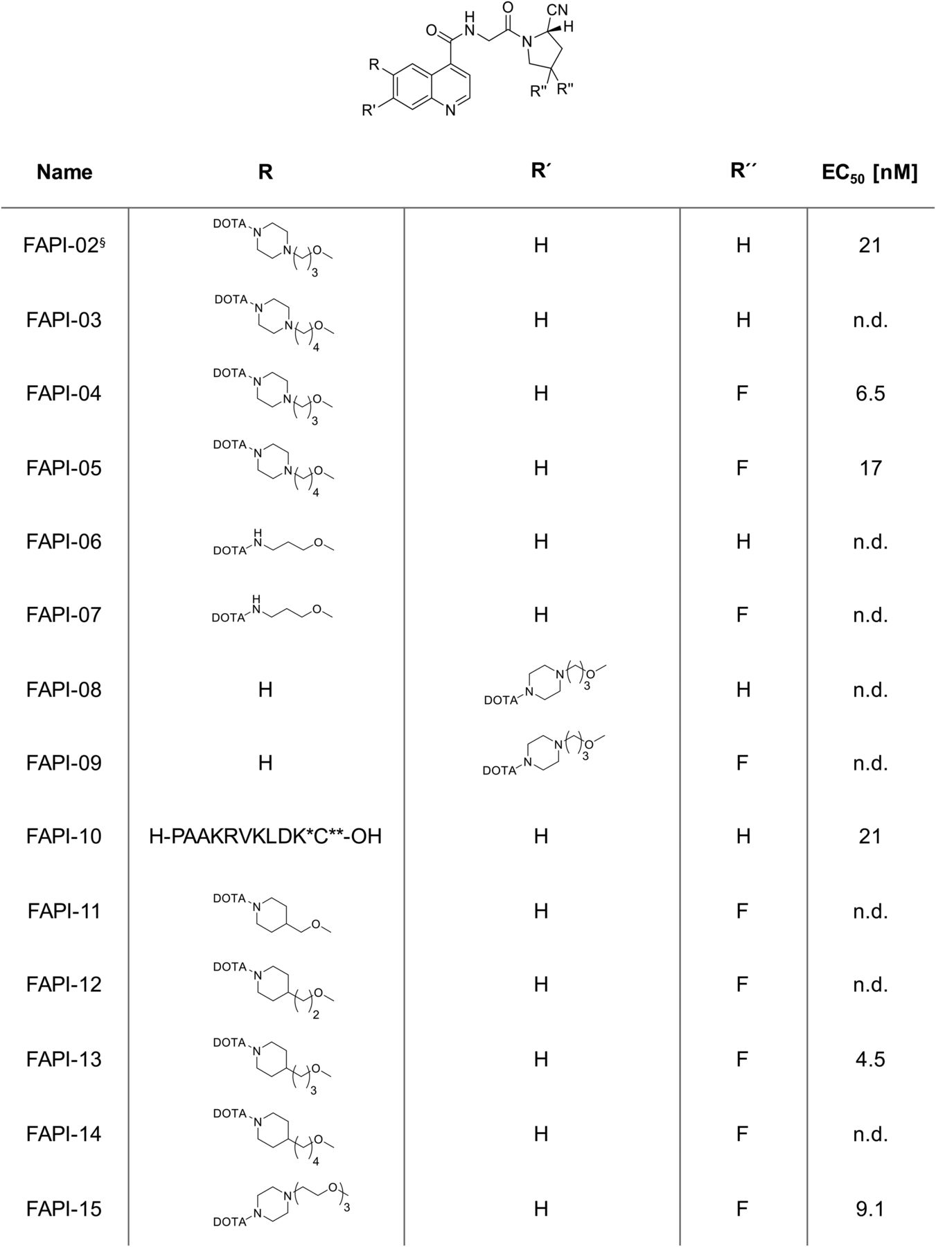
Overview of synthesized FAPI derivatives. EC50 obtained by competition experiments is shown for selected compounds. §Discussed in previous publication (28). *ε-amine modified by DOTA. **Cysteine-thiol attached to maleimide-carrying piperazinylpropoxy moiety. n.d. = not determined.
Cell-Based Experiments
To evaluate the binding and internalization properties of the FAPI derivatives, radioligand binding assays were performed using FAP-expressing HT-1080 cells (Fig. 2). All tracers showed an almost complete internalization, with values over 90%. Because of insufficient binding to human FAP after 24 h, the compounds FAPI-06, FAPI-07, FAPI-08, and FAPI-09 were not characterized further. Although they showed acceptable accumulation after 24 h, the evaluation of FAPI-11 and FAPI-12 was not pursued because of bad performance after 1 and 4 h of incubation. To verify the target specificity, binding assays were also performed using human embryonic kidney cells expressing murine FAP and dipeptidyl peptidase 4 (CD26), which show a high homology to human FAP (Supplemental Fig. 2). In these experiments, FAPI-02 and FAPI-04 showed a strong binding to murine FAP, with significantly higher values for FAPI-04, and no binding to CD26. However, FAPI-15 also had a low affinity to CD26. Calculation of the ratio of murine FAP binding to CD26 binding revealed values of 45, 750, and 38 for FAPI-02, FAPI-04, and FAPI-15, respectively (Supplemental Fig. 2).
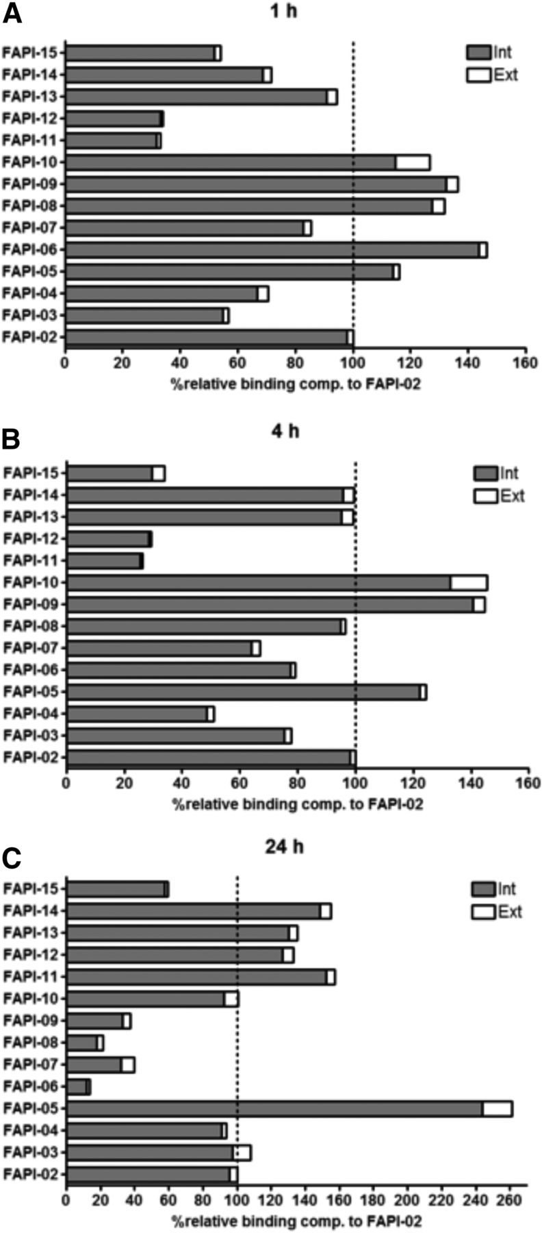
Relative binding and internalization rates of 177Lu-labeled FAPI derivatives compared with FAPI-02 (set to 100%) on FAP-expressing HT-1080 cells at 1 h (A), 4 h (B), and 24 h (C) after radiotracer administration.
In the first set of experiments, the difluoroproline analogs FAPI-04 and FAPI-05 were analyzed to investigate the influence of the presence of fluorine and the difference between a propyl linkage and a butyl linkage of the piperazine and quinoline components of the radiotracers. For this purpose, the half-maximal effective concentration (EC50) for each FAPI was determined by a competition assay, which demonstrated a higher target specificity for FAPI-04 (6.5 nM) than for FAPI-05 (17.2 nM; Supplemental Fig. 3). Efflux experiments showed that FAPI-04 and FAPI-05 were excreted considerably more slowly than FAPI-02, resulting in increased half-lives of 3.0 and 2.8 h (FAPI-02, 1.7 h; Supplemental Fig. 4). FAPI-13 was characterized in more detail because of its resemblance to the compound FAPI-04 and showed the lowest EC50 in this set of data (4.5 nM; Supplemental Fig. 3).
The compounds FAPI-10 and FAPI-15 were examined because of their unique modifications: FAPI-10 is a peptide conjugate of the FAPI-02 core with the intention of localizing the radioactivity in the nucleus, and FAPI-15 is similar to FAPI-04 but possesses a triethylene glycol spacer instead of the 1,3-propyl moiety. In both cases, EC50 was comparable to that of the parenting radiotracer.
Organ Distribution
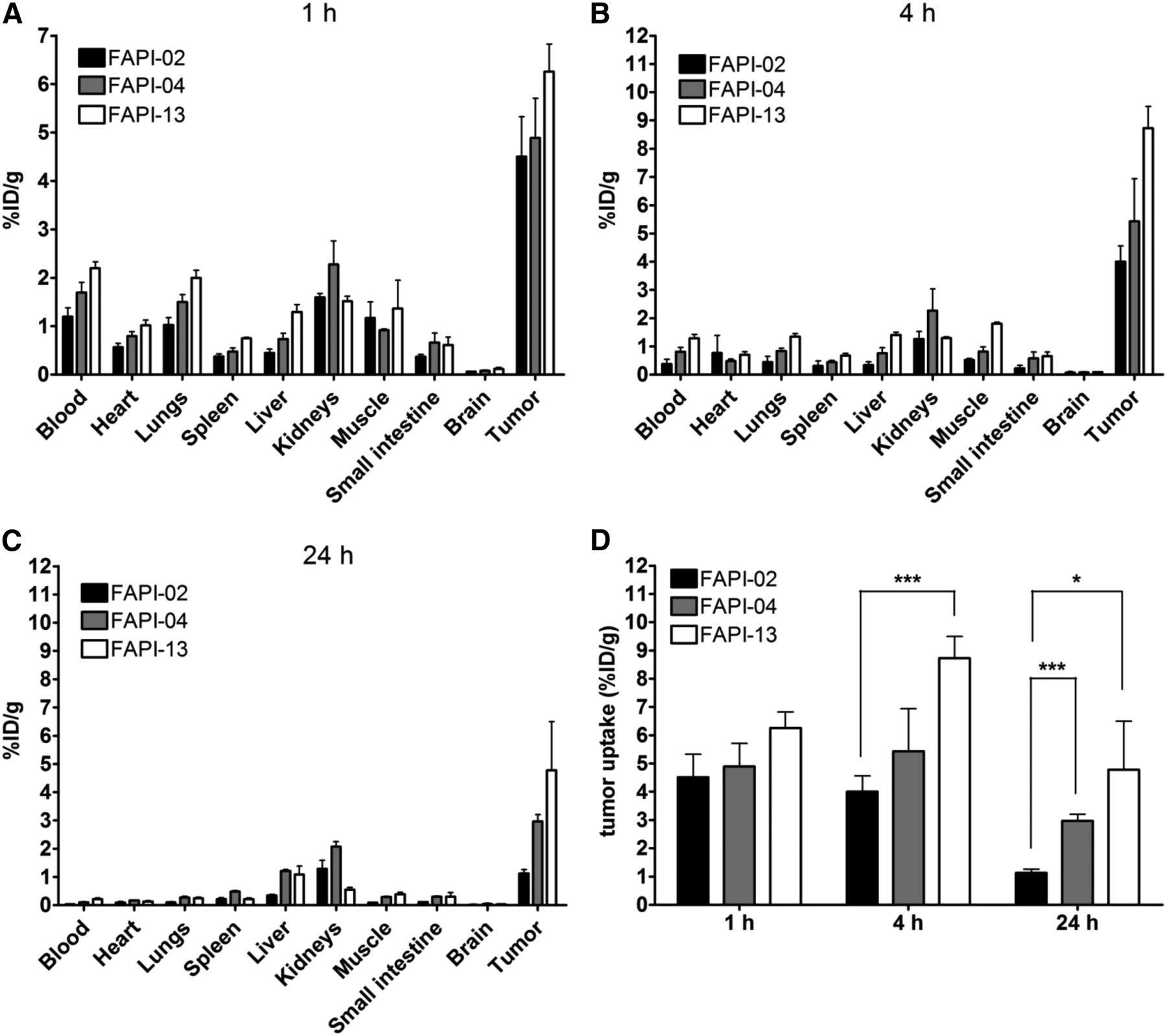
To evaluate distribution, tumor uptake, and excretion of selected FAPI derivatives, biodistribution studies were performed on mice bearing human HT-1080 FAP-expressing xenografts. Figure 3 shows the results of the biodistribution assays performed with FAPI-04 and FAPI-13 compared with the existing data for FAPI-02. FAPI-10 was discarded because of a relatively low tumor accumulation at 1 and 4 h after administration. Moreover, it accumulated strongly within the kidneys (≤10 %ID/g; FAPI-02, 1.6 %ID/g), with high kidney values even after 24 h (Supplemental Fig. 5). FAPI-05 produced results comparable to FAPI-02 but was discarded with regard to the better-performing derivatives FAPI-04 and FAPI-13. Tracer uptake in most normal tissues was slightly higher for FAPI-04 than for FAPI-02 and was further increased for FAPI-13. Tumor accumulation was higher for FAPI-04 and FAPI-13 than for FAPI-02, especially after 24 h. A calculation of the area the under curve is shown in Supplemental Table 1. The values for FAPI-02, FAPI-04, and FAPI-15 were 64, 99.4, and 157.5, respectively. However, the tumor-to-blood ratio favored FAPI-04 over FAPI-13 (Supplemental Fig. 6).

(A–C) Biodistribution of selected FAPI derivatives in HT-1080 FAP xenografts at 1 h (A), 4 h (B), and 24 h (C) after radiotracer administration. (D) Tumor uptake of selected compounds.
Small-Animal PET Imaging
For further characterization of the remaining FAPIs, small-animal PET studies were conducted with HT-1080 FAP-xenografted mice (Figs. 4 and 5). 68Ga-FAPI-02 and 68Ga-FAPI-04 demonstrated the highest tumor uptake at 1 h after injection (SUVmax, 0.88 and 1.2, respectively), with no significant decrease within 2 h (SUV, 0.71 and 1.1, respectively). In contrast, tumor accumulation of 68Ga-FAPI-13 increased steadily for up to 2 h after injection (SUV at 1 h, 0.93; at 2 h, 0.97). However, uptake was higher in background, spinal cord, and bone (Supplemental Fig. 7). Additionally, the applicability of 68Ga-FAPI-04 in a second xenograft model, SK-LMS-1 was successfully demonstrated (Fig. 4). Furthermore, target specificity was elucidated by a blocking experiment on an HT-1080 FAP xenograft. As shown in Figure 5, accumulation in the tumor was suppressed effectively by coadministration of unlabeled compound.

PET imaging of FAPI-04 in mice bearing SK-LMS-1 and HT-1080 FAP tumors, along with corresponding time–activity curves.

PET imaging of FAPI-04 in HT-1080 FAP tumor-bearing mice with and without simultaneous injection of unlabeled FAPI-04 as competitor, along with corresponding time–activity curves.
Clinical Application in Patients with Breast Cancer
Diagnostic PET/CT scans were performed 10 min, 1 h, and 3 h after intravenous administration of 68Ga-FAPI-04 in 2 patients with metastasized breast cancer (Figs. 6 and 7). In both patients, accumulation of the tracer in metastases was robust (SUVmax, 7–15.5 for patient in Fig. 6 and 15.3–29.9 for patient in Fig. 7). In contrast, tracer uptake in normal tissue was very low (Supplemental Table 2). The radioactivity cleared rapidly from the bloodstream and was excreted predominantly via the kidneys, resulting in high-contrast images. In addition to imaging, therapy with 2.9 GBq of 90Y-FAPI-04 was used in one patient. The bremsstrahlung images showed accumulation of the tracer at 3 h and even at 1 d after injection in this patient. This rather low dose led to a significant reduction of pain medication in this patient (from baseline morphine and 3–4 additional morphine administrations per day to baseline medication only).
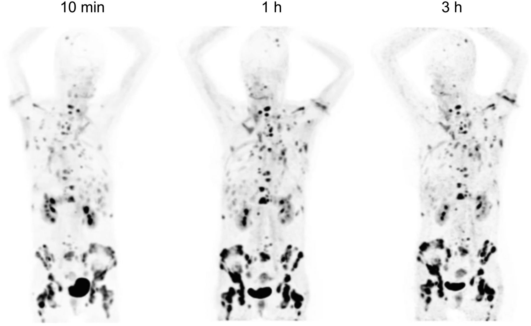
PET maximum-intensity projections of patient with metastasized breast cancer 10 min, 1 h, and 3 h after administration of 263 MBq of 68Ga-FAPI-04. Activity is seen in renal pelvis, bladder, and metastases. Normal organs show low uptake resulting in high image contrast.

(A) PET maximum-intensity projection of patient with metastasized breast cancer 1 h after administration of 270 MBq of 68Ga-FAPI-04. Robust uptake is seen in metastases. (B) Bremsstrahlung images showing uptake at 3 h and even 1 d after treatment with 90Y-FAPI-04 in same patient.
DISCUSSION
In this work, several approaches were investigated to improve the pharmacokinetic properties of the quinoline-based radiopharmaceuticals developed by our laboratory (26). Herein, particular focus was on the elaboration of a theranostic tracer (a tracer that can be used for both diagnostics and therapy). This concept is based on the linking of the FAP-targeting moiety to the chelator DOTA, which allows the incorporation of different isotopes suitable for imaging or therapeutic purposes. Using the ligand FAPI-04, we were able to demonstrate the targeted delivery of the positron-emitting 68Ga as well as the therapeutic nuclide 90Y in a clinical setting. The main challenge for potential therapeutic application of the tracer was in optimizing its tumor uptake and retention time. We addressed this task by assessing a series of novel compounds based on the initial FAP inhibitor, FAPI-02. The most striking improvement was in the use of 4,4-difluoroproline, which has already been successful with respect to enzyme inhibition as described by Jansen et al. (28). By this substitution, the EC50 of 177Lu-FAPI-04 was reduced by a factor of 3, compared with 177Lu-FAPI-02. Also, the difference in affinity between FAP and CD26 was shifted in favor of 177Lu-FAPI-04 as shown by 177Lu-FAP/CD26 binding ratios of 45 and 750 for FAPI-02 and 177Lu-FAPI-04, respectively. In addition, the efflux experiments showed a significantly slower washout for 177Lu-FAPI-04 than for 177Lu-FAPI-02, resulting in an increased half-life of 3.0 h for 177Lu-FAPI-04, versus 1.7 h for 177Lu-FAPI-02 (Supplemental Fig. 4).
Although in vitro 177Lu-FAPI-02 uptake was higher than 177Lu-FAPI-04 uptake after 1 and 4 h of incubation and equaled 177Lu-FAPI-04 uptake after 24 h, the FAPI-04 PET imaging and biodistribution studies in small animals demonstrated higher accumulation, longer dwell times, and no significant increase in background activity. Compared with 177Lu-FAPI-04, 177Lu-FAPI-13 showed higher tumor uptake leading to larger areas under the curve. However, retention of the tracer in the blood was higher, leading to lower tumor-to-blood ratios. This may lead to higher hematotoxicity during treatment, narrowing the therapeutic window. Because of these improvements, therapeutic implementation of 90Y-labeled FAPI-04 was initiated. A previous stability analysis of 177Lu-FAPI-04 in human serum revealed no degradation during 24 h, qualifying this compound for clinical translation (Supplemental Fig. 1).
A structure–activity relationship analysis was done for modifications of the heterocyclic segment and the position of the linker. The simple 3-amino-1-propyl derivatives FAPI-06 and FAPI-07 showed suitable cell binding at 1 and 4 h but were almost completely eliminated after 24 h, demonstrating that the heterocyclic segment is necessary for sufficient retention in the tumor cells. The 7-quinolyl–linked compounds FAPI-08 and FAPI-09 revealed the same pharmacokinetic profile in the course of incubation, as might be explained by the removal of the radiolabeled moiety. This indicates that the position of the linker at the quinoline moiety is equally essential. In FAPI-06 and FAPI-07, the bond between DOTA and the propylamine is far more accessible than the piperazine-bound DOTA of FAPI-02 and FAPI-04, and an enzymatic breakdown can proceed much more quickly.
The binding to albumin or other plasma proteins with a longer retention time in the circulation is seen as a knockout criterion for possible theranostics and the reason 177Lu-FAPI-13 was not considered for further development despite having the lowest EC50 and the highest tumor accumulation of the compared compounds in tumor-bearing animals. The higher lipophilicity of the piperidine moiety is presumably the cause for the higher affinity, better tissue permeation, and longer target prevalence but leads to unspecific binding to serum proteins, thus competing with the attachment to FAP and having a negative impact on the pharmacokinetic properties (29,30).
The in vitro and in vivo results led to the transfer of FAPI-04 to clinical diagnosis and therapy in 2 patients with metastasized breast cancer. As seen in the animal experiments, PET/CT imaging in these patients showed a rapid, predominantly renal washout from the body and an equally rapid accumulation in tumors (SUV, 7–29.9), leading to visualization of metastases even at 10 min after tracer administration. Besides this finding, radioactivity was seen only in the renal pelvis and the bladder, with no accumulation in the renal parenchyma or any other organ, making FAPI-04 an attractive tracer for both diagnostic and therapeutic applications. To fit the physical half-life of the isotope used for therapy to the retention in the tumor, 90Y, with a half-life of 64 h, was chosen for treatment. In a proof-of-principle approach, a patient was treated with 2.9 GBq of 90Y-FAPI-04, which resulted in visualization of the metastases in bremsstrahlung images at 24 h after tracer administration (Fig. 7). Clinically, this treatment was associated with a significant reduction in pain medication. Furthermore, no side effects were observed, especially with respect to hematotoxicity. Because tracer uptake was low in all normal organs, we expect that the dose can be significantly increased to obtain tumoricidal effects. However, any such dose increase in future approaches has to be based on dosimetric calculations and dose escalation regimens.
CONCLUSION
On the basis of our previous work on quinoline-based FAP-targeted radiopharmaceuticals, we successfully developed FAPI-04 as a theranostic tool. The pharmacokinetic profile of the compound was improved by incorporation of the 4,4-difluoroprolyl building block. Moreover, a comprehensive analysis of the synthesized compounds provided valuable insight into the structure–activity relationship of the chelator-carrying side chain.
Of all the derivatives tested, FAPI-04 is the most suitable for potential application as a theranostic tracer. Similar to its precursor, FAPI-02, FAPI-04 shows rapid internalization into FAP-positive tumors and fast clearance from the body, resulting in very fast accumulation at tumor sites (10 min after tracer administration) and comparable tumor-to-organ ratios. Moreover, the effective tumor uptake after 24 h—100% higher for FAPI-04 than for FAPI-02—is of great benefit regarding theranostic application of the tracer. A proof-of-concept approach that was applied to 2 patients gave excellent performance results for diagnosis and promising first results for therapy. Future development will concentrate on the design of compounds for labeling with isotopes that have physical characteristics different from 68Ga and 90Y, such as 188Re, 64Cu, or 212Pb.
DISCLOSURE
Uwe Haberkorn, Anastasia Loktev, Thomas Lindner, and Walter Mier have been named in a patent application (EP 18155420.5) for quinolone-based FAP-targeting agents for imaging and therapy in nuclear medicine. No other potential conflict of interest relevant to this article was reported.
Acknowledgments
We gratefully acknowledge Stefan Bauer (National Center for Tumor Diseases, Heidelberg) for supplying the FAP and CD26-transfected cell lines. We thank Christian Kleist, Susanne Krämer, Stephanie Biedenstein, Kirsten Kunze, Irina Kupin, Vanessa Kohl, Marlene Tesch, Iris Morr, Sabine Weiss, Christiane Brenner, Karin Leotta, and Ursula Schierbaum for excellent technical assistance. This work was funded in part by grant 13N 13341 from the Federal Ministry of Education and Research.
Footnotes
Published online Apr. 6, 2018.
- © 2018 by the Society of Nuclear Medicine and Molecular Imaging.
REFERENCES
- Received for publication February 23, 2018.
- Accepted for publication March 22, 2018.


Jump to section
Related Articles
Cited By...
- Are FAP Theranostics Really Happening? Will Radiochemistry or Biology Win?
- SNMMI Procedure Standard/EANM Practice Guideline for Fibroblast Activation Protein (FAP) PET
- [18F]FDG and [68Ga]Ga-FAPI-04-Directed Imaging for Outcome Prediction in Patients with High-Grade Neuroendocrine Neoplasms
- Feasibility, Tolerability, and Preliminary Clinical Response of Fractionated Radiopharmaceutical Therapy with 213Bi-FAPI-46: Pilot Experience in Patients with End-Stage, Progressive Metastatic Tumors
- Addressing Biological Questions with Preclinical Cancer Imaging
- 1,090 Publications and 5 Years Later: Is FAP-Targeted Theranostics Really Happening?
- Fibroblast Activation Protein-Directed Imaging Outperforms 18F-FDG PET/CT in Malignant Mesothelioma: A Prospective, Single-Center, Observational Trial
- Anatomical pattern of entheseal and synovial fibroblast activation in patients with psoriasis and its risk of developing psoriatic arthritis
- 68Ga-Fibroblast Activation Protein Inhibitor PET/CT Improves Detection of Intermediate and Low-Grade Sarcomas and Identifies Candidates for Radiopharmaceutical Therapy
- Diagnostic Potential of Supplemental Static and Dynamic 68Ga-FAPI-46 PET for Primary 18F-FDG-Negative Pulmonary Lesions
- 68Ga-FAPI-04 positron emission tomography/CT and laparoscopy for the diagnosis of occult peritoneal metastasis in newly diagnosed locally advanced gastric cancer: study protocol of a single-centre prospective cohort study
- 68Ga-FAPI PET/CT as an Alternative to 18F-FDG PET/CT in the Imaging of Invasive Lobular Breast Carcinoma
- Diagnostic Accuracy of 68Ga-FAPI Versus 18F-FDG PET in Patients with Various Malignancies
- Imaging of Tumor Stroma Using 68Ga-FAPI PET/CT to Improve Diagnostic Accuracy of Primary Tumors in Head and Neck Cancer of Unknown Primary: A Comparative Imaging Trial
- Preclinical Development of PNT6555, a Boronic Acid-Based, Fibroblast Activation Protein-{alpha} (FAP)-Targeted Radiotheranostic for Imaging and Treatment of FAP-Positive Tumors
- Immunohistochemical FAP Expression Reflects 68Ga-FAPI PET Imaging Properties of Low- and High-Grade Intraductal Papillary Mucinous Neoplasms and Pancreatic Ductal Adenocarcinoma
- Preclinical Evaluation of a Radiotheranostic Single-Domain Antibody Against Fibroblast Activation Protein {alpha}
- 68Ga-Labeled Fibroblast Activation Protein Inhibitor (68Ga-FAPI) PET for Pancreatic Adenocarcinoma: Data from the 68Ga-FAPI PET Observational Trial
- Impact of 68Ga-FAPI PET/CT on Staging and Oncologic Management in a Cohort of 226 Patients with Various Cancers
- [68Ga]Ga-FAPI-46 PET for Visualization of Postinfarction Renal Fibrosis
- Comparison of Baseline 68Ga-FAPI and 18F-FDG PET/CT for Prediction of Response and Clinical Outcome in Patients with Unresectable Hepatocellular Carcinoma Treated with PD-1 Inhibitor and Lenvatinib
- PET imaging of fibroblast activation protein alpha (FAP) detects incipient cardiotoxicity due to anthracycline chemotherapy
- Performance of 68Ga-Labeled Fibroblast Activation Protein Inhibitor PET/CT in Evaluation of Erdheim-Chester Disease: A Comparison with 18F-FDG PET/CT
- Tumor Characterization by [68Ga]FAPI-46 PET/CT Can Improve Treatment Selection for Pancreatic Cancer Patients: An Interim Analysis of a Prospective Clinical Trial
- [68Ga]Ga-FAPI-46 PET for Visualization of Postinfarction Renal Fibrosis
- Fibroblast Activation Protein Inhibitor-Based Radionuclide Therapies: Current Status and Future Directions
- Radiolabeled GPVI-Fc for PET Imaging of Multiple Extracellular Matrix Fibers: A New Look into Pulmonary Fibrosis Progression
- First Total-Body Kinetic Modeling and Parametric Imaging of Dynamic 68Ga-FAPI-04 PET in Pancreatic and Gastric Cancer
- 68Ga-FAPI PET/CT Interobserver Agreement on Tumor Assessment: An International Multicenter Prospective Study
- Fibroblast-Activation Protein PET and Histopathology in a Single-Center Database of 324 Patients and 21 Tumor Entities
- Clinical Translation of Targeted {alpha}-Therapy: An Evolution or a Revolution?
- Fibroblast Activation Protein-Targeted Radioligand Therapy for Treatment of Solid Tumors
- Clinical Translation of Targeted {alpha}-Therapy: An Evolution or a Revolution?
- Static and Dynamic 68Ga-FAPI PET/CT for the Detection of Malignant Transformation of Intraductal Papillary Mucinous Neoplasia of the Pancreas
- Fibroblast Activation Protein Inhibitor Imaging in Nonmalignant Diseases: A New Perspective for Molecular Imaging
- FAPI PET Opens a New Window to Understanding Immune-Mediated Inflammatory Diseases
- Head-to-Head Comparison of 68Ga-FAPI-46 and 18F-FDG PET/CT for Evaluation of Head and Neck Squamous Cell Carcinoma: A Single-Center Exploratory Study
- Synthesis, Preclinical Evaluation, and a Pilot Clinical PET Imaging Study of 68Ga-Labeled FAPI Dimer
- Albumin Binder-Conjugated Fibroblast Activation Protein Inhibitor Radiopharmaceuticals for Cancer Therapy
- The Added Value of 68Ga-FAPI PET/CT in Patients with Head and Neck Cancer of Unknown Primary with 18F-FDG-Negative Findings
- Initial Clinical Experience with 90Y-FAPI-46 Radioligand Therapy for Advanced-Stage Solid Tumors: A Case Series of 9 Patients
- Feasibility, Biodistribution, and Preliminary Dosimetry in Peptide-Targeted Radionuclide Therapy of Diverse Adenocarcinomas Using 177Lu-FAP-2286: First-in-Humans Results
- Detecting Fibroblast Activation Proteins in Lymphoma Using 68Ga-FAPI PET/CT
- Fibroblast Activation Protein-Specific PET/CT Imaging in Fibrotic Interstitial Lung Diseases and Lung Cancer: A Translational Exploratory Study
- Perspective on Fibroblast Activation Protein-Specific PET/CT in Fibrotic Interstitial Lung Diseases: Imaging Fibrosis--A New Paradigm for Molecular Imaging?
- 68Ga-FAPI as a Diagnostic Tool in Sarcoma: Data from the 68Ga-FAPI PET Prospective Observational Trial
- The Annual Journal Impact Factor Saga
- Treatment of advanced gastroenteropancreatic neuroendocrine neoplasia, are we on the way to personalised medicine?
- Impact of 68Ga-FAPI PET/CT Imaging on the Therapeutic Management of Primary and Recurrent Pancreatic Ductal Adenocarcinomas
- An ultra-high-affinity small organic ligand of fibroblast activation protein for tumor-targeting applications
- FAPI PET/CT: Will It End the Hegemony of 18F-FDG in Oncology?
- Fibroblast Activation Protein-Targeted PET/CT with 68Ga-FAPI for Imaging IgG4-Related Disease: Comparison to 18F-FDG PET/CT
- FAPI-74 PET/CT Using Either 18F-AlF or Cold-Kit 68Ga Labeling: Biodistribution, Radiation Dosimetry, and Tumor Delineation in Lung Cancer Patients
- The Latest Developments in Imaging of Fibroblast Activation Protein
- Targeting Fibroblast Activation Protein: Radiosynthesis and Preclinical Evaluation of an 18F-Labeled FAP Inhibitor
- {alpha}v{beta}6-Targeted Molecular PET/CT Imaging of the Lungs After SARS-CoV-2 Infection
- Disentangling inflammatory from fibrotic disease activity by fibroblast activation protein imaging
- Design and Development of 99mTc-Labeled FAPI Tracers for SPECT Imaging and 188Re Therapy
- Imaging Fibroblast Activation Protein Alpha Improves Diagnosis of Metastatic Prostate Cancer with Positron Emission Tomography
- The Role of 68Ga-FAPI PET/CT for Patients with Malignancies of the Lower Gastrointestinal Tract: First Clinical Experience
- Radiation Dosimetry and Biodistribution of 68Ga-FAPI-46 PET Imaging in Cancer Patients
- Theranostics Targeting Fibroblast Activation Protein in the Tumor Stroma: 64Cu- and 225Ac-Labeled FAPI-04 in Pancreatic Cancer Xenograft Mouse Models
- FAP: The Next Billion Dollar Nuclear Theranostics Target?
- Molecular Imaging of Fibroblast Activity After Myocardial Infarction Using a 68Ga-Labeled Fibroblast Activation Protein Inhibitor, FAPI-04
- A Conversation Between Uwe Haberkorn and Johannes Czernin
- Development of Fibroblast Activation Protein-Targeted Radiotracers with Improved Tumor Retention
- Future of Theranostics: An Outlook on Precision Oncology in Nuclear Medicine
- The Future of Nuclear Medicine as an Independent Specialty
- Novel Structured Reporting Systems for Theranostic Radiotracers
- 68Ga-FAPI PET/CT: Biodistribution and Preliminary Dosimetry Estimate of 2 DOTA-Containing FAP-Targeting Agents in Patients with Various Cancers