
Main menu

  • Home
  • Content
    • Current
    • Ahead of print
    • Past Issues
    • JNM Supplement
    • SNMMI Annual Meeting Abstracts
    • Continuing Education
    • JNM Podcasts
  • Subscriptions
    • Subscribers
    • Institutional and Non-member
    • Rates
    • Journal Claims
    • Corporate & Special Sales
  • Authors
    • Submit to JNM
    • Information for Authors
    • Assignment of Copyright
    • AQARA requirements
  • Info
    • Reviewers
    • Permissions
    • Advertisers
  • About
    • About Us
    • Editorial Board
    • Contact Information
  • More
    • Alerts
    • Feedback
    • Help
    • SNMMI Journals
  • SNMMI
    • JNM
    • JNMT
    • SNMMI Journals
    • SNMMI

User menu

  • Subscribe
  • My alerts
  • Log in
  • My Cart

Search

  • Advanced search
Journal of Nuclear Medicine
  • SNMMI
    • JNM
    • JNMT
    • SNMMI Journals
    • SNMMI
  • Subscribe
  • My alerts
  • Log in
  • My Cart
Journal of Nuclear Medicine

Advanced Search

  • Home
  • Content
    • Current
    • Ahead of print
    • Past Issues
    • JNM Supplement
    • SNMMI Annual Meeting Abstracts
    • Continuing Education
    • JNM Podcasts
  • Subscriptions
    • Subscribers
    • Institutional and Non-member
    • Rates
    • Journal Claims
    • Corporate & Special Sales
  • Authors
    • Submit to JNM
    • Information for Authors
    • Assignment of Copyright
    • AQARA requirements
  • Info
    • Reviewers
    • Permissions
    • Advertisers
  • About
    • About Us
    • Editorial Board
    • Contact Information
  • More
    • Alerts
    • Feedback
    • Help
    • SNMMI Journals
  • View or Listen to JNM Podcast
  • Visit JNM on Facebook
  • Join JNM on LinkedIn
  • Follow JNM on Twitter
  • Subscribe to our RSS feeds
Research ArticleAI/Advanced Image Analysis

Deep-Learning Generation of Synthetic Intermediate Projections Improves 177Lu SPECT Images Reconstructed with Sparsely Acquired Projections

Tobias Rydén, Martijn Van Essen, Ida Marin, Johanna Svensson and Peter Bernhardt
Journal of Nuclear Medicine April 2021, 62 (4) 528-535; DOI: https://doi.org/10.2967/jnumed.120.245548
Tobias Rydén
1Department of Medical Physics and Bioengineering, Sahlgrenska University Hospital, Gothenburg, Sweden
  • Find this author on Google Scholar
  • Find this author on PubMed
  • Search for this author on this site
Martijn Van Essen
2Department of Clinical Physiology, Sahlgrenska University Hospital, Gothenburg, Sweden
  • Find this author on Google Scholar
  • Find this author on PubMed
  • Search for this author on this site
Ida Marin
1Department of Medical Physics and Bioengineering, Sahlgrenska University Hospital, Gothenburg, Sweden
  • Find this author on Google Scholar
  • Find this author on PubMed
  • Search for this author on this site
Johanna Svensson
3Department of Oncology, Institution of Clinical Science, Sahlgrenska Academy, University of Gothenburg, Gothenburg, Sweden; and
  • Find this author on Google Scholar
  • Find this author on PubMed
  • Search for this author on this site
Peter Bernhardt
1Department of Medical Physics and Bioengineering, Sahlgrenska University Hospital, Gothenburg, Sweden
4Department of Radiation Physics, Institution of Clinical Science, Sahlgrenska Academy, University of Gothenburg, Gothenburg, Sweden
  • Find this author on Google Scholar
  • Find this author on PubMed
  • Search for this author on this site
  • Article
  • Figures & Data
  • Supplemental
  • Info & Metrics
  • PDF
Loading

Article Figures & Data

Figures

  • Tables
  • Additional Files
  • Figure1
    • Download figure
    • Open in new tab
    • Download powerpoint
  • FIGURE 1.
    • Download figure
    • Open in new tab
    • Download powerpoint
    FIGURE 1.

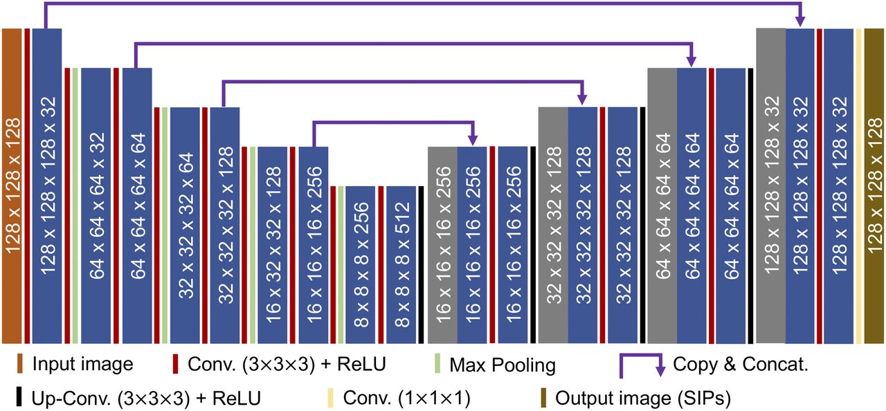
    Schematic illustration of CUSIP. Numbers indicate image size and number of features at each layer. Concat. = concatenate; Conv. = convolution; ReLU = rectified linear unit.

  • FIGURE 2.
    • Download figure
    • Open in new tab
    • Download powerpoint
    FIGURE 2.

    Comparison of acquired projections with corresponding SIPs in patients 1–4 from test group. Difference images display pixel value dissimilarities between acquired projections and SIPs. Blue indicates positive pixel values, white indicates no differences, and red indicates negative values. Unit of color bar is counts.

  • FIGURE 3.
    • Download figure
    • Open in new tab
    • Download powerpoint
    FIGURE 3.

    SPECT/CT reconstructions of Jaszczak phantom with 6 hot spheres having 25 times higher 177Lu activity concentration than background. AC-OSEM and ASCC-OSEM used 30 projections, 30–120SIPs, and 120 projections. Unit of color bar is arbitrary voxel values.

  • FIGURE 4.
    • Download figure
    • Open in new tab
    • Download powerpoint
    FIGURE 4.

    Recovery and SNR for 177Lu determined in various hot spheres in Jaszczak phantom for SPECT/CT AC-OSEM (A and C) and ASCC-OSEM (B and D) with 30 projections, 60 projections, 120 projections, 60–120SIPs, and 30–120SIPs.

  • FIGURE 5.
    • Download figure
    • Open in new tab
    • Download powerpoint
    FIGURE 5.

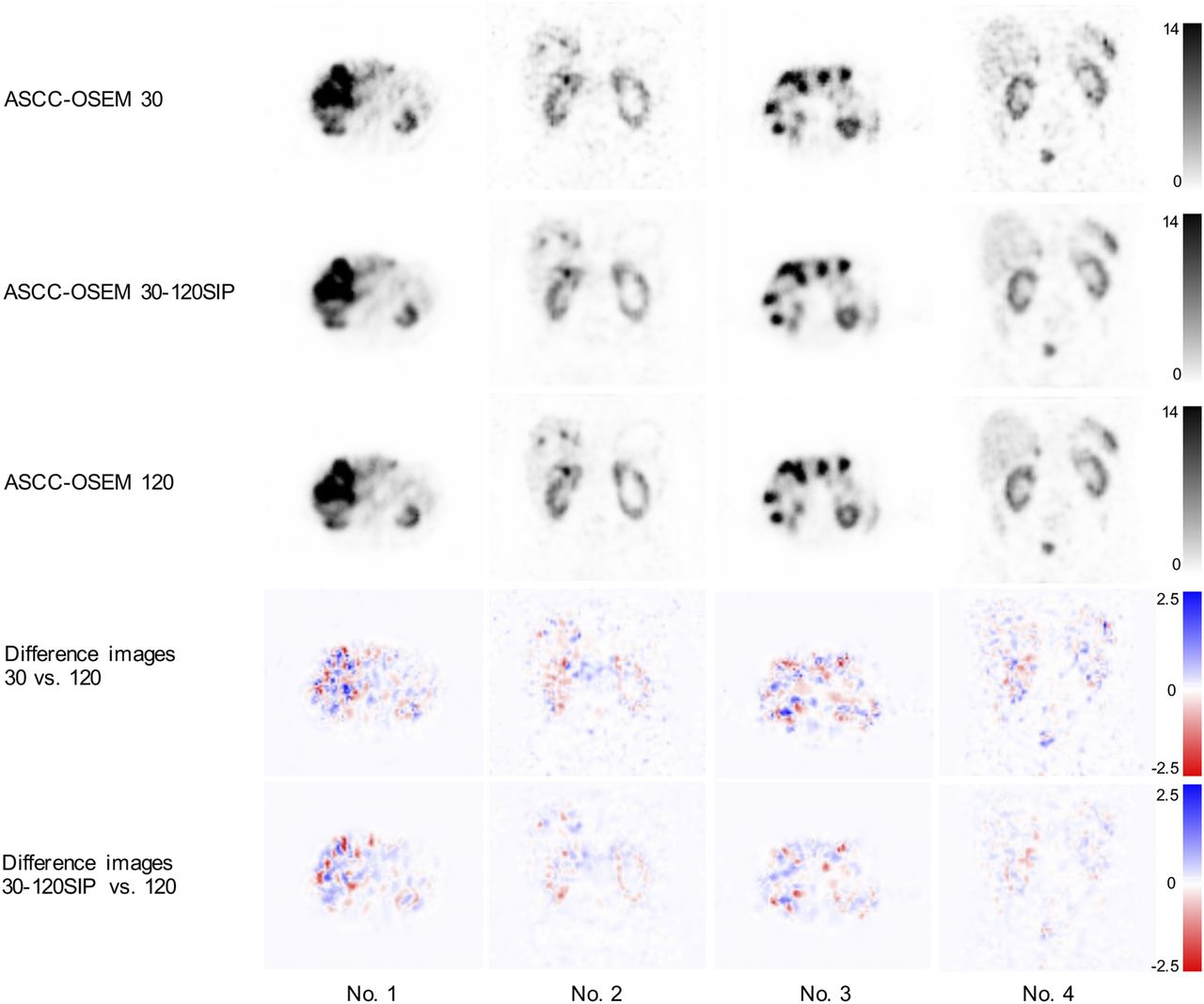
    Comparison of ASCC-OSEM with 30 projections, 30–120SIPs, and 120 projections in patients 1–4 of the 15 patients in test set. Difference images display pixel value dissimilarities between ASCC-OSEM 30 projections vs. ASCC-OSEM 120 projections and ASCC-OSEM 30–120SIPs vs. ASCC-OSEM 120 projections. Blue indicates positive pixel values, white indicates no differences, and red indicates negative values. Unit of color bar is arbitrary voxel values.

  • FIGURE 6.
    • Download figure
    • Open in new tab
    • Download powerpoint
    FIGURE 6.

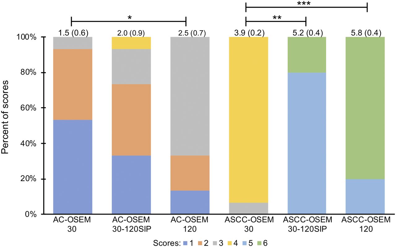
    Evaluation scores for SPECT/CT AC-OSEM and ASCC-OSEM. Mean scores and SDs are shown at top of bars. Asterisks indicate statistical significance of scores between projection sets within AC-OSEM and ASCC-OSEM. *0.01 ≤ P < 0.05. **0.001 ≤ P < 0.01. ***P < 0.001.
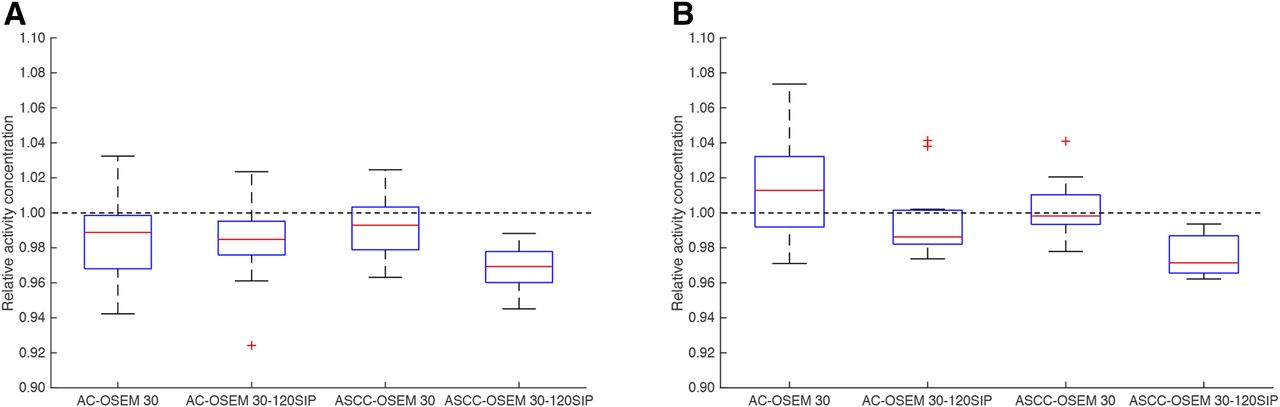
  • FIGURE 7.
    • Download figure
    • Open in new tab
    • Download powerpoint
    FIGURE 7.

    Relative kidney activity concentration for AC-OSEM and ASCC-OSEM with 30 and 30–120SIPs vs. 120 projections. Relative activity concentration was determined in left (A) and right (B) kidneys.

Tables

  • Figures
  • Additional Files
    • View popup
    TABLE 1

    Clinical SPECT Acquisition Protocols for 177Lu Dosimetry

    StudyTreatmentAcquisition time (min)Projections (n)Frame time (s)BPs (n)Measurement time after injection (h)
    Marin et al. (17)177Lu-DOTATATE21.3–42.76440–8014, 24, 144–192
    Sandström et al. (13)177Lu-DOTATATE30606011, 24, 96, 168
    Sandström et al. (14)177Lu-DOTATATE301203011, 24, 96, 168
    Hagmarker et al. (5)177Lu-DOTATATE3012030124
    Santoro et al. (15)177Lu-DOTATATE22.5604514, 24, 72, 192
    Garkavij et al. (16)177Lu-DOTATATE22.56045124/96
    Delker at al. (18)177Lu-PSMA-61721.312820124, 48, 72
    Kabasakal et al. (24)177Lu-PSMA20/BP9625224
    Hou et al. (19)177Lu-DOTATATE12–169615–2014, 24, 72
    Chicheportiche et al. (20)177Lu-DOTATATE156030120, 25, 168
    Beauregard et al. (23)177Lu-DOTATATE8–129610–1514, 24, 96
    Violet et al. (21)177Lu-PSMA-617(8–12)/BP9610–152–34, 24, 96
    Hippeläinen et al. (22)177Lu-DOTATATE10.76420124, 48, 168
    • BP = bed position; PSMA = prostate-specific membrane antigen.

    • View popup
    TABLE 2

    RMSE, PSNR, and SSIM for SIPs and SPECT Images in Test Group

    Image typeRMSEPSNRSSIM
    SIPs2.95 (0.77)39.2 (3.8)0.926 (0.061)
    AC-OSEM 30 projections0.147 (0.060)47.2 (3.5)0.989 (0.008)
    AC-OSEM 30–120SIPs0.109 (0.044)*49.5 (3.3)*0.993 (0.005)*
    ASCC-OSEM 30 projections0.259 (0.101)49.0 (3.5)0.993 (0.005)
    ASCC-OSEM 30GF projections0.273 (0.162)48.3 (2.5)0.995 (0.004)†
    ASCC-OSEM 30–120SIPs0.195 (0.091)*50.8 (3.2)*0.996 (0.003)*
    • ↵* P < 0.001.

    • ↵† P < 0.01.

    • GF = gaussian postfilter, SD of 4 mm.

    • Data in parentheses are SDs.

Additional Files

  • Figures
  • Tables
  • HTML Page - index.htslp

    Files in this Data Supplement:

    • Supplemental Data
PreviousNext
Back to top

In this issue

Journal of Nuclear Medicine: 62 (4)
Journal of Nuclear Medicine
Vol. 62, Issue 4
April 1, 2021
  • Table of Contents
  • Table of Contents (PDF)
  • About the Cover
  • Index by author
Print
Download PDF
Article Alerts
Sign In to Email Alerts with your Email Address
Email Article

Thank you for your interest in spreading the word on Journal of Nuclear Medicine.

NOTE: We only request your email address so that the person you are recommending the page to knows that you wanted them to see it, and that it is not junk mail. We do not capture any email address.

Enter multiple addresses on separate lines or separate them with commas.
Deep-Learning Generation of Synthetic Intermediate Projections Improves 177Lu SPECT Images Reconstructed with Sparsely Acquired Projections
(Your Name) has sent you a message from Journal of Nuclear Medicine
(Your Name) thought you would like to see the Journal of Nuclear Medicine web site.
Citation Tools
Deep-Learning Generation of Synthetic Intermediate Projections Improves 177Lu SPECT Images Reconstructed with Sparsely Acquired Projections
Tobias Rydén, Martijn Van Essen, Ida Marin, Johanna Svensson, Peter Bernhardt
Journal of Nuclear Medicine Apr 2021, 62 (4) 528-535; DOI: 10.2967/jnumed.120.245548

Citation Manager Formats

  • BibTeX
  • Bookends
  • EasyBib
  • EndNote (tagged)
  • EndNote 8 (xml)
  • Medlars
  • Mendeley
  • Papers
  • RefWorks Tagged
  • Ref Manager
  • RIS
  • Zotero
Share
Deep-Learning Generation of Synthetic Intermediate Projections Improves 177Lu SPECT Images Reconstructed with Sparsely Acquired Projections
Tobias Rydén, Martijn Van Essen, Ida Marin, Johanna Svensson, Peter Bernhardt
Journal of Nuclear Medicine Apr 2021, 62 (4) 528-535; DOI: 10.2967/jnumed.120.245548
Twitter logo Facebook logo LinkedIn logo Mendeley logo
  • Tweet Widget
  • Facebook Like
  • Google Plus One
Bookmark this article

Jump to section

  • Article
    • Visual Abstract
    • Abstract
    • MATERIALS AND METHODS
    • RESULTS
    • DISCUSSION
    • CONCLUSION
    • DISCLOSURE
    • Footnotes
    • REFERENCES
  • Figures & Data
  • Supplemental
  • Info & Metrics
  • PDF

Related Articles

  • This Month in JNM
  • PubMed
  • Google Scholar

Cited By...

  • Impact of the Reference Multiple-Time-Point Dosimetry Protocol on the Validity of Single-Time-Point Dosimetry for [177Lu]Lu-PSMA-I&T Therapy
  • A Deep-Learning-Based Partial-Volume Correction Method for Quantitative 177Lu SPECT/CT Imaging
  • A Pipeline for Automated Voxel Dosimetry: Application in Patients with Multi-SPECT/CT Imaging After 177Lu-Peptide Receptor Radionuclide Therapy
  • Dosimetry for Radiopharmaceutical Therapy: The European Perspective
  • Google Scholar

More in this TOC Section

AI/Advanced Image Analysis

  • Ultrashort Oncologic Whole-Body [18F]FDG Patlak Imaging Using LAFOV PET
  • Need for Objective Task-Based Evaluation of Image Segmentation Algorithms for Quantitative PET: A Study with ACRIN 6668/RTOG 0235 Multicenter Clinical Trial Data
Show more AI/Advanced Image Analysis

Clinical

  • Dual PET Imaging in Bronchial Neuroendocrine Neoplasms: The NETPET Score as a Prognostic Biomarker
  • Addition of 131I-MIBG to PRRT (90Y-DOTATOC) for Personalized Treatment of Selected Patients with Neuroendocrine Tumors
  • SUVs Are Adequate Measures of Lesional 18F-DCFPyL Uptake in Patients with Low Prostate Cancer Disease Burden
Show more Clinical

Similar Articles

Keywords

  • dosimetry
  • 177Lu
  • deep-learning
  • SPECT
SNMMI

© 2025 SNMMI

Powered by HighWire