


Article Figures & Data
Figures
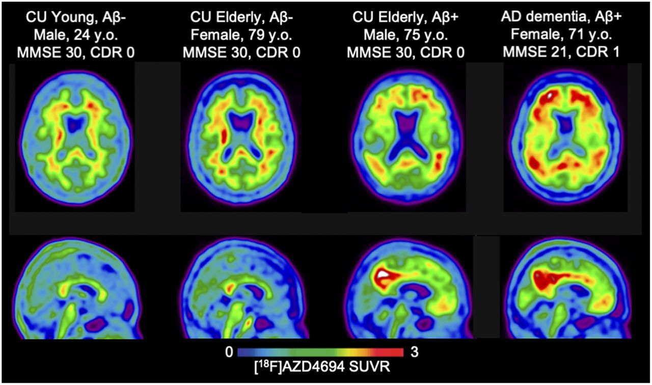
- FIGURE 1.
Transaxial (top) and midsagittal (bottom) representative 18F-AZD4694 SUVR PET images of 4 subjects representing range of binding patterns in present study. All images are presented in template space. MNI coordinates: x = 2, y = −59, z = 15. CDR = Clinical Dementia Rating; MMSE = Mini-Mental State Examination.
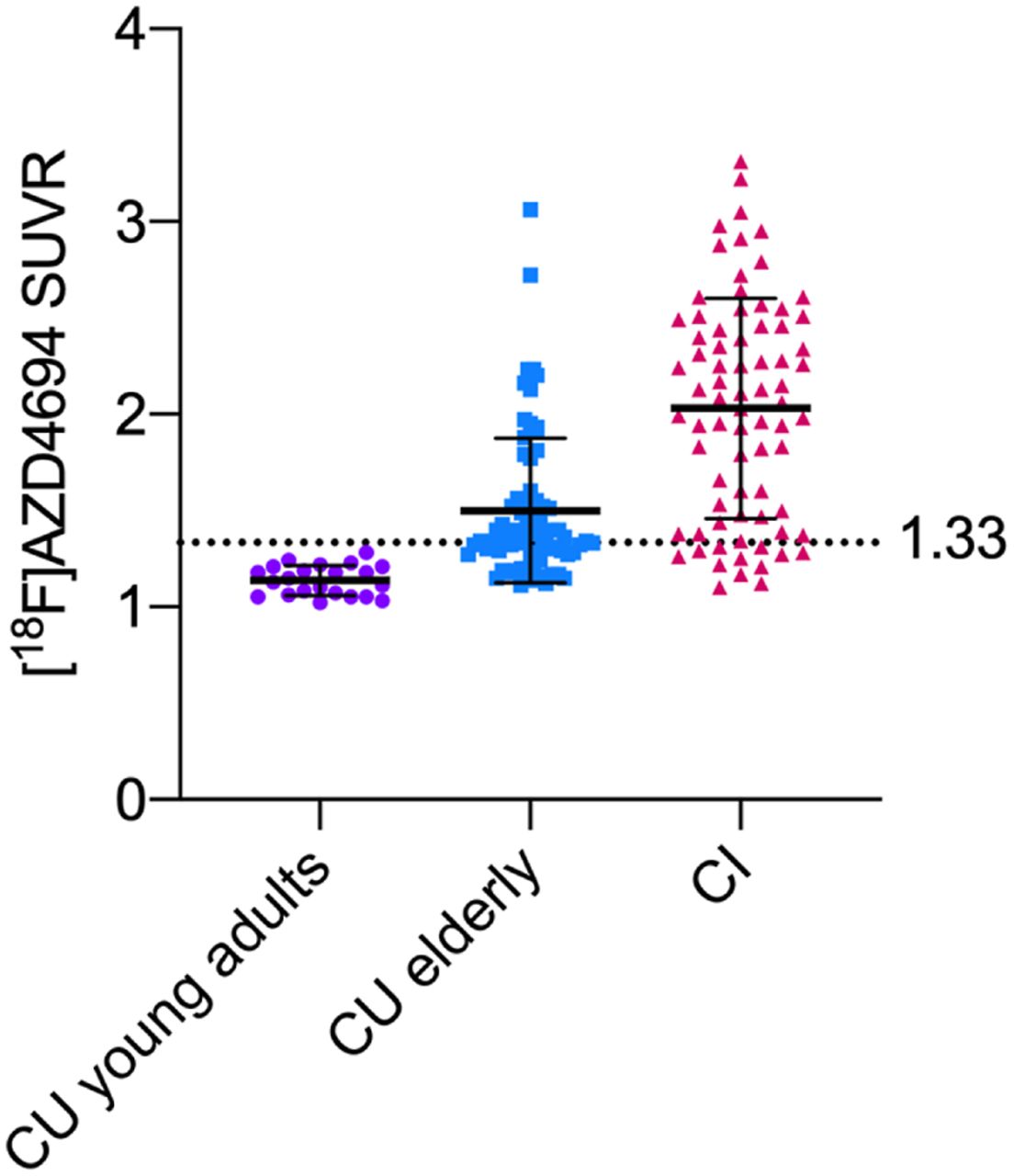
- FIGURE 2.
Means and SDs (error bars) in 18F-AZD4694 PET SUVR for CU young adults (age < 25 y), CU elderly, and CI groups. Young adults displayed minimal amyloid PET uptake (mean, 1.14; SD, 0.09). Dashed line represents 2 SDs above mean of young adults, at 1.33 18F-AZD4694 SUVR.
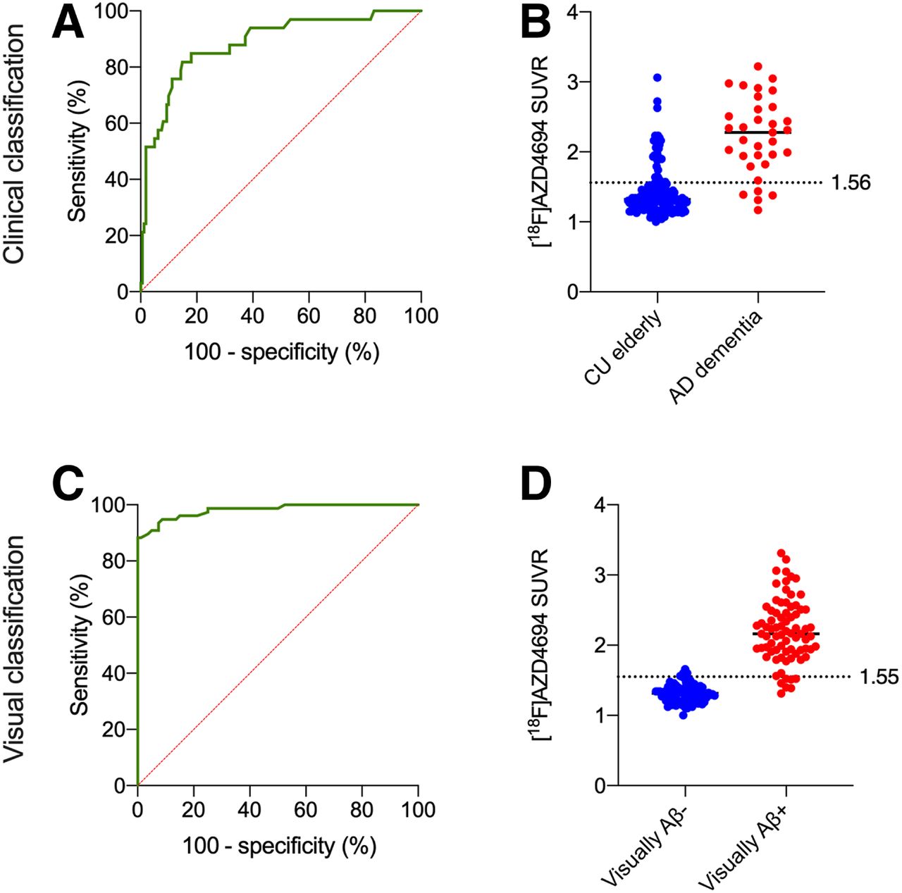
- FIGURE 3.
ROC curves contrasting visually positive vs. negative cases. (A) When contrasting CU elderly with AD dementia groups, we observed good AUC (82.5%; sensitivity, 85%; specificity, 73%). (B) Optimal threshold at this point was 1.56 SUVR, represented by dashed line. (C) Area under ROC curve contrasting visually negative vs. visually positive cases displayed excellent AUC (97%; sensitivity, 90.91%; specificity, 95%). (D) 18F-AZD4694 PET means are shown for visually positive (red) and visually negative (blue) groups, with dashed line representing optimal threshold derived from ROC curve (1.55 SUVR).
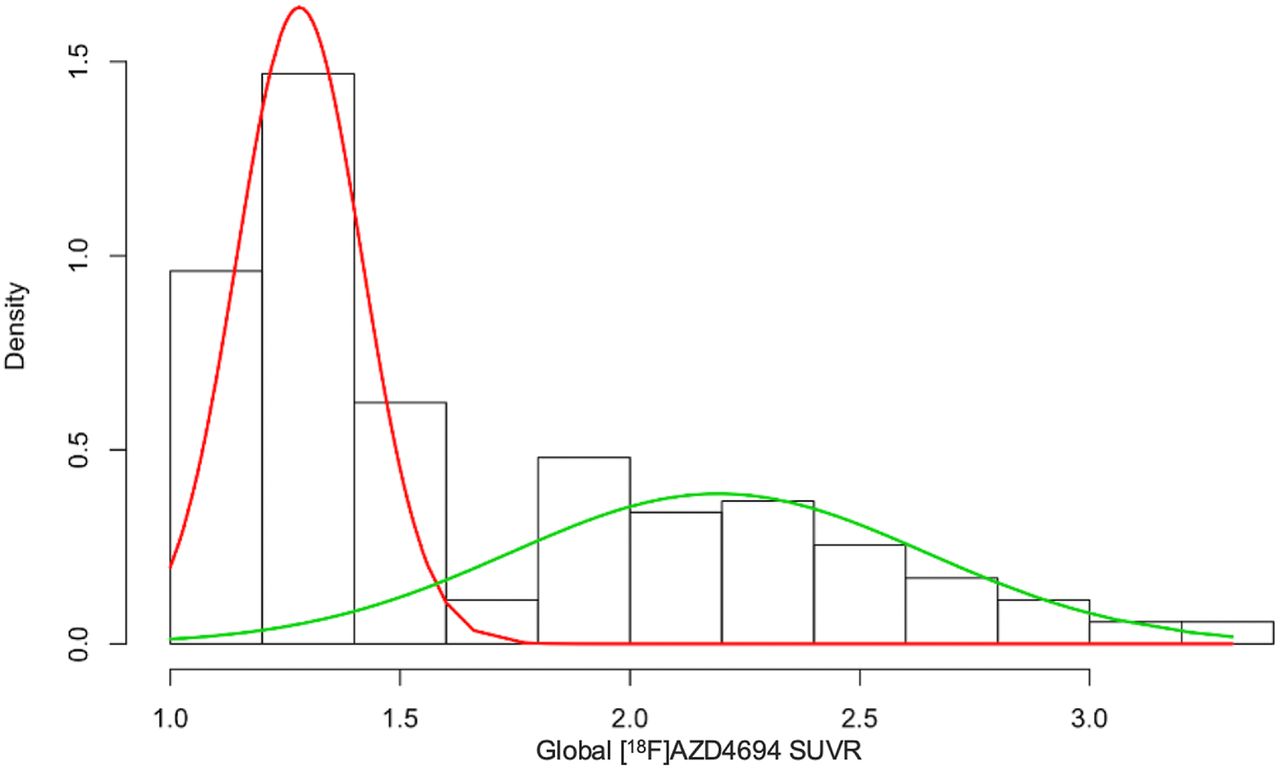
- FIGURE 4.
Gaussian mixture modeling representing 2 distributions. Low 18F-AZD4694 (red) and high 18F-AZD4694 (green) gaussian distributions are superimposed on subject density histogram for all 18F-AZD4694 PET SUVRs from CU elderly and CI populations. Optimal cut point from gaussian mixture modeling was 1.55 SUVR.
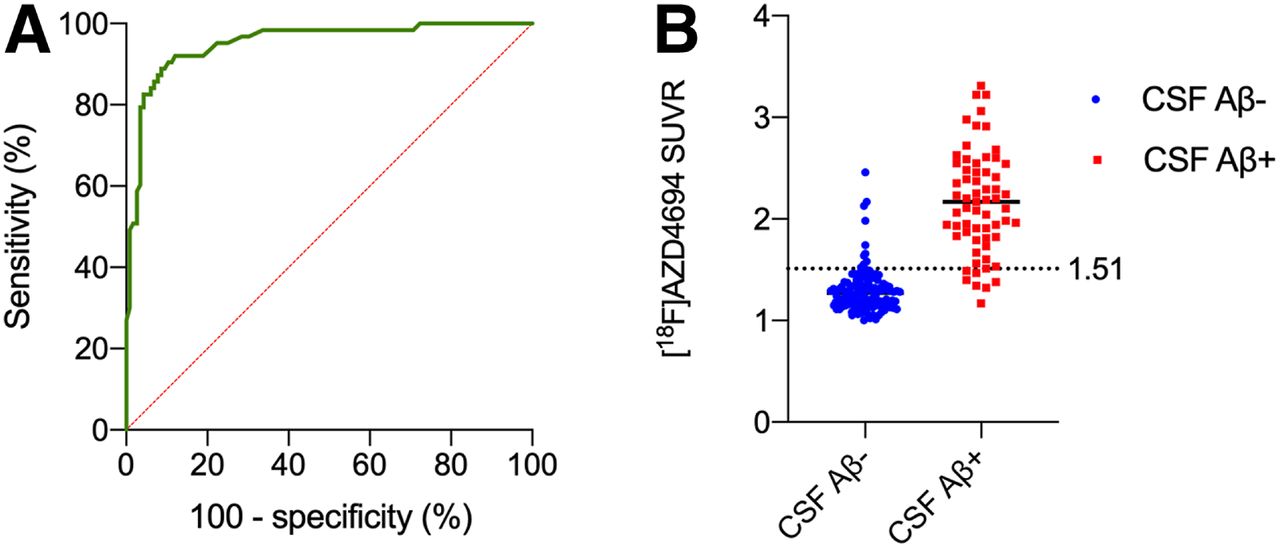
- FIGURE 5.
ROC curves contrasting CSF-positive vs. -negative individuals. (A) Area under ROC curve contrasting individuals dichotomized on basis of their cerebrospinal measure of Aβ42/Aβ40 ratio. This method resulted in area under ROC curve of 95% (sensitivity, 88.9%; specificity, 91.4%). (B) AZD4694 PET means are shown for CSF-negative (blue) and CSF-positive (red) individuals, with dashed line representing optimal threshold derived from ROC curve (1.51 SUVR).


Tables
P* Characteristic CU young CU elderly CI CU youngvs.CU elderly CU elderlyvs.CI Total patients (n) 22 89 65 — — Mean age (y) 22.7 (SD, 1.3) 72.33 (SD, 5.88) 67.91 (SD, 8.97) <0.0001 0.0004 Female (n) 14 (63%) 51 (57%) 36 (55%) 0.59 0.81 Mean education (y) 16.61 (SD, 1.33) 15.06 (SD, 3.81) 15.1 (SD, 3.34) 0.06 0.94 APOEε4 carriers (n) 6 (27%) 33 (37%) 41 (63%) 0.58 <0.0001 Mean MMSE 29.77 (SD, 0.53) 29.12 (SD, 1.07) 24.03 (SD, 6.07) 0.009 <0.0001 Mean neocortical 18F-AZD4694 SUVR 1.14 (SD, 0.09) 1.48 (0.38) 2.04 (SD, 0.57) <0.0001 <0.0001 Mean CSF Aβ42/Aβ40 0.09 (SD, 0.006) 0.07 (SD, 0.02) 0.05 (SD, 0.02) <0.0001 <0.0001 ↵* Assessed with 2-sided independent-samples t tests for each variable except sex and APOEε4 status, for which contingency χ2 tests were performed.
MMSE = Mini-Mental State Examination.
Supplemental Data
Files in this Data Supplement:


Jump to section
Related Articles
Cited By...
- Transcriptomic signatures of A{beta}- and tau-induced neuronal dysfunction reveal inflammatory processes at the core of Alzheimers disease pathophysiology
- Diagnostic accuracy of the plasma ALZpath pTau217 immunoassay to identify Alzheimers disease pathology
- The Association of Age-Related and Off-Target Retention with Longitudinal Quantification of [18F]MK6240 Tau PET in Target Regions
- Revealing the combined roles of A{beta} and tau in Alzheimers disease via a pathophysiological activity decoder
- Performance of plasma amyloid, tau, and astrocyte biomarkers to identify cerebral AD pathophysiology
- Astrocyte biomarker signatures of amyloid-{beta} and tau pathologies in Alzheimers disease