
Main menu

  • Home
  • Content
    • Current
    • Ahead of print
    • Past Issues
    • JNM Supplement
    • SNMMI Annual Meeting Abstracts
    • Continuing Education
    • JNM Podcasts
  • Subscriptions
    • Subscribers
    • Institutional and Non-member
    • Rates
    • Journal Claims
    • Corporate & Special Sales
  • Authors
    • Submit to JNM
    • Information for Authors
    • Assignment of Copyright
    • AQARA requirements
  • Info
    • Reviewers
    • Permissions
    • Advertisers
  • About
    • About Us
    • Editorial Board
    • Contact Information
  • More
    • Alerts
    • Feedback
    • Help
    • SNMMI Journals
  • SNMMI
    • JNM
    • JNMT
    • SNMMI Journals
    • SNMMI

User menu

  • Subscribe
  • My alerts
  • Log in
  • Log out
  • My Cart

Search

  • Advanced search
Journal of Nuclear Medicine
  • SNMMI
    • JNM
    • JNMT
    • SNMMI Journals
    • SNMMI
  • Subscribe
  • My alerts
  • Log in
  • Log out
  • My Cart
Journal of Nuclear Medicine

Advanced Search

  • Home
  • Content
    • Current
    • Ahead of print
    • Past Issues
    • JNM Supplement
    • SNMMI Annual Meeting Abstracts
    • Continuing Education
    • JNM Podcasts
  • Subscriptions
    • Subscribers
    • Institutional and Non-member
    • Rates
    • Journal Claims
    • Corporate & Special Sales
  • Authors
    • Submit to JNM
    • Information for Authors
    • Assignment of Copyright
    • AQARA requirements
  • Info
    • Reviewers
    • Permissions
    • Advertisers
  • About
    • About Us
    • Editorial Board
    • Contact Information
  • More
    • Alerts
    • Feedback
    • Help
    • SNMMI Journals
  • View or Listen to JNM Podcast
  • Visit JNM on Facebook
  • Join JNM on LinkedIn
  • Follow JNM on Twitter
  • Subscribe to our RSS feeds
Research ArticleTheranostics

68Ga-PSMA PET/CT for Primary Lymph Node and Distant Metastasis NM Staging of High-Risk Prostate Cancer

Søren Klingenberg, Mads R. Jochumsen, Benedicte P. Ulhøi, Jacob Fredsøe, Karina D. Sørensen, Michael Borre and Kirsten Bouchelouche
Journal of Nuclear Medicine February 2021, 62 (2) 214-220; DOI: https://doi.org/10.2967/jnumed.120.245605
Søren Klingenberg
1Department of Nuclear Medicine and PET Centre, Aarhus University Hospital, Aarhus, Denmark
2Department of Clinical Medicine, Aarhus University, Aarhus, Denmark
3Department of Molecular Medicine, Aarhus University Hospital, Aarhus, Denmark
  • Find this author on Google Scholar
  • Find this author on PubMed
  • Search for this author on this site
Mads R. Jochumsen
1Department of Nuclear Medicine and PET Centre, Aarhus University Hospital, Aarhus, Denmark
2Department of Clinical Medicine, Aarhus University, Aarhus, Denmark
  • Find this author on Google Scholar
  • Find this author on PubMed
  • Search for this author on this site
Benedicte P. Ulhøi
4Department of Pathology, Aarhus University Hospital, Aarhus, Denmark; and
  • Find this author on Google Scholar
  • Find this author on PubMed
  • Search for this author on this site
Jacob Fredsøe
2Department of Clinical Medicine, Aarhus University, Aarhus, Denmark
3Department of Molecular Medicine, Aarhus University Hospital, Aarhus, Denmark
  • Find this author on Google Scholar
  • Find this author on PubMed
  • Search for this author on this site
Karina D. Sørensen
2Department of Clinical Medicine, Aarhus University, Aarhus, Denmark
3Department of Molecular Medicine, Aarhus University Hospital, Aarhus, Denmark
  • Find this author on Google Scholar
  • Find this author on PubMed
  • Search for this author on this site
Michael Borre
2Department of Clinical Medicine, Aarhus University, Aarhus, Denmark
5Department of Urology, Aarhus University Hospital, Aarhus, Denmark
  • Find this author on Google Scholar
  • Find this author on PubMed
  • Search for this author on this site
Kirsten Bouchelouche
1Department of Nuclear Medicine and PET Centre, Aarhus University Hospital, Aarhus, Denmark
2Department of Clinical Medicine, Aarhus University, Aarhus, Denmark
  • Find this author on Google Scholar
  • Find this author on PubMed
  • Search for this author on this site
  • Article
  • Figures & Data
  • Info & Metrics
  • PDF
Loading

Article Figures & Data

Figures

  • Tables
  • FIGURE 1.
    • Download figure
    • Open in new tab
    • Download powerpoint
    FIGURE 1.

    Study flow for 68Ga-PSMA PET/CT primary staging of high-risk PCa patients.
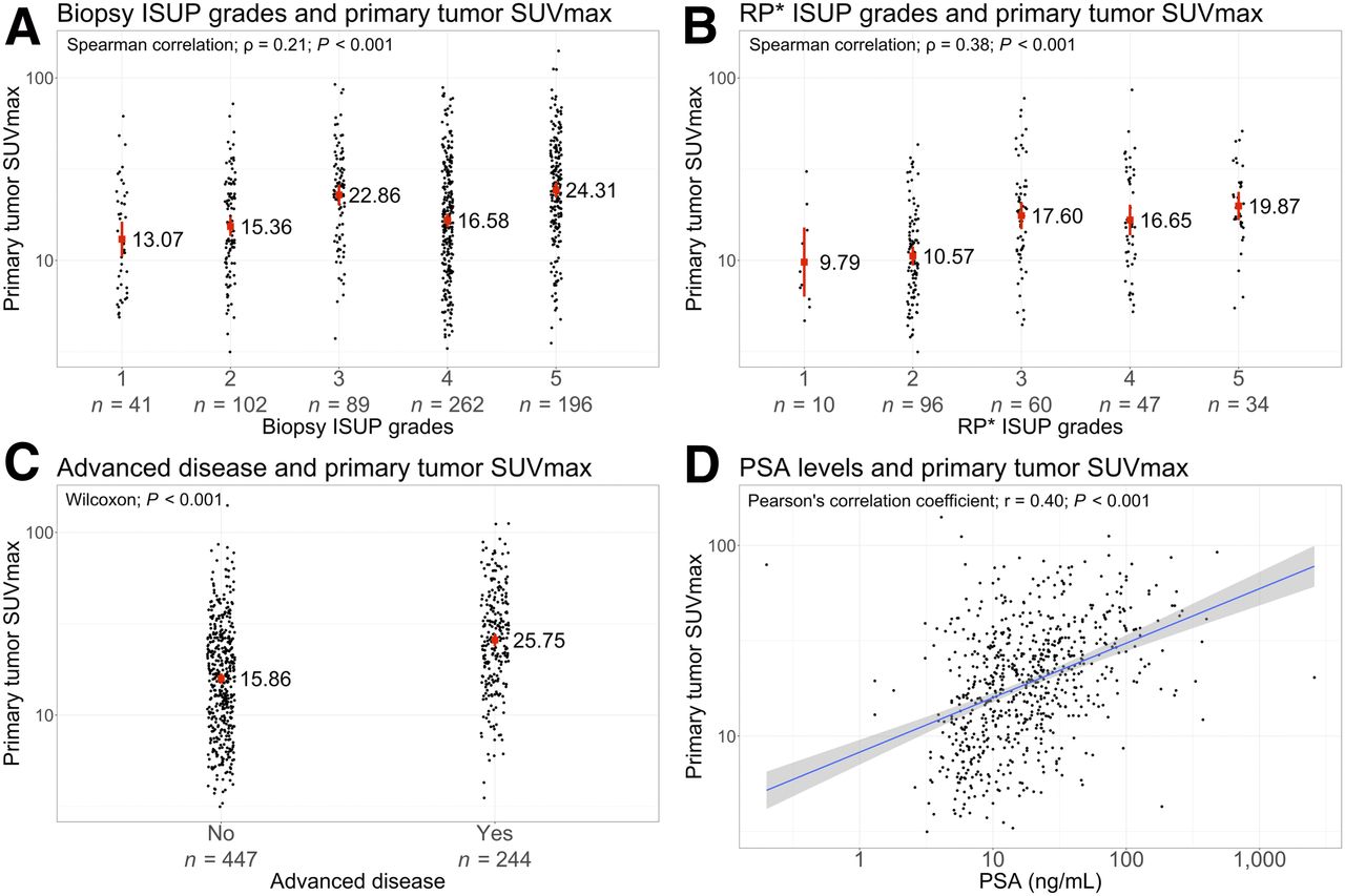
  • FIGURE 2.
    • Download figure
    • Open in new tab
    • Download powerpoint
    FIGURE 2.

    (A) Correlation between SUVmax of primary tumors and ISUP grade from transrectal ultrasound–guided or MRI-guided biopsy. Data are median. (B) Correlation between SUVmax of primary tumors and RP ISUP grade. Data are median. (C) Primary tumor SUVmax in correlation with advanced disease (N1/M1). Data are median. (D) Correlation between SUVmax of primary tumors and PSA at diagnosis. Data are line of best fit with 95% CI. *Radical prostatectomy.
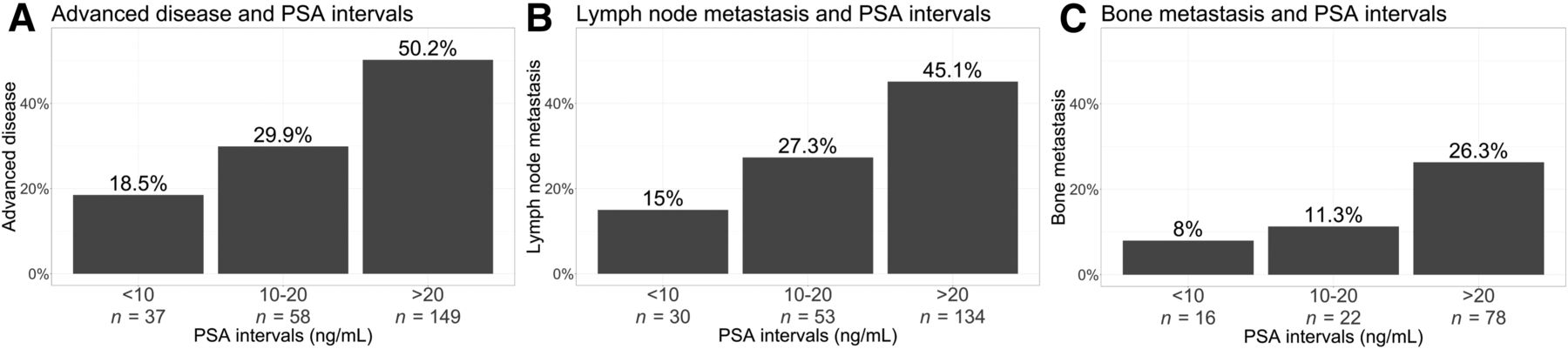
  • FIGURE 3.
    • Download figure
    • Open in new tab
    • Download powerpoint
    FIGURE 3.

    Occurrence of advanced disease (N1/M1) (A), LNMs (N1/M1a) (B), and BMs (M1b) (C) according to PSA interval.
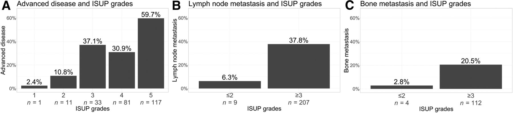
  • FIGURE 4.
    • Download figure
    • Open in new tab
    • Download powerpoint
    FIGURE 4.

    Incidence of advanced disease (N1/M1) (A), LNMs (N1/M1a) (B), and BMs (M1b) (C) according to ISUP grade.

  • FIGURE 5.
    • Download figure
    • Open in new tab
    • Download powerpoint
    FIGURE 5.

    Presence of advanced disease (N1/M1) according to clinical stage.
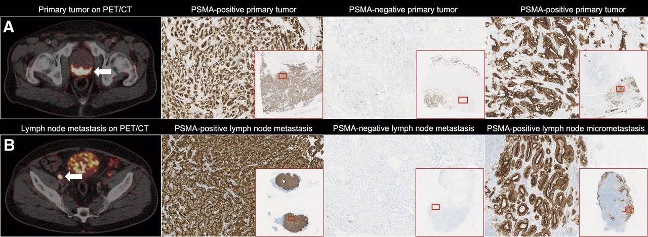
  • FIGURE 6.
    • Download figure
    • Open in new tab
    • Download powerpoint
    FIGURE 6.

    (A) Primary prostate tumors. From left: 68Ga-PSMA PET/CT (arrow), PSMA-positive immunohistochemical staining of primary tumor from previous image, PSMA-negative immunohistochemical staining of primary tumor, and PSMA-positive immunohistochemical staining of primary tumor. (B) LNMs from corresponding primary tumors in A. From left: LNMs on 68Ga-PSMA PET/CT (arrow), PSMA-positive immunohistochemical staining of true-positive LNMs from previous image, PSMA-negative immunohistochemical staining of undetected LNMs, and PSMA-positive immunohistochemical staining of undetected LN micrometastasis, characteristically located in the border of the LN cortex.

Tables

  • Figures
    • View popup
    TABLE 1

    Study Group Characteristics and Odds Ratios (ORs) for Having Advanced Disease Compared with Reference

    CharacteristicStudy group (n = 691)Advanced disease on 68Ga-PSMA PET/CTUnivariate ORs of advanced diseaseMultivariate ORs of advanced disease
    Age (y)
     Median70.4
     Range45.2–87.2
    Time from biopsy to scan (mo)
     Median0.7
     Range0.1–5.1
    Prescan PSA (ng/mL)
     <10200 (28.9%)37 (18.5%)Reference = 1Reference = 1
     10–20194 (28.1%)58 (29.9%)OR1 = 1.88 (1.17, 3.01)OR1 = 1.45 (0.86, 2.47)
     >20297 (43.0%)149 (50.2%)OR2 = 4.44 (2.90, 6.77)OR2 = 4.32 (2.66, 7.12)
     Unknown0 (0.0%)
    ISUP grade in biopsies
     141 (5.9%)1 (2.4%)Reference = 1Reference = 1
     2102 (14.8%)11 (10.8%)OR1 = 4.84 (0.89, 89.87)OR1 = 3.75 (0.67, 70.51)
     389 (12.9%)33 (37.1%)OR2 = 23.57 (4.76, 427.76)OR2 = 19.67 (3.84, 361.15)
     4262 (38.0%)81 (30.9%)OR3 = 17.90 (3.79, 320.27)OR3 = 25.55 (5.21, 462.70)
     5196 (28.4%)117 (59.7%)OR4 = 59.24 (12.47, 1061.62)OR4 = 53.17 (10.79, 964.19)
     Unknown1 (0.1%)
    Clinical stage*
     cT1123 (17.8%)13 (10.6%)Reference = 1Reference = 1
     cT2246 (35.6%)60 (24.4%)OR1 = 2.73 (1.47, 5.40)OR1 = 2.18 (1.13, 4.45)
     cT3314 (45.4%)165 (52.5%)OR2 = 9.37 (5.23, 18.11)OR2 = 7.13 (3.80, 14.29)
     cT48 (1.2%)6 (75.0%)OR3 = 25.38 (5.25, 186.23)OR3 = 17.26 (2.82, 160.69)
     Unknown0 (0.0%)
    • ↵* Digital rectal examination.

    • Qualitative data are numbers followed by percentages in parentheses; continuous data are OR followed by 95% CI in parentheses.

    • View popup
    TABLE 2

    Primary Tumor SUVmax with 95% CI in Relation to ISUP Grade and Advanced Disease

    ParameterSUVmax in primary tumors
    ISUP grade in biopsies
     1 (n = 41)13.07 (10.48, 16.31)
     2 (n = 102)15.36 (13.59, 17.36)
     3 (n = 89)22.86 (20.04, 26.07)
     4 (n = 262)16.58 (15.20, 18.08)
     5 (n = 196)24.31 (22.09, 26.75)
    ISUP grade in RP specimens
     1 (n = 10)9.79 (6.33, 15.15)
     2 (n = 96)10.57 (9.38, 11.92)
     3 (n = 60)17.60 (14.84, 20.87)
     4 (n = 47)16.65 (13.72, 20.20)
     5 (n = 34)19.87 (16.62, 23.75)
    Advanced disease
     No (n = 447)15.86 (14.87, 16.91)
     Yes (n = 244)25.75 (23.76, 27.91)
    • Data are median followed by 95% CI in parentheses.

    • View popup
    TABLE 3

    SUVmax of Different Lesion Sites

    Lesion siteLesions measured (n)Median SUVmax
    Primary tumor69119.5 (range, 3.1–140.5)
    LNs (regional and nonregional)1,22221.1 (range, 2.2–136.3)
    BMs38617.3 (range, 1.8–137.9)
    Visceral metastases408.1 (range, 3.2–48.0)
    • View popup
    TABLE 4

    Histopathologic Coherence Between PSMA-Positive or -Negative LNs and Pathologic Verification in Patients with PLND

    Histology (n = 177)
    68Ga-PSMA PET/CTPositive (n = 36)Negative (n = 141)Index
    Positive (n = 16)n = 11 (30.6%)n = 5 (3.5%)PPV 68.8%
    Negative (n = 161)n = 25 (69.4%)n = 136 (96.5%)NPV 84.5%
    IndexSensitivity 30.6%Specificity 96.5%Accuracy 83.1%
    • PPV and NPV = positive and negative predictive values, respectively.

PreviousNext
Back to top

In this issue

Journal of Nuclear Medicine: 62 (2)
Journal of Nuclear Medicine
Vol. 62, Issue 2
February 1, 2021
  • Table of Contents
  • Table of Contents (PDF)
  • About the Cover
  • Index by author
Print
Download PDF
Article Alerts
Sign In to Email Alerts with your Email Address
Email Article

Thank you for your interest in spreading the word on Journal of Nuclear Medicine.

NOTE: We only request your email address so that the person you are recommending the page to knows that you wanted them to see it, and that it is not junk mail. We do not capture any email address.

Enter multiple addresses on separate lines or separate them with commas.
68Ga-PSMA PET/CT for Primary Lymph Node and Distant Metastasis NM Staging of High-Risk Prostate Cancer
(Your Name) has sent you a message from Journal of Nuclear Medicine
(Your Name) thought you would like to see the Journal of Nuclear Medicine web site.
Citation Tools
68Ga-PSMA PET/CT for Primary Lymph Node and Distant Metastasis NM Staging of High-Risk Prostate Cancer
Søren Klingenberg, Mads R. Jochumsen, Benedicte P. Ulhøi, Jacob Fredsøe, Karina D. Sørensen, Michael Borre, Kirsten Bouchelouche
Journal of Nuclear Medicine Feb 2021, 62 (2) 214-220; DOI: 10.2967/jnumed.120.245605

Citation Manager Formats

  • BibTeX
  • Bookends
  • EasyBib
  • EndNote (tagged)
  • EndNote 8 (xml)
  • Medlars
  • Mendeley
  • Papers
  • RefWorks Tagged
  • Ref Manager
  • RIS
  • Zotero
Share
68Ga-PSMA PET/CT for Primary Lymph Node and Distant Metastasis NM Staging of High-Risk Prostate Cancer
Søren Klingenberg, Mads R. Jochumsen, Benedicte P. Ulhøi, Jacob Fredsøe, Karina D. Sørensen, Michael Borre, Kirsten Bouchelouche
Journal of Nuclear Medicine Feb 2021, 62 (2) 214-220; DOI: 10.2967/jnumed.120.245605
Twitter logo Facebook logo LinkedIn logo Mendeley logo
  • Tweet Widget
  • Facebook Like
  • Google Plus One
Bookmark this article

Jump to section

  • Article
    • Abstract
    • MATERIALS AND METHODS
    • RESULTS
    • DISCUSSION
    • CONCLUSION
    • DISCLOSURE
    • Footnotes
    • REFERENCES
  • Figures & Data
  • Info & Metrics
  • PDF

Related Articles

  • This Month in JNM
  • PubMed
  • Google Scholar

Cited By...

  • Considerations Surrounding the Sentinel Lymph Node in Prostate Cancer and Unanswered Questions
  • Reply: Considerations Surrounding the Sentinel Lymph Node in Prostate Cancer and Unanswered Questions
  • Diagnostic Performance of [18F]AlF-Thretide PET/CT in Patients with Newly Diagnosed Prostate Cancer Using Histopathology as Reference Standard
  • 68Ga-PSMA-11 PET/MRI in Patients with Newly Diagnosed Intermediate- or High-Risk Prostate Adenocarcinoma: PET Findings Correlate with Outcomes After Definitive Treatment
  • Utility of 18F-rhPSMA-7.3 PET for Imaging of Primary Prostate Cancer and Preoperative Efficacy in N-Staging of Unfavorable Intermediate- to Very High-Risk Patients Validated by Histopathology
  • Head-to-Head Comparison of 68Ga-Prostate-Specific Membrane Antigen PET/CT and Ferumoxtran-10-Enhanced MRI for the Diagnosis of Lymph Node Metastases in Prostate Cancer Patients
  • Google Scholar

More in this TOC Section

Theranostics

  • Determination of the Intralesional Distribution of Theranostic 124I-Omburtamab Convection-Enhanced Delivery in Treatment of Diffuse Intrinsic Pontine Glioma
  • Evidence-Based Clinical Protocols to Monitor Efficacy of [177Lu]Lu-PSMA Radiopharmaceutical Therapy in Metastatic Castration-Resistant Prostate Cancer Using Real-World Data
  • 177Lu-Labeled Anticlaudin 6 Monoclonal Antibody for Targeted Therapy in Esophageal Cancer
Show more Theranostics

Clinical

  • Dual PET Imaging in Bronchial Neuroendocrine Neoplasms: The NETPET Score as a Prognostic Biomarker
  • Addition of 131I-MIBG to PRRT (90Y-DOTATOC) for Personalized Treatment of Selected Patients with Neuroendocrine Tumors
  • SUVs Are Adequate Measures of Lesional 18F-DCFPyL Uptake in Patients with Low Prostate Cancer Disease Burden
Show more Clinical

Similar Articles

Keywords

  • prostate cancer
  • 68Ga-prostate-specific membrane antigen PET/CT
  • 68Ga-PSMA
  • primary staging
  • high-risk
SNMMI

© 2025 SNMMI

Powered by HighWire