
Main menu

  • Home
  • Content
    • Current
    • Ahead of print
    • Past Issues
    • JNM Supplement
    • SNMMI Annual Meeting Abstracts
    • Continuing Education
    • JNM Podcasts
  • Subscriptions
    • Subscribers
    • Institutional and Non-member
    • Rates
    • Journal Claims
    • Corporate & Special Sales
  • Authors
    • Submit to JNM
    • Information for Authors
    • Assignment of Copyright
    • AQARA requirements
  • Info
    • Reviewers
    • Permissions
    • Advertisers
  • About
    • About Us
    • Editorial Board
    • Contact Information
  • More
    • Alerts
    • Feedback
    • Help
    • SNMMI Journals
  • SNMMI
    • JNM
    • JNMT
    • SNMMI Journals
    • SNMMI

User menu

  • Subscribe
  • My alerts
  • Log in
  • My Cart

Search

  • Advanced search
Journal of Nuclear Medicine
  • SNMMI
    • JNM
    • JNMT
    • SNMMI Journals
    • SNMMI
  • Subscribe
  • My alerts
  • Log in
  • My Cart
Journal of Nuclear Medicine

Advanced Search

  • Home
  • Content
    • Current
    • Ahead of print
    • Past Issues
    • JNM Supplement
    • SNMMI Annual Meeting Abstracts
    • Continuing Education
    • JNM Podcasts
  • Subscriptions
    • Subscribers
    • Institutional and Non-member
    • Rates
    • Journal Claims
    • Corporate & Special Sales
  • Authors
    • Submit to JNM
    • Information for Authors
    • Assignment of Copyright
    • AQARA requirements
  • Info
    • Reviewers
    • Permissions
    • Advertisers
  • About
    • About Us
    • Editorial Board
    • Contact Information
  • More
    • Alerts
    • Feedback
    • Help
    • SNMMI Journals
  • View or Listen to JNM Podcast
  • Visit JNM on Facebook
  • Join JNM on LinkedIn
  • Follow JNM on Twitter
  • Subscribe to our RSS feeds
Research ArticleOncology

18F-FET PET Imaging in Differentiating Glioma Progression from Treatment-Related Changes: A Single-Center Experience

Gabriele D. Maurer, Daniel P. Brucker, Gabriele Stoffels, Katharina Filipski, Christian P. Filss, Felix M. Mottaghy, Norbert Galldiks, Joachim P. Steinbach, Elke Hattingen and Karl-Josef Langen
Journal of Nuclear Medicine April 2020, 61 (4) 505-511; DOI: https://doi.org/10.2967/jnumed.119.234757
Gabriele D. Maurer
1Dr. Senckenberg Institute of Neurooncology, Goethe University Hospital, Frankfurt am Main, Germany
2University Cancer Center Frankfurt, Goethe University Hospital, Frankfurt am Main, Germany
3German Cancer Consortium (DKTK), Partner Site Frankfurt/Mainz, Heidelberg, Germany
  • Find this author on Google Scholar
  • Find this author on PubMed
  • Search for this author on this site
Daniel P. Brucker
2University Cancer Center Frankfurt, Goethe University Hospital, Frankfurt am Main, Germany
3German Cancer Consortium (DKTK), Partner Site Frankfurt/Mainz, Heidelberg, Germany
  • Find this author on Google Scholar
  • Find this author on PubMed
  • Search for this author on this site
Gabriele Stoffels
4Institute of Neuroscience and Medicine (INM-3 and INM-4), Research Center Juelich, Juelich, Germany
  • Find this author on Google Scholar
  • Find this author on PubMed
  • Search for this author on this site
Katharina Filipski
3German Cancer Consortium (DKTK), Partner Site Frankfurt/Mainz, Heidelberg, Germany
5German Cancer Research Center (DKFZ), Heidelberg, Germany
6Institute of Neurology (Edinger Institute), Goethe University Hospital, Frankfurt am Main, Germany
  • Find this author on Google Scholar
  • Find this author on PubMed
  • Search for this author on this site
Christian P. Filss
4Institute of Neuroscience and Medicine (INM-3 and INM-4), Research Center Juelich, Juelich, Germany
7Department of Nuclear Medicine, RWTH University Hospital, Aachen, Germany
  • Find this author on Google Scholar
  • Find this author on PubMed
  • Search for this author on this site
Felix M. Mottaghy
7Department of Nuclear Medicine, RWTH University Hospital, Aachen, Germany
8Department of Radiology and Nuclear Medicine, Maastricht University Medical Center, Maastricht, The Netherlands
9Center of Integrated Oncology, Universities of Aachen, Bonn, Cologne, and Duesseldorf, Germany
  • Find this author on Google Scholar
  • Find this author on PubMed
  • Search for this author on this site
Norbert Galldiks
4Institute of Neuroscience and Medicine (INM-3 and INM-4), Research Center Juelich, Juelich, Germany
9Center of Integrated Oncology, Universities of Aachen, Bonn, Cologne, and Duesseldorf, Germany
10Department of Neurology, University Hospital Cologne, Cologne, Germany; and
  • Find this author on Google Scholar
  • Find this author on PubMed
  • Search for this author on this site
Joachim P. Steinbach
1Dr. Senckenberg Institute of Neurooncology, Goethe University Hospital, Frankfurt am Main, Germany
2University Cancer Center Frankfurt, Goethe University Hospital, Frankfurt am Main, Germany
3German Cancer Consortium (DKTK), Partner Site Frankfurt/Mainz, Heidelberg, Germany
  • Find this author on Google Scholar
  • Find this author on PubMed
  • Search for this author on this site
Elke Hattingen
11Institute of Neuroradiology, Goethe University Hospital, Frankfurt am Main, Germany
  • Find this author on Google Scholar
  • Find this author on PubMed
  • Search for this author on this site
Karl-Josef Langen
4Institute of Neuroscience and Medicine (INM-3 and INM-4), Research Center Juelich, Juelich, Germany
7Department of Nuclear Medicine, RWTH University Hospital, Aachen, Germany
9Center of Integrated Oncology, Universities of Aachen, Bonn, Cologne, and Duesseldorf, Germany
  • Find this author on Google Scholar
  • Find this author on PubMed
  • Search for this author on this site
  • Article
  • Figures & Data
  • Supplemental
  • Info & Metrics
  • PDF
Loading

Article Figures & Data

Figures

  • Tables
  • Additional Files
  • FIGURE 1.
    • Download figure
    • Open in new tab
    • Download powerpoint
    FIGURE 1.

    WHO grade, diagnosis according to WHO 2016 classification of brain tumors (2), and MGMT promoter methylation status of tumors that were later examined with 18F-FET PET; N.d. = not determined or inconclusive.
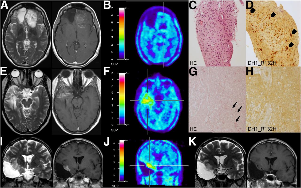
  • FIGURE 2.
    • Download figure
    • Open in new tab
    • Download powerpoint
    FIGURE 2.

    Examples of false-negative and -positive 18F-FET PET ratings. (A–D) A 45-y-old-patient had been diagnosed with IDH-mutant, MGMT promoter methylated glioblastoma in November 2010. After gross total resection, radiotherapy, and irinotecan chemotherapy, she received bevacizumab every other week. In January 2017, follow-up MRI indicated disease progression (RANO criteria). However, in February 2017, 18F-FET PET imaging was not suggestive of tumor, and so patient continued follow-up. Subsequent MRI revealed enlargement of both contrast-enhancing and non–contrast-enhancing lesions (tumor progression, RANO criteria), but 18F-FET PET remained negative. In November 2017, biopsy revealed tumor progression. Shown are axial MRI, October 2017, T2 image (A, left) and contrast-enhanced T1 image (A, right); 18F-FET PET, November 2017 (B); and histology (hematoxylin-eosin [HE]) (C) and immunohistochemistry (IDH1_R132H, arrowheads point to IDH1_R132H-positive tumor cells) (D) of biopsy samples, November 2017. (E–H) A 39-y-old patient had undergone subtotal resection of IDH1_R132H-mutant and 1p/19q-codeleted oligodendroglioma in August 2010, temozolomide chemotherapy until January 2011, proton therapy in May and June 2015, and lomustine chemotherapy from July to December 2015. In July 2017, putative recurrent tumor was resected. Neuropathology showed sequelae of radiation but no tumor. Shown are axial MRI, May 2017, T2 image (E, left) and contrast-enhanced T1 image (E, right); 18F-FET PET indicating tumor progression, June 2017 (F); and necrosis and calcification (arrows, HE) (G) without IDH1_R132H-positive tumor cells (H) in resected samples, July 2017. (I–K) IDH-mutant, MGMT promoter methylated glioblastoma of 38-y-old patient had been treated by partial resection in April 2016, radiotherapy, and temozolomide chemotherapy from April to June 2016. Against our advice, patient decided not to continue tumor-specific therapy. However, imaging alterations regressed spontaneously. Shown are coronal MRI, February 2017, T2 image (I, left) and contrast-enhanced T1 image (I, right); 18F-FET PET indicating tumor progression, April 2017 (J); and follow-up MRI, February 2018, T2 image (K, left) and contrast-enhanced T1 image (K, right).
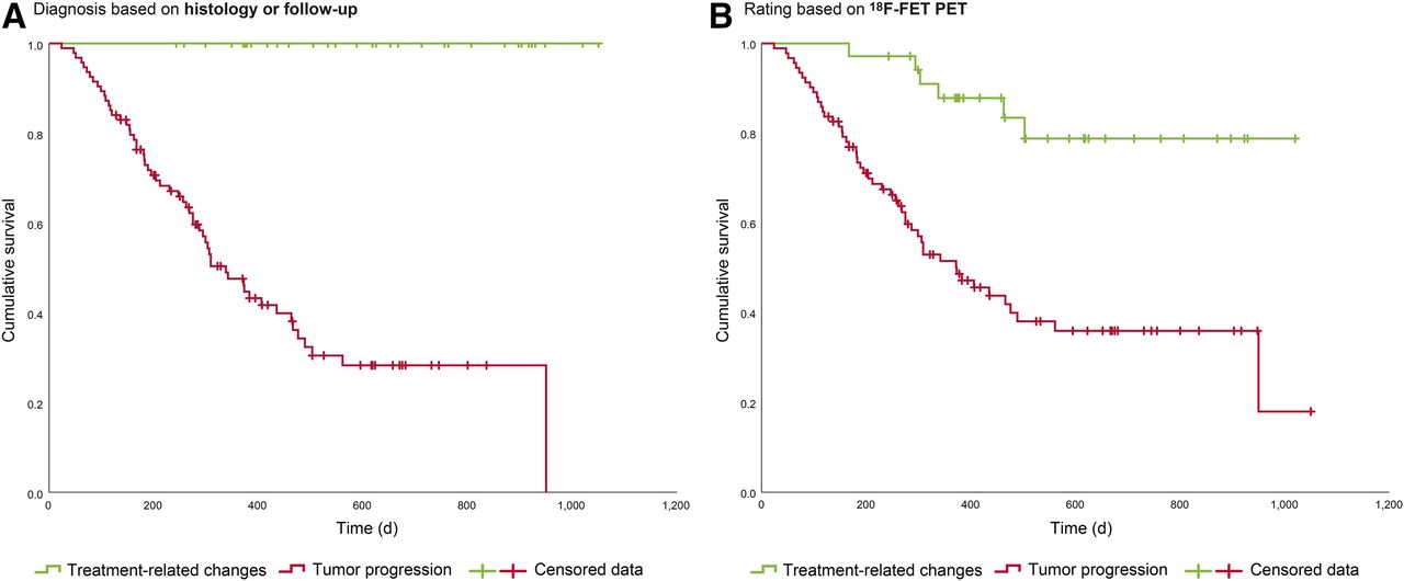
  • FIGURE 3.
    • Download figure
    • Open in new tab
    • Download powerpoint
    FIGURE 3.

    Overall survival of all 127 patients. (A) Overall survival after 18F-FET PET imaging, depending on whether TP or TRCs were present, as assessed by histology or follow-up (P [log-rank] < 0.001). (B) Overall survival after 18F-FET PET imaging, depending on 18F-FET PET results (P [log-rank] < 0.001).

Tables

  • Figures
  • Additional Files
    • View popup
    TABLE 1

    Patient and Tumor Characteristics, Part 1

    CharacteristicData%
    Sex (n)
     Male8365
     Female4435
    Age when 18F-FET PET imaging was  performed (y)
     Mean ± SD50 ± 12
     Range20–78
    KPS when 18F-FET PET imaging was  performed (n)
     100%4939
     90%4636
     80%1915
     70%119
     60%22
    Interval between last therapy and  18F-FET PET scan (d)
     Median103
     Range0–3,540
    Therapy before 18F-FET PET imaging (n)
     Radiotherapy11490
     Chemotherapy
      Temozolomide10683
      Lomustine-containing regimen2923
     Bevacizumab97
     Tumor treating fields97
     Reresection2117
     Reirradiation1915
     Nivolumab76
     Other*65
    • ↵* This section included 3 patients treated with nivolumab or placebo in context of clinical trial, 1 patient treated with sitimagene ceradenovec/ganciclovir, 1 patient treated with brachytherapy using 125I seeds, and 1 patient treated with irinotecan.

    • KPS = Karnofsky performance status.

    • View popup
    TABLE 2

    Patient and Tumor Characteristics, Part 2

    CharacteristicData%
    Diagnosis (n)
     Glioblastoma, IDH-wild-type, WHO IV5946
     Glioblastoma, IDH-mutant, WHO IV76
     Glioblastoma, not otherwise  specified, WHO IV10.8
     Astrocytoma, IDH-wild-type
      WHO II22
      WHO III76
     Astrocytoma, IDH-mutant
      WHO II108
      WHO III2117
     Astrocytoma, not otherwise specified
      WHO II10.8
      WHO III10.8
     Oligodendroglioma, IDH-mutant and  1p/19q-codeleted
      WHO II76
      WHO III65
     Diffuse midline glioma, H3 K27  M-mutant, WHO IV10.8
     Other*
      WHO II10.8
      WHO III10.8
      ND22
    MGMT promoter methylation  status (n)
     Methylated5745
     Unmethylated4031
     ND3024
    Extent of resection at initial  diagnosis (n)
     Gross total resection6753
     Subtotal resection86
     Partial resection2016
     Biopsy3024
     None22
    • ↵* This section included 1 diffuse glioma, IDH-wild-type, nuclear ATRX retained, MGMT promoter methylated; 1 anaplastic glioma, IDH-mutant, nuclear ATRX retained, MGMT promoter methylated; 1 suspected diffuse pontine glioma (treated without prior biopsy); and 1 suspected diffuse medulla oblongata glioma (treated without prior biopsy).

    • ND = not determined or inconclusive.

    • View popup
    TABLE 3

    Univariate and Multivariate Analyses of Overall Survival

    Survival analysisPatients (n)HR95% CIP
    Univariate
     Diagnosis based on 18F-FET PET1274.9972.139–11.675<0.001
     IDH status
     IDH-wild-type701.000
     IDH-mutant510.1810.091–0.363<0.001
     MGMT promoter methylation status
     Unmethylated401.000
     Methylated570.4930.278–0.8770.016
     WHO grade1253.8592.230–6.678<0.001
     Age (y)1271.0431.020–1.066<0.001
     KPS (%)1270.9650.940–0.9900.007
     Number of glioma recurrences before 18F-FET PET scan1271.0510.792–1.395NS
     Interval between last therapy and 18F-FET PET scan (d)1240.9970.996–0.9990.001
    Multivariate
     Diagnosis based on 18F-FET PET3.4241.446–8.1090.005
     WHO grade2.1431.212–3.7920.009
     IDH status0.4120.210–0.8080.010
     KPS (%)0.9750.950–1.0010.057
    • HR = hazard ratio; CI = confidence interval; KPS = Karnofsky performance status; NS = not statistically significant.

Additional Files

  • Figures
  • Tables
  • Supplemental Data

    Files in this Data Supplement:

    • Supplemental Data
PreviousNext
Back to top

In this issue

Journal of Nuclear Medicine: 61 (4)
Journal of Nuclear Medicine
Vol. 61, Issue 4
April 1, 2020
  • Table of Contents
  • Table of Contents (PDF)
  • About the Cover
  • Index by author
Print
Download PDF
Article Alerts
Sign In to Email Alerts with your Email Address
Email Article

Thank you for your interest in spreading the word on Journal of Nuclear Medicine.

NOTE: We only request your email address so that the person you are recommending the page to knows that you wanted them to see it, and that it is not junk mail. We do not capture any email address.

Enter multiple addresses on separate lines or separate them with commas.
18F-FET PET Imaging in Differentiating Glioma Progression from Treatment-Related Changes: A Single-Center Experience
(Your Name) has sent you a message from Journal of Nuclear Medicine
(Your Name) thought you would like to see the Journal of Nuclear Medicine web site.
Citation Tools
18F-FET PET Imaging in Differentiating Glioma Progression from Treatment-Related Changes: A Single-Center Experience
Gabriele D. Maurer, Daniel P. Brucker, Gabriele Stoffels, Katharina Filipski, Christian P. Filss, Felix M. Mottaghy, Norbert Galldiks, Joachim P. Steinbach, Elke Hattingen, Karl-Josef Langen
Journal of Nuclear Medicine Apr 2020, 61 (4) 505-511; DOI: 10.2967/jnumed.119.234757

Citation Manager Formats

  • BibTeX
  • Bookends
  • EasyBib
  • EndNote (tagged)
  • EndNote 8 (xml)
  • Medlars
  • Mendeley
  • Papers
  • RefWorks Tagged
  • Ref Manager
  • RIS
  • Zotero
Share
18F-FET PET Imaging in Differentiating Glioma Progression from Treatment-Related Changes: A Single-Center Experience
Gabriele D. Maurer, Daniel P. Brucker, Gabriele Stoffels, Katharina Filipski, Christian P. Filss, Felix M. Mottaghy, Norbert Galldiks, Joachim P. Steinbach, Elke Hattingen, Karl-Josef Langen
Journal of Nuclear Medicine Apr 2020, 61 (4) 505-511; DOI: 10.2967/jnumed.119.234757
Twitter logo Facebook logo LinkedIn logo Mendeley logo
  • Tweet Widget
  • Facebook Like
  • Google Plus One
Bookmark this article

Jump to section

  • Article
    • Abstract
    • MATERIALS AND METHODS
    • RESULTS
    • DISCUSSION
    • CONCLUSION
    • DISCLOSURE
    • Acknowledgments
    • Footnotes
    • REFERENCES
  • Figures & Data
  • Supplemental
  • Info & Metrics
  • PDF

Related Articles

  • This Month in JNM
  • PubMed
  • Google Scholar

Cited By...

  • Diagnostic Value of PET Tracers in Differentiating Glioma Tumor Recurrence from Treatment-Related Changes: A Systematic Review and Meta-Analysis
  • Borderline Findings in O-(2-[18F]-Fluoroethyl)-L-Tyrosine PET of Patients with Suspected Glioma Relapse: Role in Clinical Practice
  • Diagnostic Potential of Supplemental Static and Dynamic 68Ga-FAPI-46 PET for Primary 18F-FDG-Negative Pulmonary Lesions
  • [18F]-fluoroethyl-L-tyrosine (FET) in glioblastoma (FIG) TROG 18.06 study: protocol for a prospective, multicentre PET/CT trial
  • 11C-Methionine PET for Identification of Pediatric High-Grade Glioma Recurrence
  • Impact of 18F-FET PET/MRI on Clinical Management of Brain Tumor Patients
  • Google Scholar

More in this TOC Section

Oncology

  • 18F-FDG PET/CT manifestations of three Cases of Female Desmoplastic Small Round Cell Tumor
  • Utility of Fluorine-18-fluorodeoxyglucose PET/CT in Uterine sarcoma patients.
  • 18F-FDG PET/CT imaging features of parotid lesions; Case based pictorial review and its multi-modality correlation.
Show more Oncology

Clinical

  • Dual PET Imaging in Bronchial Neuroendocrine Neoplasms: The NETPET Score as a Prognostic Biomarker
  • Addition of 131I-MIBG to PRRT (90Y-DOTATOC) for Personalized Treatment of Selected Patients with Neuroendocrine Tumors
  • SUVs Are Adequate Measures of Lesional 18F-DCFPyL Uptake in Patients with Low Prostate Cancer Disease Burden
Show more Clinical

Similar Articles

Keywords

  • 18F-FET PET
  • glioma
  • Tumor progression
  • Pseudoprogression
  • treatment-related changes
SNMMI

© 2025 SNMMI

Powered by HighWire