


Article Figures & Data
Figures
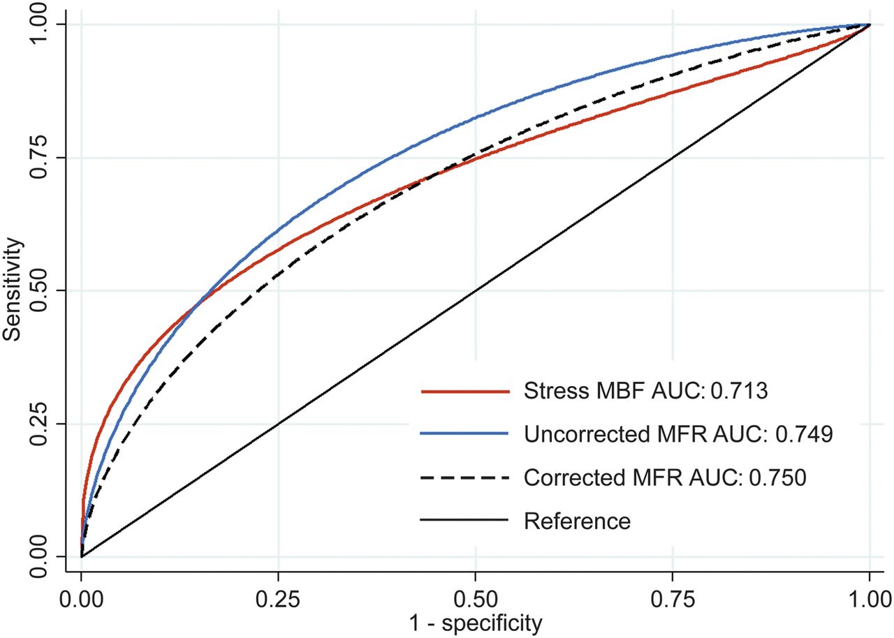
- FIGURE 1.
Receiver-operating-characteristic curves for diagnosing CAV ≥ grade 2. There was no difference between uncorrected MFR AUC and stress MBF AUC (P = 0.499) or corrected MFR AUC (P = 0.310).
- FIGURE 2.
Kaplan–Meier survival curves for all-cause mortality stratified by presence of abnormal regional perfusion. Patients with SSS ≥ 4 were more likely to experience all-cause mortality during follow-up (log-rank P = 0.017).
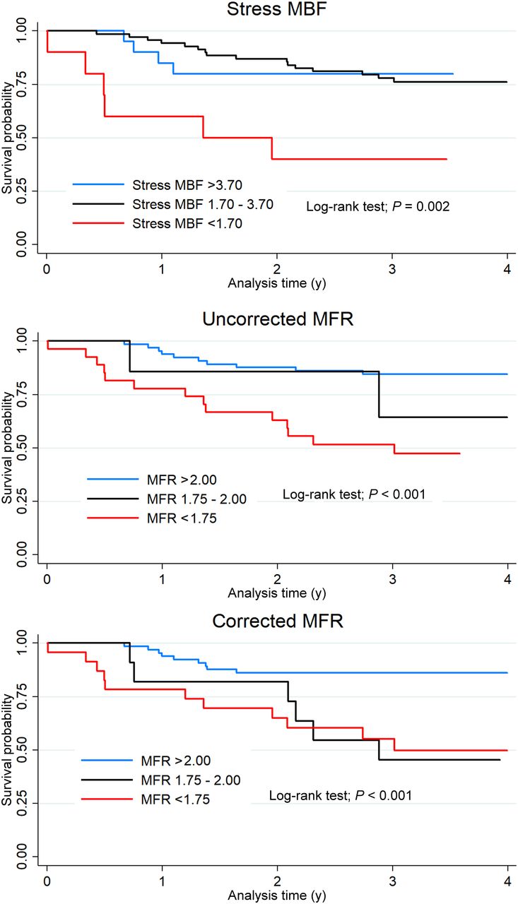
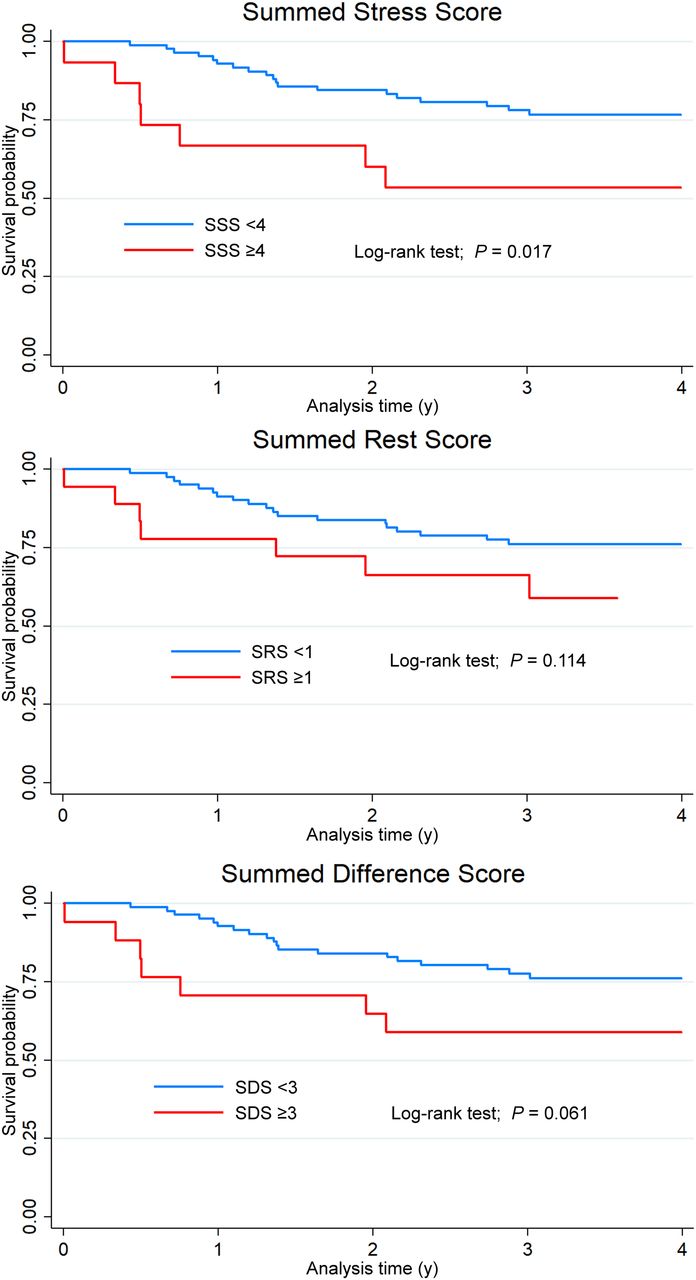
- FIGURE 3.
Kaplan–Meier survival curves for all-cause mortality stratified by quantitative PET results.
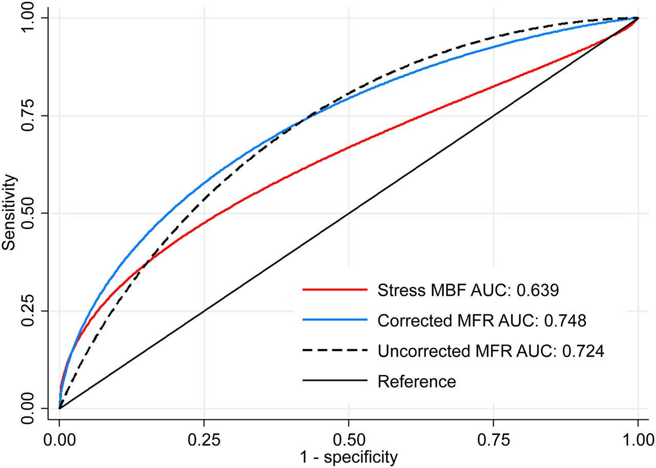
- FIGURE 4.
Receiver-operating-characteristic curves for identifying all-cause mortality during follow-up. Uncorrected MFR AUC was significantly larger than stress MBF AUC (P = 0.047). There was no significant difference between uncorrected MFR AUC and corrected MFR AUC (P = 0.681).

Tables
Characteristic No death (n = 73) Death (n = 26) P Age (y) 66.7 ± 10.5 74.0 ± 7.3 0.001 Male 54 (74.0%) 21 (80.8%) 0.599 Age at transplantation (y) 54.3 ± 11.1 61.9 ± 6.5 0.001 Donor age (y) 30.2 ± 11.9 35.4 ± 10.7 0.089 Time after transplantation (y) 12.5 ± 5.2 12.5 ± 5.4 0.977 Body mass index (kg/m2) 26.5 ± 5.6 25.8 ± 5.0 0.560 Hypertension 62 (84.9%) 19 (73.1%) 0.236 Diabetes 31 (42.5%) 15 (57.7%) 0.252 Dyslipidemia 53 (72.6%) 21 (80.8%) 0.600 Former smoker 4 (5.5%) 2 (7.7%) 0.651 Renal failure 7 (9.6%) 4 (15.4%) 0.472 CAV grade* (0/1/2/3) 46/17/5/3 13/6/2/4 0.489 Cytomegalovirus viremia 10 (13.7%) 3 (11.5%) 1.000 History of acute cellular rejection 10 (13.7%) 5 (19.2%) 0.531 History of antibody-mediated rejection 4 (5.5%) 4 (15.4%) 0.154 Medication use Aspirin 39 (53.4%) 16 (61.5%) 0.501 β-blockers 32 (43.8%) 9 (34.6%) 0.490 Angiotensin-converting inhibitor or receptor blocker 36 (49.3%) 11 (42.3%) 0.649 Diuretics 16 (21.9%) 9 (34.6%) 0.292 Statins 58 (79.5%) 16 (61.5%) 0.113 Calcineurin inhibitor 63 (86.3%) 22 (84.6%) 1.000 Mammalian target of rapamycin inhibitor 32 (43.8%) 9 (34.6%) 0.490 ↵* No death (n = 71); death (n = 25).
Data are expressed as number followed by percentage in parentheses or as mean ± SD.
Characteristic No death (n = 73) Death (n = 26) P Resting heart rate 81.88 ± 12.5 81.5 ± 12.8 0.980 Rate–pressure product (bpm × mm Hg) 10,895 ± 2,229 11,122 ± 1,627 0.635 Resting LVEF (%) 65.4 ± 9.7 56.8 ± 13.1 <0.001 82Rb semiquantitative imaging SRS 0 (0–0) 0 (0–1) 0.136 SSS 0 (0–2) 0 (0–8) 0.102 SDS 0 (0–1) 0 (0–4) 0.072 82Rb quantitative imaging Rest total perfusion deficit 0 (0–0.3) 0.2 (0.0–1.5) 0.018 Stress total perfusion deficit 1.1 (0.0–4.4) 2.1 (0.6–7.9) 0.111 Ischemic total perfusion deficit 1.1 (0.0–3.9) 1.9 (0.5–5.8) 0.200 Rest MBF (mL/min/g) 1.29 (1.06–1.44) 1.29 (1.14–1.56) 0.216 Stress MBF (mL/min/g) 2.88 (2.41–3.60) 2.54 (1.71–3.24) 0.024 MFR 2.37 (2.01–2.80) 1.69 (1.28–2.19) <0.001 Data are expressed as mean ± SD or as median followed by IQR in parentheses.
Variable Unadjusted HR P Adjusted HR P Age 1.08 (1.03–1.14) <0.001 1.10 (1.04–1.17) 0.001 Male 1.37 (0.52–3.65) 0.523 — — Body mass index 0.97 (0.80–1.05) 0.442 — — LVEF 0.95 (0.92–0.98) 0.001 0.98 (0.94–1.02) 0.232 Cardiac risk factors Hypertension 0.54 (0.23–1.28) 0.161 — — Diabetes 1.83 (0.84–3.99) 0.129 — — Dyslipidemia 1.55 (0.58–.11) 0.380 — — Renal failure 1.68 (0.58–4.88) 0.339 — — Cytomegalovirus viremia 0.77 (0.23–2.55) 0.666 — — Acute cellular rejection 1.43 (0.54–3.79) 0.474 — — Antibody-mediated rejection 1.88 (0.84–4.20) 0.124 — — PET parameters SRS 1.15 (1.01–1.31) 0.033 0.71 (0.20–2.54) 0.602 SSS 1.09 (1.03–1.15) 0.002 1.02 (0.29–3.54) 0.976 SDS 1.15 (1.06–1.26) 0.001 1.22 (0.35–4.19) 0.754 Rest MBF 1.81 (0.78–4.19) 0.166 — — Stress MBF 0.56 (0.35–0.90) 0.017 1.14 (0.64–2.05) 0.656 Uncorrected MFR* 0.34 (0.19–0.62) <0.001 0.30 (0.11–0.81) 0.017 Corrected MFR* 0.44 (0.26–0.74) 0.002 0.43 (0.20–0.90) 0.025 ↵* Multivariable analysis was performed separately with corrected and uncorrected MFR.
Data in parentheses are 95% CIs.
Diagnosis of CAV grade 2/3 Annualized all-cause mortality rate Cutoff Abnormal (n) Sensitivity Specificity Abnormal Normal MFR < 2.0 34 (34.3%) 71.4% 71.8% 17.7% 4.7% MFR < 1.75 27 (27.3%) 57.1% 77.7% 19.6% 5.2% Stress MBF < 3.7 79 (79.8%) 92.9% 22.4% 9.0% 6.7% Stress MBF < 1.7 10 (10.1%) 42.9% 95.3% 35.8% 7.0% SSS > 1 32 (32.2%) 64.3% 72.9% 12.3% 7.0% SSS > 3 15 (15.2%) 64.3% 92.9% 18.7% 7.1% LVEF ≤ 45 10 (10.1%) 42.9% 95.3% 25.0% 7.2% MBF < 1.7 and SSS > 1 or LVEF ≤ 45 8 (8.1%) 42.9% 97.7% 60.7% 6.8% SSS ≥ 4, LVEF ≤ 45 or MFR < 1.75 36 (36.4%) 71.4% 69.4% 15.4% 5.0% LVEF ≤ 45% and MFR < 1.75* 5 (5.1%) 35.7% 100.0% 51.6% 7.4% ↵* Same patients would be identified using MBF < 1.7 and LVEF < 45%.
Uncorrected MFR was used because it was numerically superior in all models.
Supplemental Data
Files in this Data Supplement:


Jump to section
Related Articles
Cited By...
- Diagnosis and management of cardiac allograft vasculopathy
- Appropriate Use Criteria for PET Myocardial Perfusion Imaging
- Reply: Clarifying the Utility of Myocardial Blood Flow and Myocardial Flow Reserve After Cardiac Transplantation
- Myocardial Blood Flow and Myocardial Flow Reserve After Cardiac Transplantation: Mistakes in Diagnostic Value and Prognosis