


EXECUTIVE SUMMARY
In the last decade, myocardial perfusion imaging (MPI) with PET has emerged to play a pivotal role in the clinical routine process for the detection of hemodynamically significant obstructive coronary artery disease (CAD) and cardiovascular risk stratification (1–5). The high spatial and contrast resolution in concert with photon attenuation-free images of PET have led to high image quality associated with the highest sensitivity and specificity of PET/CT perfusion imaging in the detection and characterization of CAD (1,2,6,7). In addition, the noninvasive evaluation and quantification of global and regional myocardial blood flow (MBF) in milliliters per gram per minute during hyperemic stress and at rest, as well as the calculation of the resulting myocardial flow reserve (MFR), extends the scope of standard MPI from the detection of advanced and flow-limiting epicardial CAD to a comprehensive assessment of ischemic burden. This improved scope results not only from the traditionally sought significant left main or multivessel disease, but also from the more recently appreciated cardiac effects of nonobstructive CAD and coronary microvascular disease (CMD), which conveys important diagnostic and incremental prognostic information (1,2,4–11).
The increased availability and high sensitivity of PET MPI in concert with concerns about missed diagnoses, however, may at times lead to an inappropriate application of this technology. Thus, to avoid unnecessary financial burden on the health-care system and, in some cases, unnecessary exposure of patients to ionizing radiation, we have established a consensus document that outlines the most appropriate and cost-effective use of PET MPI. It is hoped that this expert guidance will help to render the use of PET MPI more consistent and will improve health-care outcomes for the targeted patient population while minimizing unnecessary imaging costs. The goal of this document is to describe the appropriate use of PET MPI in patients with suspected or known CAD and in patients with suspected microvascular angina. Through these recommendations, it is expected that PET MPI will be applied to benefit these patients in the most cost-effective manner.
Representatives from the Society of Nuclear Medicine and Molecular Imaging (SNMMI), the American College of Cardiology (ACC), the American Society of Nuclear Cardiology (ASNC), the Canadian Cardiovascular Society (CCS), the Canadian Society of Cardiovascular Nuclear and CT Imaging (CSCNCTI), the Society of Cardiovascular CT (SCCT), the American Heart Association (AHA), the American College of Physicians (ACP), and the European Association of Nuclear Medicine (EANM) assembled as an autonomous workgroup to develop the following appropriate use criteria (AUC). This process was performed in accordance with the Protecting Access to Medicare Act of 2014. This legislation requires that all referring physicians consult AUC by using a clinical decision support mechanism before ordering any advanced diagnostic imaging services. Such services are defined as diagnostic MRI, CT, nuclear medicine procedures (including PET), and others as specified by the Secretary of Health and Human Services in consultation with physician specialty organizations and other stakeholders (12). The AUC in this paper are intended to aid referring medical practitioners in the appropriate use of PET MPI for the most common scenarios encountered in patients with suspected or known CAD as well as in patients with suspected microvascular angina.
INTRODUCTION
This document describes the appropriate use of MPI and concurrent MBF assessment with PET (PET MPI). The authors have strived to include the most common clinical scenarios for PET MPI in patients with suspected CAD or CMD, or with known CAD, in order to assist health-care practitioners in the cardiovascular field who are considering PET MPI. It is important to keep in mind, however, that a patient may have variations of the clinical scenarios covered in the current document, or signs or symptoms not described, for which PET MPI may still be indicated. Because each patient is unique, as is each patient’s clinical presentation, this document is not intended to replace clinical judgment. PET MPI is also applied in conjunction with 18F-FDG PET for myocardial viability and cardiac sarcoidosis assessment, for which myocardial perfusion flow characterization is integral to diagnosis and, thus, patient management.
PET affords not only the assessment of relative differences in myocardial perfusion, but also, in conjunction with tracer kinetic modeling, the calculation of regional and global MBF of the left ventricle in absolute terms in milliliters per gram per minute (1,2,6,7). This enables the calculation of MBF during pharmacologically induced hyperemic flow and at rest in milliliters per gram per minute. The assessment of hyperemic MBF and MFR (MFR = MBF during hyperemia/MBF at rest), therefore, allows the assessment of CMD as a potential substrate for angina symptoms or shortness of breath in the absence of obstructive CAD (3,13). In this respect, the noninvasive evaluation and quantification of hyperemic MBF and MFR expand the scope of conventional MPI from detection of end-stage, advanced, flow-limiting epicardial CAD to early stages of atherosclerosis or CMD. It is important to keep in mind that the identification of stress-induced myocardial perfusion deficits with conventional MPI indicates hemodynamically obstructive CAD that carries important diagnostic and prognostic information for the evaluation of patients with suspected or known CAD. Conversely, the assessment of only the “relative” distribution of the radiotracer uptake in the left ventricular (LV) myocardium with standard MPI commonly identifies the culprit or most advanced CAD lesion, and the hemodynamic effects of less severe lesions may be missed in multivessel CAD disease (2,8–10). Adding the concurrently PET-determined regional hyperemic MBF and MFR aids in the unraveling of the flow-limiting effects of the remaining CAD lesions of less or intermediate severity. Another advantage can also be seen in the identification of diffuse or balanced myocardial ischemia because of significant left main or 3-vessel disease with PET. Pronounced reductions in hyperemic MBF and MFR in all 3 major coronary artery distributions, in conjunction with transient ischemic cavity dilation at peak stress versus rest on PET imaging, identifies diffuse ischemia, otherwise likely to be missed by MPI alone (2,8–10). From a methodologic point of view, the high spatial and contrast resolution of the photon attenuation-free images of PET in conjunction with the superior properties of positron-emission blood flow radiotracers, such as 13N-ammonia or 82Rb, affords several unique advantages of cardiac PET perfusion and flow assessment. The principle of coincidence detection, together with the superior properties of MBF radiotracers, has resulted in an increase in spatial and contrast resolution of PET MPI, explaining, at least in part, the high sensitivity (mean ≈92%) and specificity (mean ≈90%) in the detection of hemodynamically obstructive CAD lesions (1,2).
A systematic review of the literature for this AUC document demonstrated that evidence was strongest for diagnostic accuracy and predictive utility of PET MPI perfusion defects in signifying hemodynamic effects of flow-limiting obstructive CAD. In addition, there was good evidence to support the diagnostic accuracy and predictive utility of abnormal MBF or flow quantification, as well as the predictive utility of reduced LV systolic function. Taken together, the evidence is excellent to good that findings on PET MPI perfusion and flow quantification precisely identify persons with CAD and, at the same time, find persons at risk for mortality and future cardiovascular events. Although the assessment of CMD in obstructive and nonobstructive CAD has incremental and independent prognostic value, further evidence is needed to clarify the diagnostic accuracy of PET for diagnosing CMD and the optimal threshold to define abnormal hyperemic MBF increases or MFR. Only a few studies describe the effects of PET MPI perfusion and flow quantification on the clinical decision-making process and clinical outcome (14–18), which thus warrants further evaluation in well-designed and large-scale clinical trials. In principle, further evidence is also needed to define the role of PET MPI perfusion and flow quantification in asymptomatic adults and children, leaving the decision of its use in these populations for the time being to best clinical judgment.
From the evidence of this systematic review, outcome data, clinical expertise, and standard clinical practice, we evaluated a total of 210 clinical scenarios for PET MPI and covered them in the following 11 sections: section 1: Symptomatic Patients with Suspected or Known CAD; section 2: Asymptomatic Patients (Without Symptoms or Ischemic Equivalent); section 3: Diagnosed Heart Failure (Resting LV Function Previously Assessed but No Prior CAD Evaluation); section 4: Evaluation of Patients with Known or Suspected Cardiac Sarcoidosis; section 5: Evaluation of Arrhythmias Without Ischemic Equivalent (No Prior Cardiac Evaluation); section 6: Syncope Without Ischemic Equivalent; section 7: Assessment of CMD in Symptomatic Patients; section 8: Specific Populations; section 9: Prior Testing or Procedures; section 10: Preoperative Evaluation for Noncardiac Surgery; and section 11: Determination of Exercise Level Before Initiation of Exercise Prescription or Cardiac Rehabilitation. This comprehensive list of elaborated clinical scenarios aims to guide health-care practitioners to an appropriate use of PET MPI, but the document does not claim to cover all potential clinical scenarios or to substitute for clinical judgment in an individual clinical scenario.
METHODOLOGY
Expert Workgroup Selection
The experts of this AUC workgroup were convened by the SNMMI to represent a multidisciplinary panel of health-care providers with substantive knowledge in the use of PET MPI. In addition to SNMMI members, representatives from the ACC, ASNC, CCS, CSCNCTI, SCCT, AHA, ACP, and EANM were included in the workgroup. Eighteen physician members were ultimately selected to participate and contribute to the AUC. A complete list of workgroup participants and external reviewers can be found in Appendix A. Appendix B is a summary of definitions of terms and acronyms, Appendix C provides the disclosures and conflicts-of-interest statements, and Appendix D describes the solicitation of public commentary.
AUC Development
The process for AUC (11) development was modeled after the RAND/UCLA Appropriateness Method (19,20) and included the development of a list of common indications for the use of PET in MPI, a systematic review of evidence related to these indications, and the development of an appropriateness score for each indication by using a modified Delphi process. This process strove to adhere to the standards of the Institute of Medicine of the National Academies for developing trustworthy clinical guidance (21). The process included a systematic synthesis of available evidence, individual and group ratings of the clinical indications by using a formal consensus process, and AUC recommendations based on final group ratings and discussions.
Scope and Development of Clinical Scenarios
To begin this process, the workgroup discussed various potential clinical scenarios for the appropriate use of MPI and concurrent MBF assessment with PET. For clinical scenarios, some of the relevant populations of interest were patients who had known or suspected CAD; who were obese; who were symptomatic; who had prior equivocal or discordant studies, suspected microvascular angina, prior revascularization, or failed medical therapy; and who were of all genders, ages, races, and geographic locations.
The workgroup identified 210 clinical scenarios for PET MPI that are evaluated and covered in 11 sections. The scenarios are intended to be as representative of the relevant patient population as possible for development of AUC. The resulting AUC are based on evidence and expert opinion regarding diagnostic accuracy and effects on clinical outcomes and clinical decision making as applied to each scenario. Other factors affecting the AUC recommendations were potential harm—including long-term harm that may be difficult to capture—costs, availability, and patient preferences.
Systematic Review
To inform the workgroup, a systematic review of the relevant evidence was commissioned by an independent group, the Pacific Northwest Evidence-Based Practice Center of Oregon Health and Science University (22). The primary purpose of the systematic review was to synthesize evidence on PET MPI for the detection of CAD and CMD, the prediction of future cardiovascular events, the effects on clinical decision making, and the effects on clinical outcomes in symptomatic adults and children. The workgroup selected the following key questions to guide the review:
In adults and children undergoing evaluation for symptoms of potential CAD, what is the accuracy of PET MPI for diagnosis of CAD or CMD?
In symptomatic adults and children, what is the utility of PET MPI for predicting future cardiovascular events?
In adults and children undergoing PET MPI for evaluation of symptoms of potential CAD, what are the effects on clinical decision making (use of treatments, subsequent testing)?
In adults and children undergoing PET MPI for evaluation of symptoms of potential CAD, what are the effects on clinical outcomes?
In adults without symptoms of CAD, what is the utility of PET MPI for predicting future cardiovascular events?
In asymptomatic adults who undergo PET MPI, what are the effects on clinical decision making (use of treatments, subsequent testing)?
Potential modifiers of effects of interest were demographic and clinical characteristics of the populations studied, use of stress techniques (pharmacologic or exercise), and use of alternative tracers. The inclusion and exclusion criteria for papers for this review were based on the study parameters established by the workgroup, using the PICOTS (population, intervention, comparisons, outcomes, timing, and setting) approach. Searches for relevant studies and systematic reviews were conducted in the following databases: Cochrane Central Register of Controlled Trials, Cochrane Database of Systematic Reviews, and Ovid MEDLINE (through November 2017). These searches were supplemented by reviewing the reference lists of relevant publications and suggestions from SNMMI workgroup members.
Two investigators independently reviewed abstracts and full-text articles against prespecified eligibility criteria, as defined by PICOTS. For diagnostic accuracy, we included studies of symptomatic adults and children undergoing imaging with PET MPI or PET/CT MPI with any tracer for suspicion of or confirmation of CAD or CMD. Studies had to assess diagnostic accuracy against a reference standard of coronary angiography, usually a >50% or >70% reduction in luminal diameter for an anatomic diagnosis of CAD, with or without a functional measure of obstruction (usually a fractional flow reserve of <0.8). The reference standard in the only study that assessed CMD was epicardial artery vasomotor dysfunction during cold pressor testing on coronary angiography; in the 2 studies that assessed cardiac allograft vasculopathy (CAV), it was the International Society for Heart and Lung Transplantation criteria, including angiographic stenosis and evidence of allograft dysfunction. For predictive utility, we included longitudinal studies of symptomatic and asymptomatic adults and children regarding the association between PET MPI findings and death and future cardiovascular events, as well as clinical decision making (use of treatments, subsequent testing) and clinical outcomes (e.g., mortality, morbidity, quality of life, and harms). PET findings included perfusion deficits at rest or with stress and measures of MBF, including response to cold pressor testing. For predictive utility, we also evaluated PET measures of left ventricular ejection fraction (LVEF). Non-English language articles and studies published only as conference abstracts were excluded.
Two investigators independently assessed the quality (risk of bias) of each study as “good,” “fair,” or “poor” by using predefined criteria that were specific for each study design. Specifically, AMSTAR (A MeaSurement Tool to Assess systematic Reviews) (23) was used for systematic reviews (except diagnostic accuracy), adapted by the US Preventive Services Task Force criteria for randomized trials and cohort studies, and the QUADAS-2 (Quality Assessment of Diagnostic Accuracy Studies-2) (24) for primary studies and systematic reviews of diagnostic accuracy. Discrepancies were resolved through a consensus process. The strength of overall evidence was graded as high, moderate, low, or very low by using GRADE (Grading of Recommendations Assessment, Development and Evaluation) methods on the basis of quality of evidence, consistency, directness, precision, and reporting bias.
Database searches resulted in 1,189 potentially relevant articles. After a dual review of abstracts and titles, we selected 206 articles for full-text dual review, and 76 studies (in 82 publications) were determined to meet inclusion criteria and were included in this review.
Rating and Scoring
In developing these AUC (11) for PET MPI, the workgroup members used the following definition of appropriateness to guide their considerations and group discussions: “The concept of appropriateness, as applied to health care, balances risk and benefit of a treatment, test, or procedure in the context of available resources for an individual patient with specific characteristics” (25).
At the beginning of the process, workgroup members convened via webinar/teleconference to develop the initial clinical indications. On evaluating the evidence summary of the systematic literature review, the workgroup further refined its draft clinical indications to ensure their accuracy and facilitate consistent interpretation when scoring each indication for appropriateness. Using the evidence summary, workgroup members were first asked individually to assess the appropriateness and provide a score for each identified indication. Workgroup members then convened in a group setting for several successive webinars to discuss each indication and associated scores from the first round of individual scoring. After deliberate discussion, a consensus score was determined and then assigned to the associated appropriate use indication. For this scoring round, the expert panel was encouraged to include their clinical expertise in addition to the available evidence in determining the final scores. All members contributed to the final discussion, and no one was forced into consensus. After the rating process was completed, the final appropriate use ratings were summarized in a format similar to that outlined by the RAND/UCLA Appropriateness Method.
The workgroup scored each indication as “appropriate,” “may be appropriate,” or “rarely appropriate” on a scale from 1 to 9. Scores 7–9 indicate that the use of the procedure is appropriate for the specific clinical indication and is generally considered acceptable. Scores 4–6 indicate that the use of the procedure may be appropriate for the specific indication. This implies that more research is needed to classify the indication definitively. Scores 1–3 indicate that the use of the procedure is rarely appropriate for the specific indication and is generally not considered acceptable.
As stated by other societies that develop AUC, the division of these scores into 3 general levels of appropriateness is partially arbitrary, and the numeric designations should be viewed as a continuum. In addition, if there was a difference in clinical opinion for an indication such that workgroup members could not agree on a common score, that indication was given a “may be appropriate” rating to indicate a lack of agreement on appropriateness based on the available literature and the members’ collective clinical opinion, indicating the need for additional research.
SECTION 1: SYMPTOMATIC PATIENTS WITH SUSPECTED OR KNOWN CAD
Introduction and Background
The accuracy and prognostic value of MPI (PET or SPECT) to define ischemia in order to detect CAD and to risk stratify patients is well established. A wealth of data for SPECT and now a large body of evidence for PET MPI support this concept, applying PET for diagnosis and prognostication. Driven by the Bayes’ theorem, defining the pretest likelihood of disease must be a step in considering a decision to order PET MPI. This, along with knowing the impact of a positive or negative test result on decision making, helps the clinician and imager determine the need for PET MPI. Guidelines indicate a role for ischemia testing with MPI (SPECT and PET can both provide this role). However, the superiority of PET over SPECT for accuracy is now well established, as is the prognostic value of PET MPI. Although not explicitly mentioned in the case scenarios, the advantage of PET for flow quantification is implicit when considering PET for symptomatic patients, given its added value in the diagnosis of obstructive disease, in particular multivessel CAD, and in the detection of CMD or even nonresponsiveness to pharmacologic stress. Moreover, the incremental prognostic value of flow quantification beyond relative MPI further empowers PET as a risk assessment tool beyond the known utility of relative perfusion imaging alone.
Clinical Scenarios and AUC Scores
Table 1 presents the following clinical scenarios and final AUC scores for the use of stress–rest perfusion PET to assess the presence of flow-limiting obstructive CAD as a potential substrate for chest pain (or equivalent) symptoms, or for risk stratification and prognostic value, in symptomatic patients with suspected or known CAD.
Clinical Scenarios for the Use of Stress–Rest Perfusion PET to Assess the Presence of Flow-Limiting Obstructive CAD as a Potential Substrate for Chest Pain (or Equivalent) Symptoms, or for Risk Stratification and Prognostic Value, in Symptomatic Patients with Suspected or Known CAD
Clinical Scenarios 1–15
The following scenarios address patients presenting a low pretest probability of CAD:
Scenario 1: Low pretest probability for CAD, with an interpretable electrocardiogram (ECG) and where the patient is able to undergo adequate exercise stress (Score 2 – Rarely Appropriate)
Scenario 2: Low pretest probability of CAD, ECG uninterpretable or unable to exercise (Score 3 – Rarely Appropriate)
Scenario 3: Low pretest probability for CAD, with an uninterpretable ECG (Score 3 – Rarely Appropriate)
Scenario 4: Low pretest probability for CAD, where the patient is unable to undergo adequate exercise stress (Score 3 – Rarely Appropriate)
The following scenarios address patients presenting an intermediate pretest probability of CAD:
Scenario 5: In symptomatic patients with intermediate pretest probability for CAD, where ECG is interpretable and where the patient is able to undergo adequate exercise stress (Score 7 – Appropriate)
Scenario 6: Intermediate pretest probability for CAD, where ECG is uninterpretable (Score 9 – Appropriate)
Scenario 7: Intermediate pretest probability for CAD, where the patient is unable to undergo adequate exercise stress (Score 9 – Appropriate)
The following scenarios address patients presenting a high pretest probability of CAD:
Scenario 8: In symptomatic patients with high pretest probability for CAD, where ECG is interpretable and where the patient is able to undergo adequate exercise stress (Score 8 – Appropriate)
Scenario 9: High pretest probability for CAD, where ECG is uninterpretable (Score 9 – Appropriate)
Scenario 10: High pretest probability for CAD, where the patient is unable to undergo adequate exercise stress (Score 9 – Appropriate)
The following scenarios address symptomatic patients presenting to the emergency department (ED) (or inpatients) with acute coronary syndrome (ACS) features:
Scenario 11: Patients presenting to the ED (or inpatients) with symptoms suspicious for CAD without (or with) known CAD who have features consistent with non–ST elevation myocardial infarction (non-STEMI)/ACS (increase in troponin (Tn) level(s) or dynamic ECG changes) (Score 1 – Rarely Appropriate)
Scenario 12: Patients presenting to the ED (or inpatients) with symp toms suspicious for CAD without (or with) known CAD who have features consistent with unstable angina/ACS (no increase in Tn level(s), no dynamic ECG changes) (Score 7 – Appropriate)
The following scenario addresses patients presenting to the ED (or inpatients) with chest pain or equivalent symptoms without (or with) known CAD who have equivocal Tn level(s) or a temporal Tn pattern not consistent with ACS:
Scenario 13: Patients presenting to the ED (or inpatients) with chest pain or equivalent symptoms without (or with) known CAD who have equivocal Tn level(s) or a temporal Tn pattern not consistent with ACS (Score 8 – Appropriate)
The following scenario addresses patients presenting to the ED (or inpatients) with symptoms of chest pain that may be due to ACS, with normal Tn level(s) and no dynamic ECG changes:
Scenario 14: Patients presenting to the ED (or inpatients) with symptoms of chest pain that may be due to ACS, normal Tn level(s) and no dynamic ECG changes (Score 8 – Appropriate)
The following scenario addresses patients presenting to the ED (or inpatients) with symptoms of chest pain that is unlikely to be due to ACS, with normal Tn level(s) and no dynamic ECG changes:
Scenario 15: Patients presenting to the ED (or inpatients) with symp toms of chest pain that is unlikely to be due to ACS, normal Tn level(s) and no dynamic ECG changes (Score 7 – Appropriate)
The preceding scenarios evaluate the role of MPI PET in the management of symptomatic patients with suspected or known CAD (scenarios 1–10) or who present to the ED (scenarios 11–15).
In symptomatic patients with suspected or known CAD, scintigraphic MPI such as with PET (PET MPI) is best performed for risk stratification with a clinically intermediate risk of subsequent cardiac events (1,4,5,26–28). This also relates to the optimal application of PET MPI to patients with an intermediate likelihood of having CAD. Critical determinants of outcome in patients with CAD can be determined by stress-related myocardial perfusion and function (26,27,29,30). Although indices that determine the extent of LV dysfunction (LVEF, the extent of infarcted myocardium, transient ischemic dilation of the left ventricle, and increased radiotracer uptake in the lungs) are relevant predictors of cardiac mortality, parameters related to ischemia induction (exertional symptoms, ECG alterations, extent and severity of reversible perfusion deficits, and ventricular wall motion abnormalities) are independent predictors of acute ischemic syndromes (26,28–32). PET MPI, in particular, affords the advantage of high diagnostic accuracy, concurrent evaluation of myocardial perfusion (radiotracer uptake) and regional MFR, and wall motion assessment at peak pharmacologic stress (2,8,10,26,33–39). This enables the accurate identification and description of the extent and severity of stress-induced ischemia in individuals with suspected CAD and intermediate and high pretest probability. In addition, the concurrent assessment of regional and global hyperemic MBF and MFR enables the detection and characterization of flow-limiting effects of each single vessel in multivessel disease (2,4–8,10,11,40–46), and reductions in global MFR also convey important and incremental prognostic information in patients with and without obstructive CAD (2,3,7,14,26,39,46–48).
In symptomatic patients with suspected CAD but low pretest probability in scenarios 1–4, the application of PET MPI in general is rarely appropriate after standard clinical practice guidelines (27,28). Conversely, for scenarios 5–7 in the category of intermediate pretest probability for CAD, the performance of PET MPI has been judged as appropriate in accordance with standard clinical practice in scintigraphic MPI (27,28). In patients with high pretest probability for CAD, who have an uninterpretable ECG and are able or unable to exercise, PET MPI was seen as appropriate (scenarios 8–10) according to standard clinical practice in MPI (27,28).
In symptomatic patients who present to the ED (or inpatients) with ACS features that are consistent with non-STEMI or ACS (increase in Tn levels or dynamic ECG changes) (scenario 11), MPI PET is not indicated, as these patients are at high risk and, thus, they are commonly referred to further triage with invasive coronary angiography and potential intervention according to standard clinical practice (49,50). In scenario 12, this group of patients with some type of unstable angina or ACS and with positive Tn results but with neither an increase in Tn levels later nor dynamic ECG changes, PET MPI was seen as appropriate for further cardiovascular risk stratification and triage of the patient (27,28,32,49–54).
Scenarios 13–15 address the role of PET MPI in patients who present to the ED (or inpatients) with chest pain or equivalent symptoms with or without known CAD who have equivocal Tn levels or a temporal Tn pattern not consistent with ACS (scenario 13); patients who present to the ED (or inpatients) with symptoms of chest pain that may be due to ACS, with normal Tn levels and no dynamic ECG changes (scenario 14); and patients who present to the ED (or inpatients) with symptoms of chest pain that are unlikely to be due to ACS, with normal Tn levels and no dynamic ECG changes (scenario 15). Given the clinical constellation and standard clinical practice (27,28,32,49–54), these scenarios were seen as appropriate.
Summary of Recommendations
Rest–stress PET MPI is considered an appropriate test in patients with or without known CAD who have symptoms with an intermediate-to-high pretest likelihood of disease regardless of whether the patient has normal ECG results or can (or cannot) exercise. Both diagnostic accuracy and prognostic value are considerations. PET MPI is not appropriate when patients have a low pretest likelihood of disease. For patients in the ED or for inpatients with chest pain or symptoms related potentially to ischemia, when there are clear markers or high risk (elevated Tn levels, dynamic ECG changes), PET MPI is rarely appropriate. On the other hand, when such markers are not present, PET MPI is generally considered appropriate.
SECTION 2: ASYMPTOMATIC PATIENTS (WITHOUT SYMPTOMS OR ISCHEMIC EQUIVALENT)
Background
Confirmation of the effectiveness of systematic screening for CAD in asymptomatic patients has been elusive. The evidence to support the use of stress testing and imaging in general and PET MPI in particular is limited. The guidelines suggest that only high-risk subjects should be considered for further noninvasive or invasive testing. The clinical cardiovascular risk evaluation is commonly based on the patient’s 10-y atherosclerotic cardiovascular disease (ASCVD) risk estimate according to the ACC/AHA guidelines (low ASCVD risk < 7.5%, intermediate ASCVD risk ≥ 7.5%–20%, high ASCVD risk > 20%) (55).
Although the data are limited on how to manage asymptomatic patients who have a positive test result, the principles of risk stratification used in other patient groups described in this document also apply to asymptomatic individuals. The scenarios identified below represent common clinical situations in which imaging tests are often considered. In all of these categories, the use of quantitative MBF and flow reserve in combination with semiquantitative PET MPI has generally been considered to be a unique advantage of PET MPI over SPECT MPI. The combination of PET MPI and flow quantification can help improve the sensitivity for detecting obstructive CAD or CMD and refine the cardiovascular risk assessment.
Clinical Scenarios and AUC Scores
Table 2 presents the following clinical scenarios and final AUC scores for the use of PET MPI in asymptomatic patients (without symptoms or ischemic equivalent).
Clinical Scenarios for the Use of PET MPI in Asymptomatic Patients (Without Symptoms or Ischemic Equivalent)
Clinical Scenarios 16–25: Asymptomatic Patients (Without Symptoms or Ischemic Equivalent) by Pretest CAD Risk
Scenario 16: Asymptomatic patients with a low ASCVD risk (<7.5%) (Score 1 – Rarely Appropriate)
Scenario 17: Asymptomatic patients with an intermediate ASCVD risk (7.5%–20%), who have an interpretable resting ECG and are able to exercise (Score 2 – Rarely Appropriate)
Scenario 18: Asymptomatic patients with an intermediate ASCVD risk (7.5%–20%), who have an uninterpretable resting ECG or are unable to exercise (Score 5 – May be Appropriate)
Scenario 19: Asymptomatic patients with a high ASCVD risk (>20%) (Score 5 – May be Appropriate)
Scenario 20: Asymptomatic patients with an intermediate ASCVD risk (7.5%–20%), who have a calcium score of 400–1,000 (Score 6 – May be Appropriate)
Scenario 21: Asymptomatic patients with a high ASCVD risk (>20%), who have a calcium score of 400–1,000 (Score 8 – Appropriate)
Scenario 22: Asymptomatic patients who have a calcium score of >1,000 (Score 8 – Appropriate)
Scenario 23: Asymptomatic patients with peripheral vascular disease (Score 5 – May be Appropriate)
Scenario 24: Asymptomatic patients with a family history of premature CAD (Score 3 – Rarely Appropriate)
Scenario 25: Asymptomatic patients with familial hyperlipidemia (Score 5 – May be Appropriate)
PET was rated as rarely appropriate in asymptomatic patients with a low (<7.5%) or intermediate ASCVD risk (7.5%–20%) if they have an interpretable resting ECG (e.g., left bundle branch block [LBBB], paced rhythm, Wolff-Parkinson-White changes) and are able to exercise (scenarios 16 and 17). In keeping with other modalities, the use of PET was rated as may be appropriate in patients with an intermediate ASCVD risk who have an uninterpretable resting ECG or who are unable to exercise, as well as in those with a high ASCVD risk (>20%) (scenarios 18 and 19). Regarding asymptomatic patients with prior coronary artery calcium (CAC) scoring, the use of PET was rated as may be appropriate in patients with an intermediate ASCVD risk and a CAC score of 400–1,000 (scenario 20). However, PET MPI was rated as appropriate in patients with a high ASCVD risk (>20%) and a CAC score of 400–1,000 or a very high CAC score (>1,000) regardless of clinical risk (scenarios 21 and 22). This rating was based on the fact that the presence of an elevated CAC score (>400) increases the likelihood of obstructive CAD and risk of events, especially in patients with multiple coronary risk factors with a high ASCVD risk. The use of PET in patients with a history of peripheral vascular disease (e.g., stroke) or familial hyperlipidemia was rated as may be appropriate because their clinical ASCVD risk is high (scenarios 23 and 25). However, a family history of premature CAD without any other risk qualifier was rated as rarely appropriate (scenario 24) (28,56–66).
Clinical Scenarios 26–33: Asymptomatic Patients with Equivocal or Abnormal Prior Test Results
Scenario 26: Asymptomatic patients with equivocal or abnormal prior test results from coronary CT angiography (CCTA) or invasive coronary angiography (ICA) (Score 8 – Appropriate)
Scenario 27: Asymptomatic patients with recent (<90 d) equivocal or abnormal prior test results from stress testing (imaging or nonimaging) (Score 6 – May be Appropriate)
Scenario 28: Asymptomatic patients with regional or global LV systolic dysfunction (Score 8 – Appropriate)
Scenario 29: Asymptomatic patients with new LBBB (Score 8 – Appropriate)
Scenario 30: Asymptomatic patients with atrial fibrillation (AF) (Score 5 – May be Appropriate)
Scenario 31: Asymptomatic patients with abnormal resting ECG results that show abnormal or pathologic Q waves (Score 6 – May be Appropriate)
Scenario 32: Asymptomatic patients with abnormal resting ECG results that show ST-T segment abnormalities (Score 5 – May be Appropriate)
Scenario 33: Asymptomatic patients with known stable CAD with out prior revascularization (Score 6 – May be Appropriate)
The use of PET was considered appropriate in patients with equivocal or abnormal coronary angiography (CCTA or ICA) results, regional or global LV dysfunction, or new LBBB (scenarios 26, 28, and 29). PET imaging was rated as may be appropriate in asymptomatic patients with recent (<90 d) abnormal or equivocal stress testing with or without imaging, as well as in those with atrial fibrillation (AF), abnormal resting ECG results showing pathologic Q waves or ST-T segment abnormalities, or stable CAD without prior revascularization (scenarios 27, 30–33) (28,67–71).
Clinical Scenarios 34–37: Asymptomatic Patients with Prior Revascularization (Percutaneous Coronary Intervention [PCI] or Coronary Artery Bypass Graft [CABG])
The following scenarios address asymptomatic patients with a history of PCI:
Scenario 34: Asymptomatic patients with a history of PCI of <2 y (Score 2 – Rarely Appropriate)
Scenario 35: Asymptomatic patients with a history of PCI of >2 y (Score 6 – May be Appropriate)
The following scenarios address asymptomatic patients with a prior CABG.
Scenario 36: Asymptomatic patients with a prior CABG of <5 y (Score 2 – Rarely Appropriate)
Scenario 37: Asymptomatic patients with a prior CABG of >5 y (Score 6 – May be Appropriate)
In keeping with the prior multimodality AUC document, the use of PET in asymptomatic patients with a history of prior PCI (<2 y) or CABG (<5 y) was considered rarely appropriate. However, PET MPI was rated as may be appropriate for asymptomatic patients >2 y after PCI or >5 y after CABG (scenarios 34–37) (28,61).
Clinical Scenario 38: Asymptomatic Patients with Prior Heart Transplantation
Scenario 38: Evaluation for CAV in asymptomatic patients with prior heart transplantation (Score 8 – Appropriate)
Annual surveillance to assess the presence and severity of CAV is the current standard of care after orthotopic heart transplantation. Although the current standard for CAV screening is ICA, it is considered inadequate because of its low yield and limited sensitivity to detect diffuse CAV. In addition, it is a costly, invasive procedure with a low but measurable risk for complications that is associated with relative contraindications in many patients transferred from outside the hospital because of the high prevalence of renal dysfunction. Noninvasive imaging modalities such as dobutamine stress echocardiography and SPECT MPI are relatively insensitive for the detection of CAV. Recent evidence supports a unique role for quantitative PET imaging for CAV screening with high accuracy. In keeping with this evidence, the workgroup rated the use of PET MPI for CAV screening as appropriate (scenario 38) (68,72–75).
Clinical Scenario 39: Asymptomatic Patients Being Considered for Solid Organ Transplantation (Kidney, Lung, Liver)
Scenario 39: Evaluation for CAD in patients being considered for solid organ transplantation (e.g., kidney, lung, liver) (Score 8 – Appropriate)
Although coronary revascularization before noncardiac surgery has not been shown to reduce the risk of cardiac events, preoperative noninvasive screening for obstructive CAD is commonly performed among patients who are candidates for solid organ transplantation. The rationale for such testing in these candidates includes the need to determine the perioperative risk and whether the patient’s cardiovascular risk is high enough that organ transplantation would be futile and lead to an inappropriate use of a scarce organ. Given the high sensitivity of quantitative PET MPI for detection of obstructive CAD, the workgroup rated its use in this group of patients as appropriate (scenario 39) (76).
Clinical Scenarios 40 and 41: Asymptomatic Patients Undergoing Cancer Treatment
Scenario 40: Asymptomatic patients during or after chemotherapy or radiation therapy for cancer who have a reduced LVEF (Score 5 – May be Appropriate)
Scenario 41: Asymptomatic patients during or after chemotherapy or radiation therapy for cancer (Score 1 – Rarely Appropriate)
The use of PET MPI in patients with cancer before or during chemotherapy or radiation therapy was rated as may be appropriate in asymptomatic patients with new LV dysfunction (scenario 40). This was predicated on the fact that radiotherapy and newer chemotherapeutic drugs may cause accelerated progression of atherosclerosis. However, its use in patients with normal LV function was rated as rarely appropriate (scenario 41) (68).
Clinical Scenarios 42 and 43: Asymptomatic Patients with a History of Coronary Vasculitis or High-Risk Coronary Anomalies
Scenario 42: Asymptomatic patients with a history of coronary vasculitis and evidence of structurally abnormal coronary arteries (e.g., aneurysms) (Score 7 – Appropriate)
Scenario 43: Asymptomatic patients with a history of high-risk coronary anomalies (Score 5 – May be Appropriate)
The use of PET MPI in asymptomatic patients with a history of coronary vasculitis (e.g., Kawasaki disease) and evidence of structurally abnormal coronary arteries (e.g., coronary obstruction or aneurysms) was rated as appropriate (scenario 42). The use of PET MPI in patients with high-risk coronary anomalies was rated as may be appropriate, especially if it can be performed with exercise stress testing (scenario 43) (77–79).
Summary of Recommendations
The evidence to support the use of stress–rest PET MPI in asymptomatic patients is commonly limited to indications such as an intermediate-to-high probability of the presence of CAD, an uninterpretable resting ECG, coronary artery calcifications of ≥400 HU, or the presence of familial hyperlipidemia in the appropriate clinical setting. Further, in asymptomatic patients with equivocal or abnormal prior test results, further testing with PET MPI is commonly needed for risk stratification and to further triage the patient’s care. In asymptomatic patients with prior revascularization procedures, the recommendation is to follow the SPECT guidelines with potential application of PET MPI with a history of PCI of >2 y and CABG of >5 y. Other important indications for PET MPI are seen in asymptomatic patients with CAV who are being considered for solid organ transplantation, who have cancer and LV dysfunction during or after chemotherapy or radiation therapy, who have a history of coronary vasculitis and evidence of structurally abnormal coronary arteries (e.g., aneurysms), or who have a history of high-risk coronary anomalies.
SECTION 3: DIAGNOSED HEART FAILURE (RESTING LV FUNCTION PREVIOUSLY ASSESSED BUT NO PRIOR CAD EVALUATION)
Introduction
Heart failure is a common and highly morbid condition affecting 20% of Americans over their lifetimes (71). CAD is among the most common causes of heart failure. Consequently, evaluation for CAD is an important part of the workup of most patients who are diagnosed with heart failure. Because surgical revascularization of patients with ischemic heart failure with reduced ejection fraction is associated with improved survival (80), it is critical to identify this modifiable cause of heart failure. Depending on clinical risk factors and symptoms, suspicion of CAD may be sufficiently high that traditional noninvasive testing modalities, such as SPECT MPI, may have insufficient negative predictive value to avoid further testing in many patients. Conversely, because of the greater sensitivity and specificity of PET MPI, a PET-based testing strategy may be sufficient to avoid invasive angiography. Selection of appropriate patients for such a strategy requires assessment of the clinical risk of CAD on the basis of symptoms, history, and risk factors, as well as consideration of whether the patient has reduced or preserved ejection fraction. Furthermore, although conflicting clinical data have been reported for other viability testing modalities (81), most evidence supports the use of PET MPI combined with 18F-FDG PET viability assessment for selecting patients for whom the benefits of revascularization outweigh the risks (82,83).
Clinical Scenarios and AUC Scores
Table 3 presents the following clinical scenarios and final AUC scores for the use of stress and rest perfusion PET in patients with diagnosed heart failure (resting LV function previously assessed but no prior CAD evaluation).
Clinical Scenarios for the Use of Rest or Stress Perfusion PET in Patients with Diagnosed Heart Failure (Resting LV Function Previously Assessed but No Prior CAD Evaluation)
Clinical Scenarios 44–46: Patients with Diagnosed Heart Failure with Reduced Ejection Fraction
Scenario 44: Patients with diagnosed heart failure with reduced ejection fraction, no established history of CAD, and a low clinical risk of CAD: stress and rest perfusion PET (Score 7 – Appropriate)
Scenario 45: Patients with diagnosed heart failure with reduced ejection fraction, no established history of CAD, and an intermediate clinical risk of CAD: stress and rest perfusion PET (Score 9 – Appropriate)
Scenario 46: Patients with diagnosed heart failure with reduced ejection fraction, no established history of CAD, and a high clinical risk of CAD: stress and rest perfusion PET (Score 8 – Appropriate)
Ischemic heart disease is an important cause of heart failure with reduced ejection fraction. Patients without established CAD may, in many cases, warrant workup for ischemic causes, although this will vary depending on the clinical risk of CAD, which includes the presence of angina (71). In some cases, stress testing has been felt to be insufficiently sensitive to rule out ischemic causes for heart failure in patients with a higher clinical risk (i.e., insufficient negative predictive value). However, the greater sensitivity of stress perfusion PET, which is always combined with some form of attenuation correction, leading to a higher specificity, led the panel to rate this test as appropriate across the entire spectrum of risk. Notably, the addition of quantification of MBF and flow reserve will further improve the sensitivity and negative predictive value.
Clinical Scenarios 47–49: Patients with Newly Diagnosed Heart Failure with Preserved Ejection Fraction (HFpEF)
Scenario 47: Patients with diagnosed HFpEF, no established history of CAD, and a low clinical risk of CAD: stress and rest perfusion PET (Score 5 – May be Appropriate)
Scenario 48: Patients with diagnosed HFpEF, no established history of CAD, and an intermediate clinical risk of CAD: stress and rest perfusion PET (Score 9 – Appropriate)
Scenario 49: Patients with diagnosed HFpEF, no established history of CAD, and a high clinical risk of CAD: stress and rest perfusion PET (Score 9 – Appropriate)
HFpEF is a complex syndrome with contributions from myocardial ischemia, LV hypertrophy and stiffening, vascular stiffening, renal dysfunction, and altered pulmonary and skeletal muscle function, among other systemic alterations. Several previous studies have demonstrated a link between abnormalities in microvascular function and myocardial strain, as well as with clinical HFpEF (84,85). In patients with low clinical risk of CAD, the yield of stress PET will generally be suboptimal, although in select patients, testing may be helpful. Conversely, in patients with intermediate to high clinical risk, evaluation for myocardial ischemia as a cause of or contributor to heart failure symptoms is appropriate.
Clinical Scenarios 50 and 51: Patients with Diagnosed Heart Failure Who Are Undergoing Assessment of Viability and Hibernation
Scenario 50: Patients with diagnosed heart failure who are un dergoing assessment of viability and hibernation with 18F-FDG PET: rest perfusion PET (Score 9 – Appropriate)
Scenario 51: Patients with diagnosed heart failure who are un dergoing assessment of viability and hibernation: rest or stress perfusion PET (Score 9 – Appropriate)
A detailed discussion of the role of rest and stress perfusion PET in the assessment of viability and hibernation can be found in scenario 164.
Summary of Recommendations
Rest–stress PET MPI is an appropriate test for the evaluation of CAD in many patients with heart failure, although careful consideration of clinical risk and ejection faction are warranted to optimize testing efficiency and minimize complications.
SECTION 4: EVALUATION OF PATIENTS WITH KNOWN OR SUSPECTED CARDIAC SARCOIDOSIS
Introduction
This section addresses the value of rest and stress PET MPI, along with 18F-FDG PET imaging, for the evaluation of patients with known or suspected cardiac sarcoidosis.
Background
18F-FDG PET imaging has emerged as a powerful tool for the evaluation of myocardial inflammation in patients with known or suspected cardiac sarcoidosis. MPI is an integral part of the study protocol for 18F-FDG PET imaging for cardiac sarcoidosis. An abnormal resting myocardial perfusion pattern in a typical or atypical location, in conjunction with 18F-FDG PET, increases diagnostic accuracy and enhances the accuracy of clinical outcome prediction in these patients. Adding stress to rest perfusion imaging may be indicated to identify flow-limiting CAD lesions in symptomatic patients with known or suspected CAD, as outlined in section 1. Although 2 recent multisocietal consensus documents (86,87) discussed the clinical indications and procedural aspects of 18F-FDG PET for cardiac sarcoidosis, the appropriate use of MPI in the context of 18F-FDG PET for cardiac sarcoidosis has not been previously described.
Clinical Scenarios and AUC Scores
Table 4 presents the following clinical scenarios and final AUC scores for the use of rest or stress perfusion PET in the evaluation of patients with known or suspected cardiac sarcoidosis.
Clinical Scenarios for the Use of Rest or Stress Perfusion PET in the Evaluation of Patients with Known or Suspected Cardiac Sarcoidosis
Clinical Scenarios 52–55: Evaluation of Patients with Known or Suspected Cardiac Sarcoidosis
Scenario 52: Patients undergoing assessment of myocardial in flammation with 18F-FDG PET: rest perfusion PET (Score 9 – Appropriate)
Scenario 53: Patients with suspected cardiac sarcoidosis who have not been previously evaluated for CAD, in whom myocardial inflammation imaging with 18F-FDG PET is planned: stress and rest perfusion PET (Score 5 – May be Appropriate)
Scenario 54: Patients with suspected cardiac sarcoidosis who have been previously evaluated for CAD in whom myocardial inflammation imaging with 18F-FDG PET is planned: stress and rest perfusion PET (Score 1 – Rarely Appropriate)
Scenario 55: Patients undergoing reevaluation for response to therapy or recurrent inflammation with 18F-FDG PET: rest perfusion PET (Score 9 – Appropriate)
Rest MPI along with 18F-FDG PET imaging is essential for the evaluation of cardiac sarcoidosis. A pattern of perfusion metabolic mismatch may be seen in active myocardial sarcoidosis, as well as in hibernating myocardium. Hence, knowledge of the presence or absence of coronary artery obstruction is essential to the appropriate interpretation of the perfusion and metabolic patterns in patients with suspected sarcoidosis. For diagnosing CAD, imaging of the coronary arteries with either invasive or CT-based imaging is preferred over stress perfusion imaging; stress–rest MPI is an alternative to angiography. Some experts have expressed concern over same-day 18F-FDG and stress testing, as ischemia may promote glycolysis in noninflamed myocardium. Others, however, felt that this effect could be surmounted by use of vasodilator stress, careful comparison of inducible perfusion defects with 18F-FDG uptake, or temporal separation of stress testing and 18F-FDG injection.
Because of the variable success rates in the suppression of physiologic 18F-FDG uptake by normal myocardium, rest perfusion imaging is helpful and may improve specificity, especially during reevaluation for response to therapy. When myocardial 18F-FDG uptake is present on the follow-up scan (persistent inflammation), a decrease in perfusion defect size may indicate partial response. When myocardial 18F-FDG uptake is absent on the follow-up scan, the presence of a perfusion defect may indicate fibrosis (worsening), whereas the absence of a perfusion defect may indicate improvement with resolution of myocardial inflammation.
Detailed discussion of the role of rest and stress perfusion PET in the assessment of cardiac sarcoidosis can be found in scenarios 162 and 163.
Summary of Recommendations
Rest PET MPI was rated by the experts as appropriate in patients undergoing assessment of myocardial inflammation with 18F-FDG PET at baseline and during reevaluation for response to therapy or recurrent inflammation. In contrast, stress MPI was rated as may be appropriate in the evaluation of patients with suspected sarcoidosis who have not been previously evaluated for CAD, and as rarely appropriate in patients with suspected sarcoidosis who have been previously evaluated for CAD.
SECTION 5: EVALUATION OF ARRHYTHMIAS WITHOUT ISCHEMIC EQUIVALENT (NO PRIOR CARDIAC EVALUATION)
Introduction
Ischemia is a critical causal factor in a relatively small subset of arrhythmias. Most common arrhythmias such as AF are not believed to primarily be a consequence of myocardial ischemia. Nonetheless, evaluation for ischemia is often clinically necessary for patients with AF accompanied by ischemic symptoms, in patients treated with antiarrhythmic drugs, and in patients being considered for some invasive therapies. Furthermore, ischemia is a primary cause of ventricular fibrillation (VF), and prior infarctions may provide a substrate for ventricular tachycardia (VT) and premature ventricular contractions (PVCs). Consequently, many patients with arrhythmia may be referred for evaluation of CAD either invasively or noninvasively. Some of these patients may be appropriately evaluated with PET MPI, depending on clinical stability, clinical risk of CAD, and type of arrhythmia.
Clinical Scenarios and AUC Scores
Table 5 presents the following clinical scenarios and final AUC scores for the use of stress perfusion PET in the evaluation of patients with arrhythmias without an ischemic equivalent (no prior cardiac evaluation).
Clinical Scenarios for the Use of Stress Perfusion PET in the Evaluation of Arrhythmias Without Ischemic Equivalent (No Prior Cardiac Evaluation)
Clinical Scenarios 56–59: Clinically Stable Patients with Sustained VT
Scenario 56: Clinically stable patients with an episode of sus tained VT who have a low global clinical risk of CAD (Score 6 – May be Appropriate)
Scenario 57: Clinically stable patients with an episode of sus tained VT who have an intermediate clinical risk of CAD (Score 9 – Appropriate)
Scenario 58: Clinically stable patients with an episode of sus tained VT who have a high clinical risk of CAD (Score 8 – Appropriate)
Scenario 59: Clinically unstable patients with an episode of sus tained VT (Score 1 – Rarely Appropriate)
Transient myocardial ischemia is more commonly a trigger of polymorphic rather than monomorphic sustained VT. Monomorphic VT in the setting of prior myocardial infarction (MI) is typically due to scar-related reentry and not to acute ischemia. For patients suspected of having myocardial ischemia, stress testing or coronary angiography along with subsequent revascularization should be performed before catheter ablation when possible to avoid significant ischemia during VT induction, mapping, and ablation. In clinically stable patients with an episode of sustained VT, the use of stress perfusion PET is thought to be dependent on the global clinical risk of CAD in the patient. Patients with a low global clinical risk are given an AUC score of 6, may be appropriate. However, in patients considered to be at intermediate or high clinical risk, stress perfusion PET is considered appropriate. It is important to realize that in clinically unstable patients, stress perfusion PET may occasionally contribute to assessment but is more likely to delay definitive clinical care. Consequently, it was considered rarely appropriate, irrespective of the clinical risk of CAD.
Clinical Scenarios 60 and 61: Patients with a Recent Episode of VF
Scenario 60: Clinically stable patients with a recent episode of VF who have a low clinical risk of CAD (Score 1 – Rarely Appropriate)
Scenario 61: Clinically unstable patients with a recent episode of VF (Score 1 – Rarely Appropriate)
In clinically stable and unstable patients with a recent episode of VF who have a low clinical risk of CAD, stress perfusion PET is thought to be rarely appropriate, as it may result in delay of definitive therapy and will only rarely change the clinical management.
Clinical Scenarios 62–67: Patients with Exercise-Induced VT or Nonsustained VT
Scenario 62: Patients with nonsustained VT who have a low clinical risk of CAD (Score 4 – May be Appropriate)
Scenario 63: Patients with exercise-induced VT who have a low clinical risk of CAD (Score 5 – May be Appropriate)
Scenario 64: Patients with nonsustained VT who have an in termediate clinical risk of CAD (Score 7 – Appropriate)
Scenario 65: Patients with exercise-induced VT who have an intermediate clinical risk of CAD (Score 8 – Appropriate)
Scenario 66: Patients with nonsustained VT who have a high clinical risk of CAD (Score 8 – Appropriate)
Scenario 67: Patients with exercise-induced VT who have a high clinical risk of CAD (Score 4 – May be Appropriate)
Exercise-induced VT rarely occurs in the absence of structural heart disease. In its absence, idiopathic monomorphic VT has a relatively benign prognosis. The main aspect of the risk stratification process is recognizing subtle forms of organic heart disease such as arrhythmogenic right ventricular cardiomyopathy. Exercise-induced polymorphic VT is often malignant and may be related to myocardial ischemia. Exercise-induced polymorphic VT has also been seen in mitral valve prolapse (88). Patients with stable coronary disease may have short bursts of polymorphic VT during exercise tests that are not reproducible during repeated testing and that have unknown long-term clinical significance. Therefore, the appropriateness of stress perfusion PET increases with increasing risk of clinical CAD. However, among those with high clinical risk and exercise-induced VT, appropriateness is somewhat lower, as noninvasive testing may result in delay of definitive therapy.
Clinical Scenarios 68–70: Patients with Frequent PVCs
Scenario 68: Patients with frequent PVCs who have a low clin ical risk of CAD (Score 7 – Appropriate)
Scenario 69: Patients with frequent PVCs who have an inter mediate clinical risk of CAD (Score 8 – Appropriate)
Scenario 70: Patients with frequent PVCs who have a high clinical risk of CAD (Score 2 – Rarely Appropriate)
PVCs are the most common ventricular arrhythmia. Their prognostic significance cannot be interpreted without considering the presence or absence of any associated underlying cardiac condition. In the absence of structural heart disease, PVCs were generally considered to be benign. In the 1970s and 1980s, it was postulated that frequent PVCs could be a trigger for VT, VF, and sudden cardiac death after an MI, and therefore PVC suppression was thought to be warranted in this context. In a normal healthy population, PVCs have been observed in up to 75% of subjects during 48-h Holter monitoring (89), with >60 PVCs/h detected in up to 4% of individuals (90). This latter prevalence increases progressively with age, comorbidity burden, and duration of monitoring, ranging from 1% to 69% (91,92). The adverse impact of frequent PVCs on the prognosis in patients with underlying or structural cardiac disease, such as a previous MI, is well established (93). In the late 1990s, Duffee et al. demonstrated that pharmacologic suppression of PVCs in patients with presumed idiopathic dilated cardiomyopathy subsequently improved LVEF (94). Recent studies have demonstrated the potential detrimental effects of frequent PVCs in patients with structurally normal hearts and the development and reversibility of PVC-induced cardiomyopathy (95,96). Frequent PVCs can also worsen a preexisting cardiomyopathy, in which case PVC suppression may only lead to partial recovery of LV dysfunction (97). A PVC burden > 24% has been suggested to have the highest sensitivity and specificity (79% and 78%, respectively) in predicting the occurrence of PVC-induced cardiomyopathy (96). However, a recent study has shown that heart failure may be caused by a much lower PVC burden than that traditionally associated with PVC-induced cardiomyopathy (98). In the presence of LVEF impairment or regional wall motion abnormalities, stress imaging may be performed in patients with impaired LV systolic function, depending on their cardiovascular risk profile. Therefore, in patients with frequent PVCs with low or intermediate clinical risk of CAD, stress perfusion is appropriate. Stress perfusion in patients with frequent PVCs and a high clinical risk of CAD is rarely appropriate, as it may delay definitive therapy.
Clinical Scenarios 71–73: Patients with Infrequent PVCs
Scenario 71: Patients with infrequent PVCs who have a low clinical risk of CAD (Score 1 – Rarely Appropriate)
Scenario 72: Patients with infrequent PVCs who have an intermediate clinical risk of CAD (Score 5 – May be Appropriate)
Scenario 73: Patients with infrequent PVCs who have a high clinical risk of CAD (Score 5 – May be Appropriate)
As stated earlier, the prognostic significance of PVCs cannot be interpreted without considering the presence or absence of any associated underlying cardiac condition. However, a low PVC burden has no prognostic significance. Therefore, patients with infrequent PVCs are to be evaluated according their clinical risk of CAD; that is, stress PET perfusion in patients with infrequent PVCs and a low clinical risk of CAD is rarely appropriate, and stress PET perfusion in patients with an intermediate to high clinical risk may be appropriate in certain circumstances, depending on associated clinical factors.
Clinical Scenarios 74–76: Patients with New-Onset AF
Scenario 74: Patients with new-onset AF who have a low global clinical risk of CAD (Score 2 – Rarely Appropriate)
Scenario 75: Patients with new-onset AF who have an intermediate global clinical risk of CAD (Score 5 – May be Appropriate)
Scenario 76: Patients with new-onset AF who have a high global clinical risk of CAD (Score 6 – May be Appropriate)
CAD is only one of many risk factors associated with the development of AF. Patients with recent-onset AF should be evaluated according to their clinical risk of CAD and in relation to their LVEF. Therefore, in patients with new-onset AF who have a low global clinical risk of CAD, stress perfusion PET is rarely appropriate. However, in patients with new-onset AF who have an intermediate to high global clinical risk of CAD, stress perfusion PET may be appropriate, depending on the presence of LV systolic or diastolic dysfunction (99).
Clinical Scenarios 77–79: Evaluation of Patients Before the Initiation of Antiarrhythmic Medications
Scenario 77: Patients with a low global CAD risk before initiation of antiarrhythmic medications (Score 5 – May be Appropriate)
Scenario 78: Patients with an intermediate global CAD risk before initiation of antiarrhythmic medications (Score 5 – May be Appropriate)
Scenario 79: Patients with a high global CAD risk before initi ation of antiarrhythmic medications (Score 7 – Appropriate)
The safety of antiarrhythmic drug therapy typically determines the initial choice of antiarrhythmic drugs. The 2016 ESC Guidelines for the Management of Atrial Fibrillation (99) emphasize the assessment of risks of harm from ventricular arrhythmogenesis before initiation of long-term rhythm control therapy, the goal being to improve symptoms in AF safely. Appropriate options are related to specific patient characteristics, including the global clinical risk of CAD among others, with appropriateness of stress perfusion PET increasing with increasing clinical CAD risk (99).
Summary of Recommendations
Appropriateness of PET MPI for the evaluation of CAD in patients with arrhythmias varies greatly, depending on the clinical risk of CAD, patient stability, and type of arrhythmia. Consequently, careful consideration is required for optimal patient selection.
SECTION 6: SYNCOPE WITHOUT ISCHEMIC EQUIVALENT
Introduction
In patients with syncope, it is important to differentiate cardiovascular causes (i.e., bradycardia, tachycardia, hypotension due to poor cardiac output, obstructions to blood flow, or arterial dissection) from noncardiac causes (e.g., volume depletion, blood loss, neurally mediated syncope) (100). Syncope that occurs in the setting of heart disease or during exertion is more likely to be of cardiovascular etiology. It is important to understand that many cardiovascular causes of syncope are serious and carry a high risk of life-threatening or life-altering complications. MI and ischemia are uncommon causes of syncope, especially outside of aortic dissection. Consequently, the appropriateness of stress perfusion PET varies with the patient’s global clinical risk of CAD and other related clinical factors, such as whether the syncope occurred during exertion.
Clinical Scenarios and AUC Scores
Table 6 presents the following clinical scenarios and final AUC scores for the use of stress perfusion PET in patients with syncope without an ischemic equivalent.
Clinical Scenarios for the Use of Stress Perfusion PET in Patients With Syncope Without an Ischemic Equivalent
Clinical Scenarios 80–82: Patients With Syncope Without an Ischemic Equivalent
Scenario 80: Patients with syncope and a low global clinical risk of CAD (Score 2 – Rarely Appropriate)
Scenario 81: Patients with syncope and an intermediate global clinical risk of CAD (Score 5 – May be Appropriate)
Scenario 82: Patients with syncope and a high global clinical risk of CAD (Score 7 – Appropriate)
Summary of Recommendations
Outside of a massive MI, CAD is a rare cause of syncope and consequently the diagnostic focus should be elsewhere in patients with a low clinical risk of CAD. However, in subsets of patients with intermediate and higher clinical risk, PET MPI evaluation for ischemic contributions to patients with ischemia may be appropriate.
SECTION 7: ASSESSMENT OF CMD IN SYMPTOMATIC PATIENTS
Introduction
This section evaluates the role of PET MPI in the assessment of CMD in patients with angina or chest pain symptoms who have obstructive or nonobstructive hypertrophic cardiomyopathy (HCM), known LV hypertrophy, diabetes mellitus, obesity, or syndrome X. The role of PET MPI is also evaluated in the assessment of CMD in postmenopausal women with these symptoms.
ICA and noninvasive coronary angiography are commonly performed for the evaluation of obstructive CAD in patients with angina pectoris (chest tightness) or shortness of breath. In the United States, approximately 4 million invasive coronary angiograms are performed each year for diagnostic purposes (101). In these symptomatic patients undergoing invasive diagnostic coronary angiography, up to 60% may have no CAD or nonobstructive epicardial CAD (defined as a lesion with a stenosis diameter of <50%) (101,102). A substantial portion of these symptomatic patients with nonobstructive CAD may have underlying CMD as the functional substrate of their symptoms (102–107). This large subgroup of patients with chest pain symptoms and CMD (microvascular angina or vasospastic angina) commonly presents a substantial increase in morbidity (13,101,108) and impaired quality of life (109) and thus poses an important health-care concern (110). Invasive testing of CMD, with intracoronary acetylcholine application, for example, is time-consuming and not risk free (111). For this reason, such an invasive approach is rarely applied in a busy catheterization laboratory and commonly reserved for research purposes. Cardiac PET with various positron-emitting flow radiotracers (82Rb or 13N-ammonia) affords the unique advantage of concurrently assessing myocardial perfusion and MBF in milliliters per gram per minute (2). Assessing hyperemic MBF during pharmacologic vasodilation and at rest enables the calculation of the MFR (MFR = MBF hyperemia/MBF rest) and thus the noninvasive assessment of microvascular function. Notably, normal stress perfusion (or absence of regional ischemia), as determined with PET, widely signifies the absence of obstructive epicardial CAD, and the concurrent determination of hyperemic MBF and MFR affords the evaluation of the CMD. Consequently, cardiac PET perfusion and flow assessment may be applied not only to patients without evidence of obstructive CAD on coronary angiography, but also directly to patients with a likelihood of CMD without having to undergo coronary angiography. In this respect, it is important to note that the identification and characterization of microvascular disease by PET flow studies has been recognized to carry important diagnostic and prognostic information that likely affects the treatment decision process and treatment options (2,13,15,39,46,112,113).
Background
Given the wide variety of clinical manifestations in patients with microvascular angina pectoris, the Coronary Vasomotion Disorders International Study Group (COVADIS) recently established standardized criteria for microvascular angina pectoris related to CMD (102). According to COVADIS, the criteria for diagnosing microvascular angina pectoris requires the presence of (1) symptoms of myocardial ischemia, (2) absence of obstructive CAD, (3) objective evidence of myocardial ischemia, and (4) evidence of CMD. Proof of impaired hyperemic MBF or MFR, indicative of CMD, has therefore become an integral part of the diagnosis of microvascular angina pectoris in patients with chest pain syndrome. An important consideration is that CMD can be present either as a consequence of detrimental effects of classic cardiovascular risk factors, such as hypercholesterolemia, arterial hypertension, diabetes mellitus, or smoking, or as a result of myocardial disease, such as occurs in obstructive and nonobstructive HCM or secondary cardiomyopathy because of valvular dysfunction with hemodynamic alterations. In particular, LV hypertrophy or structural alterations, such as increased interstitial and perivascular fibrosis, decreased capillary density, and increased arterial stiffness, may be induced by arterial hypertension, diabetes mellitus, or obesity, potentially leading to CMD. Overall, PET assessment of CMD in these symptomatic patients has been demonstrated to convey important diagnostic and prognostic information about future outcomes (3,16,29,39,46,48,70,102,104,105,114–120), which emphasizes the central role of a disturbance in microvascular function in affecting the cardiovascular outcome. As CMD is amenable to various treatment options (2,3,121), its noninvasive detection in symptomatic patients seems to be pivotal in the treatment decision process, which not only may improve symptoms, but likely improves cardiovascular outcome as well, which warrants further clinical evaluation. A variety of medical treatment options are available to address symptomatic CMD (2,3,114,121). In patients with traditional cardiovascular risk factors, starting treatment or enhancing its intensity or dose by using, for example, angiotensin-converting enzyme inhibitors (ACE-Is) or statins, or achieving tight glucose control in diabetic patients, may be the best first step in improving CMD and thereby flow, likely also improving symptoms (13). If these attempts are without success, ranolazine or calcium-channel blockers may offer additional treatment options. For more detailed information and recommendations, see reference 121.
Clinical Scenarios and AUC Scores
Table 7 presents the following clinical scenarios and final AUC scores for the use of PET MPI in the assessment of CMD in symptomatic patients.
Clinical Scenarios for the Use of PET MPI in the Assessment of CMD in Symptomatic Patients
Clinical Scenarios 83–87: Symptomatic Patients with Known Obstructive or Nonobstructive HCM
In patients with obstructive or nonobstructive HCM (scenarios 83–87), CMD is frequently encountered that accounts for angina or chest pain symptoms and that also conveys important prognostic information (13,102,104,105,115–117,119). PET-determined CMD is helpful here to identify those HCM patients who are likely to benefit, at least in part, from intensified medical treatment with vasoactive medications such as ACE-I, angiotensin type 2 receptor blocker (ARB), calcium-channel blockers, or ranolazine in order to reduce symptoms and improve outcome; this approach does, however, still require large-scale evaluation (3,121–124). Commonly, classic CAD is less likely to be present in this relatively young population with a lower burden of cardiovascular risk factors. Nonetheless, obstructive CAD lesions may be present in up to 26% of these patients, which may manifest as regional perfusion deficit on PET MPI images and require further evaluation with either noninvasive coronary angiography or ICA (125,126). Overall, from the available prevalence and outcome data for CMD in patients with HCM, and, in particular, from standard clinical practice regarding the clinical decision-making process and treatment options for controlling symptoms, these scenarios were commonly deemed appropriate.
The following scenarios address symptomatic patients with positive or negative results of an exercise ECG stress test.
Scenario 83: Symptomatic patients with positive results of an exercise ECG stress test (Score 8 – Appropriate)
Symptomatic patients with HCM and CMD have an increased risk for cardiac arrhythmia and for a worse outcome. Positive results of an ECG stress test are nonspecific because stress-related disturbances can be related not only to ischemia, but also to wall thickening or interstitial fibrosis. In these symptomatic patients with HCM, stress–rest perfusion PET can be performed to assess the presence of flow-limiting obstructive CAD and, at the same time, to identify CMD as a potential substrate for angina symptoms (102,105). Such symptomatic patients without obstructive CAD but with CMD may benefit from ACE-I, ARB, or ranolazine to improve hyperemic flow and symptoms (2,85,121–124,126,127). From the available outcome data for CMD in this population (115,117,119) and, in particular, from the point of view of the clinical decision-making process and treatment options, this scenario was scored as appropriate (score 8).
Scenario 84: Symptomatic patients with negative results of an exercise ECG stress test (Score 8 – Appropriate)
Symptomatic patients with HCM and CMD have an increased risk for cardiac arrhythmia and for a worse outcome. Negative results of an ECG stress test may not exclude ischemia or CMD in symptomatic patients with HCM. In these patients, stress–rest perfusion PET can be performed to assess the presence of flow-limiting obstructive CAD and, at the same time, to identify CMD as a potential substrate for angina symptoms (102,105). Such symptomatic patients without obstructive CAD but with CMD may benefit from ACE-I, ARB, or ranolazine to improve hyperemic flow and symptoms (2,85,121–124,126,127). From the available outcome data for CMD in this population (115–117,119) and, in particular, from the point of view of the clinical decision-making process and treatment options, this scenario was scored as appropriate (score 8).
Scenario 85: Symptomatic patients with positive or negative results of an exercise ECG stress test and exclusion of obstructive CAD by angiography (Score 8 – Appropriate)
Symptomatic patients with HCM and CMD have an increased risk for cardiac arrhythmia and for a worse outcome. In symptomatic patients with positive or negative results of an exercise ECG stress test and exclusion of obstructive CAD by invasive or noninvasive angiography, stress–rest perfusion PET can be performed to identify CMD as a potential substrate for angina symptoms (102,105). Such symptomatic patients without obstructive CAD but with CMD may benefit from ACE-I, ARB, or ranolazine to improve hyperemic flow and symptoms (2,3,121–124,126,127). From the available outcome data for CMD in this population (115–117,119) and, in particular, from the point of view of the clinical decision-making process and treatment options, this scenario was scored as appropriate (score 8).
Scenario 86: Symptomatic patients with positive or negative results of an exercise ECG stress test and normal SPECT perfusion findings (Score 7 – Appropriate)
Symptomatic patients with HCM and CMD have an increased risk for cardiac arrhythmia and for a worse outcome. In symptomatic patients with positive or negative results of an exercise ECG stress test and normal SPECT perfusion findings, stress–rest perfusion PET can be performed to identify CMD as a potential substrate for angina symptoms (102,105). Such symptomatic patients without obstructive CAD but with CMD may benefit from ACE-I, ARB, or ranolazine to improve hyperemic flow and symptoms (2,3,121–124,126,127). From the available outcome data for CMD in this population (115–117,119) and, in particular, from the point of view of the clinical decision-making process and treatment options, this scenario was scored as appropriate (score 7).
Scenario 87: Asymptomatic patients with positive or negative results of an exercise ECG stress test (Score 3 – Rarely Appropriate)
In asymptomatic patients with HCM and positive or negative results of an exercise ECG stress test, stress–rest perfusion PET can be performed to assess CMD for cardiovascular risk stratification. Nonetheless, no outcome studies are available and there is no diagnostic value with stress–rest myocardial perfusion studies in these patients. In certain high-risk constellations, for example, related to familial risk of sudden cardiac death, stress–rest perfusion PET may be indicated for cardiovascular risk stratification. Thus, this scenario was scored as rarely appropriate (score 3).
Clinical Scenarios 88–107: Symptomatic Patients with Known LV Hypertrophy Related to Arterial Hypertension, Diabetes Mellitus, or Obesity
In symptomatic patients with known LV hypertrophy related to adverse effects of arterial hypertension, diabetes mellitus, or obesity (scenarios 88–107), PET MPI affords concurrent assessment of the presence of hemodynamically obstructive CAD (perfusion imaging) and microvascular function (MBF quantification) (2,3,102,104,105). In a substantial number of these patients without obstructive CAD (normal perfusion images), PET-determined CMD (reduced hyperemic MBF and MFR) may account for reported angina symptoms, which also conveys important prognostic information (29,39,46,48,114,118,120). Thus, apart from the identification of hemodynamically obstructive CAD (stress-related regional perfusion deficit) with PET MPI, it also adds important diagnostic and prognostic information with the concurrent assessment of CMD (MBF quantification). Given the numerous outcome data on CMD in these individuals with cardiovascular risk, with the possibility of intensifying preventive medical care to improve or even normalize CMD with improved control of symptoms (2,3,121) and likely improved outcome, these scenarios were commonly deemed appropriate.
The following scenarios address patients with arterial hypertension.
Scenario 88: Symptomatic patients with positive results of an exercise ECG stress test (Score 9 – Appropriate)
In symptomatic patients with positive results of an exercise ECG stress test, stress–rest perfusion PET can be performed to assess the presence of flow-limiting obstructive CAD and, at the same time, to identify CMD as a potential substrate for angina symptoms (2,3,102,104,105). Positive results of an ECG stress test are nonspecific because stress-related disturbances can be related not only to ischemia, but also to wall thickening or interstitial fibrosis. The advantage of stress–rest perfusion PET is the concurrent evaluation of the presence of obstructive CAD (perfusion deficit) and of CMD. The concurrent assessment of CMD adds diagnostic and prognostic value in these high-risk populations (3,29,39,48,114,118,120). Such symptomatic patients without obstructive CAD but with CMD may also benefit from ACE-I, ARB, or ranolazine to improve hyperemic flow and symptoms (2,3,121,123,124,126,127). From the available outcome data for CMD and from the point of view of the clinical decision-making process and treatment options, this scenario was scored as appropriate (score 9).
Scenario 89: Symptomatic patients with negative results of an exercise ECG stress test (Score 9 – Appropriate)
In symptomatic patients with negative results of an exercise ECG stress test, stress–rest perfusion PET can be performed to assess the presence of flow-limiting obstructive CAD and, at the same time, to identify CMD as a potential substrate for angina symptoms (2,3,102,104,105). Negative results of an ECG stress test may not exclude ischemia or CMD in these patients. The advantage of stress–rest perfusion PET is the concurrent evaluation of the presence of obstructive CAD (perfusion deficit) and of CMD. The concurrent assessment of CMD adds diagnostic and prognostic value in these high-risk populations (3,29,39,46,48,114,118,120). Such symptomatic patients without obstructive CAD but with CMD may also benefit from ACE-I, ARB, or ranolazine to improve hyperemic flow and symptoms (2,3,121,123,124,126,127). From the available outcome data of CMD and the point of view of the clinical decision-making process and treatment options, this scenario was scored as appropriate (score 9).
Scenario 90: Symptomatic patients with positive or negative results of an exercise ECG stress test without evidence of CAD by angiography (Score 9 – Appropriate)
In symptomatic patients with positive or negative results of an exercise ECG stress test without evidence of CAD by angiography, stress–rest perfusion PET can be performed to identify CMD as a potential substrate for angina symptoms (2,3,102,104,105). Such symptomatic patients without obstructive CAD but with CMD may benefit from ACE-I, ARB, or ranolazine to improve hyperemic flow and symptoms (2,3,121,123,124,126,127). From the available outcome data for CMD (3,29,39,46,48,114,118,120) and the point of view of the clinical decision-making process and treatment options, this scenario was scored as appropriate (score 9).
Scenario 91: Asymptomatic patients with positive results of an exercise ECG stress test (Score 4 – May be Appropriate)
In asymptomatic patients with positive results of an exercise ECG stress test, stress–rest perfusion PET can be performed to assess for macro- and microvascular CAD for cardiovascular risk stratification (2,3). Positive results of an ECG stress test are seen as nonspecific because stress-related disturbances of repolarization can be related not only to ischemia, but also to wall thickening or interstitial fibrosis. No outcome studies are available and there is no diagnostic value with stress–rest myocardial perfusion studies in these asymptomatic patients. Conversely, positive results of an ECG stress test may give rise to concern for silent ischemia that could affect the cardiovascular outcome. From the clinical point of view, stress–rest perfusion PET may be indicated for further diagnostic evaluation and cardiovascular risk stratification in such high-risk populations. Thus, this scenario was scored as may be appropriate (score 4).
Scenario 92: Asymptomatic patients with negative results of an exercise ECG stress test (Score 1 – Rarely Appropriate)
In asymptomatic patients with negative results of an exercise ECG stress test, stress–rest perfusion PET can be performed to assess for macro- and microvascular CAD for cardiovascular risk stratification. Nonetheless, no outcome studies are available and there is no added diagnostic value with stress–rest myocardial perfusion studies in these asymptomatic patients. Thus, this scenario was scored as rarely appropriate (score 1).
Scenario 93: Symptomatic patients with positive or negative results of an exercise ECG stress test and exclusion of coronary atherosclerosis as shown by angiography (Score 2 – Rarely Appropriate)
In symptomatic patients with positive or negative results of an exercise ECG stress test and exclusion of coronary atherosclerosis by angiography, stress–rest perfusion PET can be performed to identify CMD as a potential substrate for angina symptoms. In such patients with completely normal coronary angiogram results, the presence of CMD is very unlikely. Thus, this scenario was scored as rarely appropriate (score 2).
Scenario 94: Symptomatic patients with positive or negative results of an exercise ECG stress test who have atherosclerosis and nonobstructive CAD as shown by angiography (Score 8 – Appropriate)
In symptomatic patients with positive or negative results of an exercise ECG stress test who have atherosclerosis and nonobstructive CAD as shown by angiography, stress–rest perfusion PET can be performed to identify CMD as a potential substrate for angina symptoms. CMD may be present in up to 60% of such patients (102,104,105). Symptomatic patients without obstructive CAD but with CMD may benefit from ACE-I, ARB, or ranolazine to improve hyperemic flow and symptoms (2,3,121,123,124,126,127). From the available outcome data for CMD (29,39,46,48,114,118,120) and in particular from the point of view of the clinical decision-making process and treatment options, this scenario was scored as appropriate (score 8).
The following scenarios address patients with diabetes mellitus.
Scenario 95: Symptomatic patients with positive results of an exercise ECG stress test (Score 8 – Appropriate)
In symptomatic patients with diabetes mellitus and positive results of an exercise ECG stress test, stress–rest perfusion PET can be performed to assess the presence of flow-limiting obstructive CAD and, at the same time, to identify CMD as a potential substrate for angina symptoms. Positive results of an ECG stress test are nonspecific because stress-related disturbances can be related not only to ischemia, but also to wall thickening or interstitial fibrosis. The advantage of stress–rest perfusion PET is the concurrent evaluation of the presence of obstructive CAD (perfusion deficit) and of CMD. The concurrent assessment of CMD adds diagnostic and prognostic value in these high-risk populations (3,48). Such symptomatic patients without obstructive CAD but with CMD may also benefit from ACE-I, ARB, or ranolazine to improve hyperemic flow and symptoms (2,3,121,123,124,126,127). From the available outcome data for CMD (29,39,46,48,114,118,120) and the point of view of the clinical decision-making process and treatment options, this scenario was scored as appropriate (score 8).
Scenario 96: Symptomatic patients with negative results of an exercise ECG stress test (Score 8 – Appropriate)
In symptomatic patients with diabetes mellitus and negative results of an exercise ECG stress test, stress–rest perfusion PET can be performed to assess the presence of flow-limiting obstructive CAD and, at the same time, to identify CMD as a potential substrate for angina symptoms. Negative results of an ECG stress test may not exclude ischemia or CMD in these patients. The advantage of stress–rest perfusion PET is the concurrent evaluation of the presence of obstructive CAD (perfusion deficit) and of CMD. The concurrent assessment of CMD adds diagnostic and prognostic value in these high-risk populations (3,48). Such symptomatic patients without obstructive CAD but with CMD may also benefit from ACE-I, ARB, or ranolazine to improve hyperemic flow and symptoms (2,3,121,123,124,126,127). From the available outcome data for CMD (29,39,46,48,114,118,120) and the point of view of the clinical decision-making process and treatment options, this scenario was scored as appropriate (score 8).
Scenario 97: Symptomatic patients with positive or negative results of an exercise ECG stress test who have atherosclerosis and nonobstructive CAD as shown by angiography (Score 8 – Appropriate)
In symptomatic patients with diabetes mellitus and positive or negative results of an exercise ECG stress test who have atherosclerosis and nonobstructive CAD as shown by angiography, stress–rest perfusion PET can be performed to identify CMD as a potential substrate for angina symptoms. CMD may be present in up to 60% of such patients (102,104,105). Symptomatic patients without obstructive CAD but with CMD may benefit from ACE-I, ARB, or ranolazine to improve hyperemic flow and symptoms (2,3,121,123,124,126,127). From the available outcome data for CMD (29,39,46,48,114,118,120) and in particular from the point of view of the clinical decision-making process and treatment options, this scenario was scored as appropriate (score 8).
Scenario 98: Symptomatic patients with positive or negative results of an exercise ECG stress test who have normal SPECT perfusion findings (Score 8 – Appropriate)
In symptomatic patients with diabetes mellitus and positive or negative results of an exercise ECG stress test who have normal SPECT perfusion findings, stress–rest perfusion PET can be performed to identify CMD as a potential substrate for angina symptoms. CMD may be present in up to 60% of such patients (102,104). Symptomatic patients without obstructive CAD but with CMD may benefit from ACE-I, ARB, or ranolazine to improve hyperemic flow and symptoms (2,3,121,123,124,126,127). From the available outcome data for CMD (29,39,46,48,114,118,120) and in particular from the point of view of the clinical decision-making process and treatment options, this scenario was scored as appropriate (score 8).
Scenario 99: Asymptomatic patients with positive results of an exercise ECG stress test (Score 7 – Appropriate)
In asymptomatic patients with diabetes mellitus and positive results of an exercise ECG stress test, stress–rest perfusion PET can be performed to assess for macro- and microvascular CAD for cardiovascular risk stratification (3,48). Positive results of an ECG stress test are nonspecific because stress-related disturbances of the repolarization can be related to ischemia and to interstitial fibrosis. The value of stress–rest myocardial perfusion studies in these asymptomatic but high-risk patients is controversial (128–133). Positive results of an ECG stress test may give rise to concern for silent ischemia that could affect the cardiovascular outcome (48,130,131). In particular, the presence of CMD may predict a worse cardiovascular outcome (29,39,46,48,114,118,120). From the clinical point of view, stress–rest perfusion PET may be indicated for further diagnostic evaluation and cardiovascular risk stratification in such a high-risk population. Thus, this scenario was scored as appropriate (score 7).
Scenario 100: Asymptomatic patients with negative results of an exercise ECG stress test (Score 1 – Rarely Appropriate)
In asymptomatic patients with diabetes mellitus and negative results of an exercise ECG stress test, stress–rest perfusion PET can be performed to assess for macro- and microvascular CAD for cardiovascular risk stratification (128–133). Nonetheless, no specific outcome studies are available and there is no added diagnostic value with stress–rest myocardial perfusion and flow studies in these asymptomatic patients. Thus, this scenario was scored as rarely appropriate (score 1).
The following scenarios address patients with obesity.
Scenario 101: Symptomatic patients with positive results of an exercise ECG stress test (Score 9 – Appropriate)
In symptomatic patients with positive results of an exercise ECG stress test, stress–rest perfusion PET can assess the presence of flow-limiting obstructive CAD and, at the same time, identify CMD as a potential substrate for angina symptoms. Positive results of an ECG stress test are nonspecific because stress-related disturbances can be related not only to ischemia, but also to wall thickening or interstitial fibrosis. The advantage of stress–rest perfusion PET is the concurrent evaluation of the presence of obstructive CAD (perfusion deficit) with high diagnostic accuracy and of CMD (29,114,120,134). The concurrent assessment of CMD adds diagnostic and prognostic value in these high-risk populations (114). Such symptomatic patients without obstructive CAD but with CMD may also benefit from ACE-I, ARB, or ranolazine to improve hyperemic flow and symptoms (2,3,121,123,124,126,127). From the available outcome data for microvascular disease and, in particular, from the point of view of the clinical decision-making process and treatment options, this scenario was scored as appropriate (score 9).
Scenario 102: Symptomatic patients with negative results of an exercise ECG stress test (Score 9 – Appropriate)
In symptomatic patients with negative results of an exercise ECG stress test, stress–rest perfusion PET can be performed to assess the presence of flow-limiting obstructive CAD and, at the same time, to identify CMD as a potential substrate for angina symptoms. Negative results of an ECG stress test may not exclude ischemia or CMD in these patients. The advantage of stress–rest perfusion PET is the concurrent evaluation of the presence of obstructive CAD (perfusion deficit) with high diagnostic accuracy and of CMD (29,114,120). The concurrent assessment of CMD adds diagnostic and prognostic value in these high-risk populations (114). Such symptomatic patients without obstructive CAD but with CMD may also benefit from ACE-I, ARB, or ranolazine to improve hyperemic flow and symptoms (2,3,121,123,124,126,127). From the available outcome data for CMD and, in particular, from the point of view of the clinical decision-making process and treatment options, this scenario was scored as appropriate (score 9).
Scenario 103: Symptomatic patients with positive or negative results of an exercise ECG stress test who have atherosclerosis and nonobstructive CAD as shown by angiography (Score 8 – Appropriate)
In symptomatic patients with positive or negative results of an exercise ECG stress test who have atherosclerosis and nonobstructive CAD as shown by angiography, stress–rest perfusion PET can be performed to identify CMD as a potential substrate for angina symptoms. CMD may be present in up to 60% of such patients (102,104,105). Symptomatic patients without obstructive CAD but with CMD may benefit from ACE-I, ARB, or ranolazine to improve hyperemic flow and symptoms (2,3,121,123,124,126,127). From the available outcome data for CMD and, in particular, from the point of view of the clinical decision-making process and treatment options, this scenario was scored as appropriate (score 8).
Scenario 104: Symptomatic patients with positive or negative results of an exercise ECG stress test who do not have atherosclerotic CAD as shown by angiography (Score 2 – Rarely Appropriate)
In symptomatic patients with positive or negative results of an exercise ECG stress test who do not have atherosclerotic CAD as shown by angiography, stress–rest perfusion PET can be performed to identify CMD as a potential substrate for angina symptoms. In such patients with completely normal results of a coronary angiogram, the presence of CMD is very unlikely. Thus, this scenario was scored as rarely appropriate (score 2).
Scenario 105: Symptomatic patients with positive or negative results of an exercise ECG stress test who have normal SPECT perfusion findings (Score 8 – Appropriate)
In symptomatic patients with positive or negative results of an exercise ECG stress test who have normal SPECT perfusion findings, stress–rest perfusion PET can be performed to identify CMD as a potential substrate for angina symptoms. The prevalence of CMD may be as high as 60% (102,104,105). Such symptomatic patients without obstructive CAD but with CMD may benefit from ACE-I, ARB, or ranolazine to improve hyperemic flow and symptoms (2,3,121,123,124,126,127). From the available outcome data for CMD (114) and, in particular, from the point of view of the clinical decision-making process and treatment options, this scenario was scored as appropriate (score 8).
Scenario 106: Asymptomatic patients with positive results of an exercise ECG stress test (Score 5 – May be Appropriate)
In asymptomatic patients with positive results of an exercise ECG stress test, stress–rest perfusion PET can be performed to assess for macro- and microvascular CAD. Positive results of an ECG stress test are nonspecific because stress-related disturbances of repolarization can be related not only to ischemia, but also to wall thickening or interstitial fibrosis. No outcome studies are available and there is no diagnostic value with stress–rest myocardial perfusion studies in these asymptomatic patients. Conversely, positive results of an ECG stress test may give rise to concern for silent ischemia that could affect the cardiovascular outcome (29,114,120). From the clinical point of view, stress–rest perfusion PET may be indicated for further diagnostic evaluation and cardiovascular risk stratification in such a high-risk population (114). Thus, this scenario was scored as may be appropriate (score 5).
Scenario 107: Asymptomatic patients with negative results of an exercise ECG stress test (Score 1 – Rarely Appropriate)
In asymptomatic patients with negative results of an exercise ECG stress test, stress–rest perfusion PET can be performed to assess for macro- and microvascular CAD for cardiovascular risk stratification. Nonetheless, no outcome studies are available and there is no added diagnostic value with stress–rest myocardial perfusion studies in these asymptomatic patients. Thus, this scenario was scored as rarely appropriate (score 1).
Clinical Scenarios 108–114: Postmenopausal Women
In symptomatic postmenopausal women (scenarios 108–114), the application of PET MPI may be critical, as it concurrently determines the presence of hemodynamically obstructive CAD (perfusion imaging) and microvascular function (MBF quantification) (2,3,102,104,105). Postmenopausal women in the presence of cardiovascular risk and significant reduction in estrogen plasma levels are apt to initiate and rapidly develop CAD or CMD. In particular, there is a high prevalence of CMD in women with recurrent chest pain but nonobstructive CAD. PET MPI offers a comprehensive assessment of the presence of both hemodynamically obstructive CAD and CMD, which conveys important diagnostic and prognostic information (39,108,113,135–143). From the available outcome data for CMD and standard clinical practice regarding the clinical decision-making process and treatment options (2,3,121), these scenarios were commonly deemed appropriate.
Scenario 108: Symptomatic postmenopausal women with positive results of an exercise ECG stress test (Score 9 – Appropriate)
In symptomatic postmenopausal women with positive results of an exercise ECG stress test, stress–rest perfusion PET can be performed to assess the presence of flow-limiting obstructive CAD and, at the same time, to identify CMD as a potential substrate for angina symptoms (2,85,102,104). Positive results of an ECG stress test are nonspecific because stress-related disturbances can be related not only to ischemia, but also to wall thickening or interstitial fibrosis. The advantage of stress–rest perfusion PET is the concurrent evaluation of the presence of obstructive CAD (perfusion deficit) and of CMD. The concurrent assessment of CMD adds important diagnostic and prognostic value in this population (34,108,113,135–143). Such symptomatic patients without obstructive CAD but with CMD may also benefit from ACE-I, ARB, or ranolazine to improve hyperemic flow and symptoms (2,85,121,123,124,126,127,135–139). From the available outcome data for CMD and, in particular, from the point of view of the clinical decision-making process and treatment options, this scenario was scored as appropriate (score 9).
Scenario 109: Symptomatic postmenopausal women with negative results of an exercise ECG stress test (Score 9 – Appropriate)
In symptomatic postmenopausal women with negative results of an exercise ECG stress test, stress–rest perfusion PET can be performed to assess the presence of flow-limiting obstructive CAD and, at the same time, to identify CMD as a potential substrate for angina symptoms (2,85,102,104,105). Negative results of an ECG stress test may not exclude ischemia or CMD in these patients. The advantage of stress–rest perfusion PET is the concurrent evaluation of the presence of obstructive CAD (perfusion deficit) and of CMD. The concurrent assessment of CMD adds important diagnostic and prognostic value in this population (2,85,121,123,124,126,127,135–139). Such symptomatic patients without obstructive CAD but with CMD may also benefit from ACE-I, ARB, or ranolazine to improve hyperemic flow and symptoms (2,85,121,123,124,126,127). From the available outcome data of CMD and, in particular, from the point of view of the clinical decision-making process and treatment options, this scenario was scored as appropriate (score 9).
Scenario 110: Symptomatic postmenopausal women with positive or negative results of an exercise ECG stress test and exclusion of coronary atherosclerosis by angiography (Score 1 – Rarely Appropriate)
In symptomatic postmenopausal women with positive or negative results of an exercise ECG stress test and exclusion of coronary atherosclerosis by angiography, stress–rest perfusion PET can be performed to identify CMD as a potential substrate for angina symptoms (2,85,102,104). Because the presence of CMD in such patients without evidence of any atherosclerotic process in the coronary vessels is very unlikely, this scenario was scored as rarely appropriate (score 1).
Scenario 111: Symptomatic postmenopausal women with positive or negative results of an exercise ECG stress test who have atherosclerosis and nonobstructive CAD as shown by angiography (Score 9 – Appropriate)
In symptomatic postmenopausal women with positive or negative results of an exercise ECG stress test who have atherosclerosis and nonobstructive CAD as shown by angiography, stress–rest perfusion PET can be performed to identify CMD as a potential substrate for angina symptoms (2,85,102,104). The prevalence of CMD may be as high as 60% (102,104,105). Such symptomatic patients without obstructive CAD but with CMD may benefit from ACE-I, ARB, or ranolazine to improve hyperemic flow and symptoms (2,85,121,123,124,126,127). From the available outcome data for CMD and, in particular, from the point of view of the clinical decision-making process and treatment options, this scenario was scored as appropriate (score 9).
Scenario 112: Symptomatic postmenopausal women with positive or negative results of an exercise ECG stress test and normal SPECT perfusion findings (Score 9 – Appropriate)
In symptomatic postmenopausal women with positive or negative results of an exercise ECG stress test and normal SPECT perfusion findings, stress–rest perfusion PET can be performed to identify CMD as a potential substrate for angina symptoms (2,85,102,104). The prevalence of CMD may be as high as 60% (102,104). Such symptomatic patients without obstructive CAD but with CMD may benefit from ACE-I, ARB, or ranolazine to improve hyperemic flow and symptoms (2,3,121,123,124,126,127). From the available outcome data for CMD and, in particular, from the point of view of the clinical decision-making process and treatment options, this scenario was scored as appropriate (score 9).
Scenario 113: Asymptomatic postmenopausal women with positive results of an exercise ECG stress test (Score 5 – May be Appropriate)
In asymptomatic postmenopausal women with positive results of an exercise ECG stress test, stress–rest perfusion PET can be performed to assess for macro- or microvascular CAD (2,85,102,104). Positive results of an ECG stress test are nonspecific because stress-related disturbances of repolarization can be related not only to ischemia, but also to wall thickening or interstitial fibrosis. No outcome studies are available and there is no diagnostic value with stress–rest myocardial perfusion studies in these asymptomatic patients. Conversely, positive results of an ECG stress test may give rise to concern for silent ischemia that could affect the cardiovascular outcome (108,113,144). From the clinical point of view, stress–rest perfusion PET may be indicated for further diagnostic evaluation and cardiovascular risk stratification in such a high-risk population. Thus, this scenario was scored as may be appropriate (score 5).
Scenario 114: Asymptomatic postmenopausal women with negative results of an exercise ECG stress test (Score 1 – Rarely Appropriate)
In asymptomatic postmenopausal women with negative results of an exercise ECG stress test, stress–rest perfusion PET can be performed to assess CMD for cardiovascular risk stratification. Nonetheless, no outcome studies are available and there is no diagnostic value with stress–rest myocardial perfusion studies in these asymptomatic patients. Thus, this scenario was scored as rarely appropriate (score 1).
Clinical Scenarios 115–118: Patients with Syndrome X: No Obstructive CAD but Ongoing Chest Pain Syndrome
In patients with so-called syndrome X, implying nonobstructive CAD but ongoing chest pain syndrome with recurrent hospitalizations (scenarios 115–118), the application of PET MPI is pivotal in the identification of CMD as a potential substrate for recurrent chest pain (2,85,102,105,145–148) that may be amenable to intensified preventive and vasoactive medical care of cardiovascular risk factors with statins, ACE-I, or ARB. In the absence of cardiovascular risk factors, symptoms may be controlled with calcium antagonists or ranolazine medication (2,85,121). Flow outcome data are limited and controversial in this subset of patients with CMD (140,143,149,150). Previously, the clinical term syndrome X was reserved for symptomatic patients with normal results of coronary angiograms. The clinical classification of syndrome X has been extended today to include patients with classic cardiovascular risk factors and nonobstructive CAD. This may explain, at least in part, a certain variability in clinical outcome data in these patients. Overall, the identification of CMD as a potential substrate for recurrent symptoms is central to the diagnosis and clinical decision-making process and for the treatment options and medical care for these patients (2,3,13,121,148). Thus, these scenarios were commonly deemed appropriate.
Scenario 115: Patients with ongoing chest pain syndrome and positive results of an exercise ECG stress test who have nonobstructive CAD as shown by coronary angiography (Score 9 – Appropriate)
In patients with ongoing chest pain syndrome and with positive results of an exercise ECG stress test who have nonobstructive CAD as shown by coronary angiography, stress–rest perfusion PET can be performed to identify CMD as a potential substrate for angina symptoms (2,85,102,105,145–148). Positive results of an ECG stress test are nonspecific because stress-related disturbances can be related not only to ischemia, but also to wall thickening or interstitial fibrosis. The advantage of stress–rest perfusion PET is the concurrent evaluation of the presence of obstructive CAD (perfusion deficit) and of CMD. The concurrent assessment of CMD adds important diagnostic and prognostic value in this population. The prevalence of CMD may be as high as 60% (102,105). Such symptomatic patients without obstructive CAD but with CMD may also benefit from ACE-I, ARB, or ranolazine to improve hyperemic flow and symptoms (2,85,121,123,124,126,127). From the available outcome data for CMD and, in particular, from the point of view of the clinical decision-making process and treatment options, this scenario was scored as appropriate (score 9).
Scenario 116: Patients with ongoing chest pain syndrome and negative results of an exercise ECG stress test who have no evidence of obstructive CAD as shown by coronary angiography (Score 8 – Appropriate)
In patients with ongoing chest pain syndrome and with negative results of an exercise ECG stress test who have no evidence of obstructive CAD as shown by coronary angiography, stress–rest perfusion PET can be performed to identify CMD as a potential substrate for angina symptoms (2,85,102,105,145–148). Negative results of an ECG stress test may not exclude ischemia or CMD in these patients. The advantage of stress–rest perfusion PET is the concurrent evaluation of the presence of obstructive CAD (perfusion deficit) and of CMD. The concurrent assessment of CMD adds important diagnostic and prognostic value in this population. The prevalence of CMD may be as high as 60% (102,105). Such symptomatic patients without obstructive CAD but with CMD may also benefit from ACE-I, ARB, or ranolazine to improve hyperemic flow and symptoms (2,85,121,123,124,126,127). From the available outcome data for CMD and, in particular, from the point of view of the clinical decision-making process and treatment options, this scenario was scored as appropriate (score 8).
Scenario 117: Patients with ongoing chest pain syndrome, positive results of an exercise ECG stress test, and normal SPECT perfusion findings (Score 9 – Appropriate)
In patients with ongoing chest pain syndrome, positive results of an exercise ECG stress test, and normal SPECT perfusion findings, stress–rest perfusion PET can be performed to identify CMD as a potential substrate for angina symptoms (2,85,102,105,145–148). The prevalence of CMD may be as high as 60% (102,105). Such symptomatic patients without obstructive CAD but with CMD may benefit from ACE-I, ARB, or ranolazine to improve hyperemic flow and symptoms (2,85,121,123,124,126,127). From the available outcome data for CMD and, in particular, from the clinical point of view, decision-making process, and treatment consequences, this scenario was scored as appropriate (score 9).
Scenario 118: Patients with ongoing chest pain syndrome, neg ative results of an exercise ECG stress test, and normal SPECT perfusion findings (Score 8 – Appropriate)
In patients with ongoing chest pain syndrome, negative results of an exercise ECG stress test, and normal SPECT perfusion findings, stress–rest perfusion PET can be performed to identify CMD as a potential substrate for angina symptoms (2,85,102,105,145–148). Negative results of an exercise ECG stress test and normal SPECT findings may not exclude CMD. The prevalence of CMD may be as high as 60% (102,105). Such symptomatic patients without obstructive CAD but with CMD may benefit from ACE-I, ARB, or ranolazine to improve hyperemic flow and symptoms (2,85,121,123,124,126,127). From the available outcome data of CMD and, in particular, from the point of view of the clinical decision-making process and treatment options, this scenario was scored as appropriate (score 8).
Summary of Recommendations
Microvascular angina pectoris with underlying CMD has a broad variety of clinical manifestations, as has been outlined by the COVADIS group (102). In the differential diagnosis, it is critical to evaluate or exclude other underlying causes of a chest pain syndrome such as costochondritis, musculoskeletal disturbance, or gastroesophageal reflux. Most of the compiled clinical scenarios imply recurrent chest pain in individuals without evidence of obstructive CAD or the presence of myocardial diseases (e.g., HCM, LV hypertrophy). Thus, the evaluation of CMD with PET perfusion and flow quantification is targeted to a specific clinical scenario that implies that it will convey important diagnostic and prognostic information. Because microvascular angina pectoris constitutes a frequently encountered clinical problem that is amenable to a variety of treatment options (2,9,85,121), PET perfusion, and flow quantification, most of these clinical scenarios have been scored as appropriate. Conversely, PET perfusion and flow quantification should not be applied to asymptomatic individuals or in an uncritical fashion to evaluate for the presence of CMD.
SECTION 8: SPECIFIC POPULATIONS
Introduction and Background
This section evaluates the role of PET MPI in specific populations such as those with advanced obesity (body mass index [BMI] ≥ 35 kg/m2), congenital heart disease, or familial hypercholesterolemia; women with large breasts or dense breast tissue; young women (age < 45 y); and young men (age < 40 y).
Clinical Scenarios and AUC Scores
Table 8 presents the following clinical scenarios and final AUC scores for the use of PET MPI in specific populations.
Clinical Scenarios for the Use of PET MPI in Specific Populations
Clinical Scenarios 119–133: Patients with Advanced Obesity or Women with Large Breasts or Dense Breast Tissue
In symptomatic patients with advanced obesity (BMI > 35 kg/m2) and women with large breasts or dense breast tissue (scenarios 119–133), PET MPI offers the highest diagnostic accuracy for CAD detection and the lowest radiation exposure in scintigraphic MPI (2,144,151). In addition, the identification of CMD as a potential substrate for recurrent chest pain may be amenable to intensified preventive and vasoactive medical care of cardiovascular risk factors with statins, ACE-I, or ARB (2,3,121). In the absence of cardiovascular risk factors, symptoms may be controlled with calcium antagonists (121) or ranolazine medication (126,127). Recent large-scale PET MPI investigations in obesity (29,39,114,120) also emphasize incremental adverse outcomes in the presence of CMD in obesity. Given the high diagnostic accuracy of CAD detection, the relatively low radiation exposure in these 2 specific populations, the concurrent assessment of CMD that conveys important diagnostic and prognostic information, and the possibility of intensifying preventive medical care to improve or even normalize CMD with improved control of symptoms and likely improved outcome, these scenarios were commonly deemed appropriate.
The following scenarios address advanced obesity (BMI > 35 kg/m2).
Scenario 119: Symptomatic patients with advanced obesity (BMI > 35 kg/m2) (Score 9 – Appropriate)
In symptomatic patients with advanced obesity (BMI > 35 kg/m2), stress–rest PET MPI can be performed to assess macro- and microvascular CAD disease. The reasoning behind this is that it has the highest diagnostic accuracy in the detection of flow-limiting CAD among all noninvasive imaging modalities (134,144,151). In addition, the concurrent assessment of MFR provides important information about the presence of CMD as a potential substrate for angina symptoms (2,3,102,104). Such symptomatic patients without obstructive CAD but with CMD may benefit from ACE-I, ARB, or ranolazine to improve hyperemic flow and symptoms (2,3,121,123,124,126,127). From the available outcome data for CMD (29,39,114,120) and, in particular, from the point of view of the clinical decision-making process and treatment consequences, this scenario was scored as appropriate (score 9).
Scenario 120: Symptomatic patients with advanced obesity (BMI > 35 kg/m2) who have atherosclerosis or nonobstructive CAD as shown by coronary angiography (invasive or noninvasive) (Score 8 – Appropriate)
In symptomatic patients with advanced obesity (BMI > 35 kg/m2) who have atherosclerosis or nonobstructive CAD as shown by coronary angiography (invasive or noninvasive), stress–rest PET MPI can be performed to identify CMD as a potential substrate for angina symptoms (2,3,102,104). The prevalence of CMD may be as high as 60% (102,104). Such symptomatic patients without obstructive CAD but with CMD may benefit from ACE-I, ARB, or ranolazine to improve hyperemic flow and symptoms (2,3,121,123,124,126,127). From the available outcome data for CMD (39,114) and, in particular, from the point of view of the clinical decision-making process and treatment consequences, this scenario was scored as appropriate (score 8).
Scenario 121: Symptomatic patients with advanced obesity (BMI > 35 kg/m2) and normal coronary angiography results (invasive or noninvasive) (Score 7 – Appropriate)
In symptomatic patients with advanced obesity (BMI > 35 kg/m2) and normal coronary angiography results (invasive or noninvasive), stress–rest PET MPI can be performed to identify CMD as a potential substrate for angina symptoms (2,3,102,104). The prevalence of CMD in such patients appears to be marked (102,104), even without evidence of any atherosclerotic process in the coronary vessels, which conveys important prognostic information (114). From the available outcome data for CMD (39,114), the high diagnostic accuracy of PET perfusion imaging in this specific population (29,144), and the point of view of the clinical decision-making process and treatment consequences, this scenario was scored as appropriate (score 7).
Scenario 122: Symptomatic patients with advanced obesity (BMI > 35 kg/m2) and equivocal findings on stress–rest myocardial perfusion SPECT (Score 9 – Appropriate)
In symptomatic patients with advanced obesity (BMI > 35 kg/m2) and equivocal findings on stress–rest myocardial perfusion SPECT, an additional stress–rest PET MPI can be performed for definitive evaluation of the presence or absence of obstructive CAD because PET has the highest diagnostic accuracy in the detection of flow-limiting CAD in this specific population (29,134,144). The concurrent assessment of MFR provides important information about the presence of CMD as a potential substrate for angina symptoms (2,3,102,104). From the available outcome data (39,114), clinical decision-making process, and treatment consequences, this scenario was scored as appropriate (score 9).
Scenario 123: Asymptomatic patients with advanced obesity (BMI > 35 kg/m2) who are at high cardiovascular risk when undergoing noncardiac surgery (Score 7 – Appropriate)
In asymptomatic patients with advanced obesity (BMI > 35 kg/m2) who are at high cardiovascular risk when undergoing noncardiac surgery, stress–rest cardiac perfusion PET can be seen as the best noninvasive perfusion imaging modality for preoperative cardiac risk stratification. From the available outcome data (39,114), standard clinical practice (28), and treatment consequences, this scenario was scored as appropriate (score 7).
Scenario 124: Asymptomatic patients with advanced obesity (BMI > 35 kg/m2) who are at intermediate cardiovascular risk when undergoing noncardiac surgery (Score 5 – May be Appropriate)
In asymptomatic patients with advanced obesity (BMI > 35 kg/m2) who are at intermediate cardiovascular risk when undergoing noncardiac surgery, stress–rest cardiac perfusion PET can be seen as the best noninvasive perfusion imaging modality for preoperative cardiac risk stratification. From the available outcome data (39,114), standard clinical practice (28), and treatment consequences, this scenario was scored as may be appropriate (score 5).
Scenario 125: In asymptomatic patients with advanced obesity (BMI > 35 kg/m2) who are at low cardiovascular risk when undergoing noncardiac surgery (Score 2 – Rarely Appropriate)
In asymptomatic patients with advanced obesity who are at low cardiovascular risk when undergoing noncardiac surgery, stress–rest cardiac perfusion PET can be seen as the best noninvasive perfusion imaging modality for preoperative cardiac risk stratification but is rarely appropriate (score 2).
The following scenarios address women with large breasts or with dense breast tissue that causes attenuation artifacts.
Scenario 126: Symptomatic women with large breasts or dense breast tissue (Score 8 – Appropriate)
In symptomatic women with large breasts or dense breast tissue, stress–rest PET MPI can be performed, as it has the highest diagnostic accuracy in the detection of flow-limiting CAD among all noninvasive imaging modalities and the lowest radiation exposure, in particular for the breast (29,144). In addition, the concurrent assessment of MFR provides important information about the presence of CMD as a potential substrate for angina symptoms. From the available outcome data (39,108,113,135–143), clinical decision-making process, and treatment consequences (2,3,121,123,124,126,127), this scenario was scored as appropriate (score 8).
Scenario 127: Symptomatic women with large breasts or dense breast tissue who have atherosclerosis or nonobstructive CAD as shown by angiography (invasive or noninvasive) (Score 8 – Appropriate)
In symptomatic women with large breasts or dense breast tissue who have atherosclerosis or nonobstructive CAD as shown by angiography (invasive or noninvasive), stress–rest PET MPI and PET/CT can be performed, as the concurrent assessment of MFR provides important information about the presence of CMD as a potential substrate for angina symptoms (2,3,102,104). The prevalence of CMD may be as high as 60% (102,104). Such symptomatic patients without obstructive CAD but with CMD may benefit from ACE-I, ARB, or ranolazine to improve hyperemic flow and symptoms (2,3,121,123,124,126,127). From the available outcome data of CMD (39,108,113,135–143) and in view of the clinical decision-making process and treatment consequences, this scenario was scored as appropriate (score 8).
Scenario 128: Symptomatic women with large breasts or dense breast tissue and normal coronary angiography results (invasive or noninvasive) (Score 7 – Appropriate)
In symptomatic women with large breasts or dense breast tissue and normal coronary angiography results (invasive or noninvasive), stress–rest PET MPI and PET/CT can be performed, as the concurrent assessment of MFR provides important information about the presence of CMD as a potential substrate for angina symptoms (2,3,102,104). Microvascular disease in postmenopausal women is common, even with completely normal coronary angiogram results. Such symptomatic patients with normal coronary angiogram results but with CMD may benefit from ACE-I, ARB, or ranolazine to improve hyperemic flow and symptoms (2,3,121,123,124,126,127). From the available outcome data for CMD (39,108,113,135–143) and in view of the clinical decision-making process and treatment consequences, this scenario was scored as appropriate (score 7).
Scenario 129: Symptomatic women with large breasts or dense breast tissue who have normal results on stress–rest myocardial perfusion SPECT (Score 7 – Appropriate)
In symptomatic women with large breasts or dense breast tissue who have normal results on stress–rest myocardial perfusion SPECT, an additional stress–rest PET MPI/CT can be performed, as the concurrent assessment of MFR provides important information about the presence of CMD as a potential substrate for angina symptoms (2,3,102,104). Normal results on perfusion SPECT cannot rule out CMD. Given the outcome data (39,108,113,135–143), clinical decision-making process, and treatment options (2,3,121,123,124,126,127), this scenario was scored as appropriate (score 7).
Scenario 130: Symptomatic women with large breasts or dense breast tissue who have equivocal findings on stress–rest myocardial perfusion SPECT (Score 9 – Appropriate)
In symptomatic women with large breasts or dense breast tissue who have equivocal findings on stress–rest myocardial perfusion SPECT, an additional stress–rest PET MPI can be performed for definitive evaluation of the presence or absence of obstructive CAD because PET has the highest diagnostic accuracy in the detection of flow-limiting CAD in such conditions (29,144). The concurrent assessment of MFR provides important information about the presence of CMD as a potential substrate for angina symptoms (2,3,102,104). Given the outcome data (39,108,113,135–143), clinical decision-making process, and treatment options (2,3,121,123,124,126,127), this scenario was scored as appropriate (score 9).
Scenario 131: Asymptomatic women with large breasts or dense breast tissue who are at high cardiovascular risk when undergoing noncardiac surgery (Score 7 – Appropriate)
In asymptomatic women with large breasts or dense breast tissue who are at high cardiovascular risk when undergoing noncardiac surgery, stress–rest cardiac perfusion PET can be seen as the best noninvasive perfusion imaging modality for preoperative cardiac risk stratification. From the available outcome data (39,108,113,135–143), standard clinical practice (28), and treatment consequences, this scenario was scored as appropriate (score 7).
Scenario 132: Asymptomatic women with large breasts or dense breast tissue who are at intermediate cardiovascular risk when undergoing noncardiac surgery (Score 5 – May be Appropriate)
In asymptomatic women with large breasts or dense breast tissue who are at intermediate cardiovascular risk when undergoing noncardiac surgery, stress–rest cardiac perfusion PET can be seen as the best noninvasive perfusion imaging modality for preoperative cardiac risk stratification. From the available outcome data (39,108,113,135–143), standard clinical practice (28), and treatment consequences, this scenario was scored as may be appropriate (score 5).
Scenario 133: Asymptomatic women with large breasts or dense breast tissue who are at low cardiovascular risk when undergoing noncardiac surgery (Score 2 – Rarely Appropriate)
In asymptomatic women with large breasts or dense breast tissue who are at low cardiovascular risk when undergoing noncardiac surgery, stress–rest cardiac perfusion PET can be seen as the best noninvasive perfusion imaging modality for preoperative cardiac risk stratification. From the available outcome data (39,108,113,135–143), standard clinical practice (28), and treatment consequences, this scenario was scored as rarely appropriate (score 2).
Clinical Scenarios 134–140: Symptomatic Young Women and Young Men
In symptomatic young women (age < 45 y) and young men (age < 40 y) (scenarios 134–140), the application of PET MPI may be seen as helpful for the identification of CMD in the absence of obstructive and flow-limiting CAD. In such patients who have undergone a traditional exercise ECG stress test in conjunction with a SPECT or echocardiography stress test or noninvasive CTA without evidence of obstructive CAD, PET MPI seems to be appropriate for the identification of CMD as a potential substrate for recurrent chest pain that may be amenable to intensified preventive and vasoactive medical care of cardiovascular risk factors with statins, ACE-I, or ARB (2,3,121). In the absence of cardiovascular risk factors, symptoms may be controlled with calcium antagonists (121) or ranolazine (125,127). Given the outcome data for CMD about cardiovascular risk in individuals at this younger age (3,29,39,46,48,114,118,120), the relatively low radiation burden with PET MPI, and the possibility of intensifying preventive medical care to improve or even normalize CMD with improved control of symptoms and likely improved outcome, these scenarios were commonly deemed appropriate but only when obstructive CAD has been excluded. A specific assessment of CMD appears to be needed in the appropriate clinical context.
The following scenarios address young women (age < 45 y).
Scenario 134: Symptomatic young women (age < 45 y) with positive or negative results of an exercise ECG stress test or negative results of stress imaging testing with SPECT or echocardiography, but persistent chest pain syndrome (Score 8 – Appropriate)
In symptomatic young women (age < 45 y) with positive or negative results of an exercise ECG stress test or negative results of stress imaging testing with SPECT or echocardiography but persistent chest pain syndrome, stress–rest MPI is seen as the first choice for the evaluation of CAD because PET is commonly done with pharmacologic vasodilator stress or, in rare instances, with dobutamine stress provocation. In the case of normal perfusion findings on SPECT images and persistent chest pain syndrome, an additional stress–rest PET MPI can be done to assess MFR and thus the presence of CMD as a potential substrate for angina symptoms (2,3,102,104). Such symptomatic patients without obstructive CAD but with CMD may benefit from ACE-I, ARB, or ranolazine to improve hyperemic flow and symptoms. From the available outcome data for CMD (39,108,113,135–143), clinical decision-making process, and treatment consequences (2,3,121,123,124,126,127), this scenario was scored as appropriate (score 8).
Scenario 135: Symptomatic young women (age < 45 y) with positive or negative results of an exercise ECG stress test, who have atherosclerosis or nonobstructive CAD as shown by angiography, but persistent chest pain syndrome (Score 8 – Appropriate)
In symptomatic young women (age < 45 y) with positive or negative results of an exercise ECG stress test, who have atherosclerosis or nonobstructive CAD as shown by angiography but persistent chest pain syndrome, stress–rest PET MPI can be performed to assess MFR and thus the presence of CMD as a potential substrate for angina symptoms (2,3,102,104). Such symptomatic patients without evidence of CAD but with CMD may benefit from ACE-I, ARB, or ranolazine to improve hyperemic flow and symptoms (2,3,121,123,124,126,127). From the available outcome data for CMD (39,108,113,135–143), clinical decision-making process, and treatment consequences, this scenario was scored as appropriate (score 8).
Scenario 136: Symptomatic young women (age < 45 y) with positive or negative results of an exercise ECG stress test and normal coronary angiography results, but persistent chest pain syndrome (Score 7 – Appropriate)
In symptomatic young women (age < 45 y) with positive or negative results of an exercise ECG stress test and normal coronary angiography results but persistent chest pain syndrome, stress–rest PET MPI can be performed to assess MFR and thus the presence of CMD as a potential substrate for angina symptoms (2,3,102,104). Despite normal coronary angiogram results, CMD in this population is frequently encountered (102,104). Such symptomatic patients may benefit from ACE-I, ARB, or ranolazine to improve hyperemic flow and symptoms (2,3,121,123,124,126,127). From the available outcome data for CMD (39,108,113,135–143), clinical decision-making process, and treatment consequences, this scenario was scored as appropriate (score 7).
Scenario 137: Symptomatic young women (age < 45 y) with positive or negative results of an exercise ECG stress test and normal SPECT perfusion findings, but persistent chest pain syndrome (Score 8 – Appropriate)
In symptomatic young women (age < 45 y) with positive or negative results of an exercise ECG stress test and normal SPECT perfusion findings but persistent chest pain syndrome, stress–rest perfusion PET can be performed to identify CMD as a potential substrate for angina symptoms (2,3,102,104). Findings of the exercise ECG stress test are nonspecific and normal perfusion findings with SPECT do not rule out CMD. Such symptomatic patients without obstructive CAD but with CMD may benefit from ACE-I, ARB, or ranolazine to improve hyperemic flow and symptoms (2,3,121,123,124,126,127). From the available outcome data for CMD (39,108,113,135–143) and in view of the clinical decision-making process and treatment consequences, this scenario was scored as appropriate (score 8).
The following scenarios address young men (age < 40 y).
Scenario 138: Symptomatic young men (age < 40 y) with negative results of stress–rest imaging testing with SPECT or echocardiography, but persistent chest pain syndrome (Score 8 – Appropriate)
In symptomatic young men (age < 40 y) with negative results of stress–rest imaging testing with SPECT or echocardiography but persistent chest pain syndrome, an additional stress–rest PET MPI is seen as first choice for the evaluation of CAD. An additional stress–rest PET MPI to assess MFR enables the identification of CMD that may reflect a functional substrate for chest pain amenable to medical treatment (2,3,102,104). Such symptomatic patients without obstructive CAD but with CMD may benefit from ACE-I, ARB, or ranolazine to improve hyperemic flow and symptoms (2,3,121,123,124,126,127). From the available outcome data for CMD (29,39,46,48,114,118,120) and in view of the clinical decision-making process and treatment consequences, this scenario was scored as appropriate (score 8).
Scenario 139: Symptomatic young men (age < 40 y) with positive or negative results of an exercise ECG stress test, who have atherosclerosis, nonobstructive CAD, and persistent chest pain syndrome (Score 8 – Appropriate)
In symptomatic young men (age < 40 y) with positive or negative results of an exercise ECG stress test, who have atherosclerosis, nonobstructive CAD, and persistent chest pain syndrome, stress–rest PET MPI can be performed to assess MFR and thus the presence of CMD as a potential substrate for angina symptoms (2,3,102,104). The prevalence of CMD may be as high as 60% (102). Such symptomatic patients without obstructive CAD but with CMD may benefit from ACE-I, ARB, or ranolazine to improve hyperemic flow and symptoms (2,3,121,123,124,126,127). From the available outcome data for CMD (29,39,46,48,114,118,120), and in view of the clinical decision-making process and treatment consequences, this scenario was scored as appropriate (score 8).
Scenario 140: Symptomatic young men (age < 40 y) with positive or negative results of an exercise ECG stress test and normal results on a coronary angiogram, but persistent chest pain syndrome (Score 7 – Appropriate)
In symptomatic young men (age < 40 y) with positive or negative results of an exercise ECG stress test and normal coronary angiogram results, but persistent chest pain syndrome, stress–rest PET MPI can be performed to assess MFR and thus the presence of CMD as a potential substrate for angina symptoms (2,85,102,104,105). Despite normal coronary angiogram results (102), CMD may be frequently encountered in this population. Such symptomatic patients without any evidence of CAD but with CMD may benefit from ACE-I, ARB, or ranolazine to improve hyperemic flow and symptoms (2,3,121,123,124,126,127). From the available outcome data for CMD (29,39,46,48,114,118,120), and in view of the clinical decision-making process and treatment consequences, this scenario was scored as appropriate (score 7).
Clinical Scenario 141: Children and Adolescents with Congenital Heart Disease
In symptomatic children and adolescents patients with congenital heart disease (scenario 141), exercise stress testing with imaging is seen as a first option to verify whether the congenital heart disease may affect blood flow to the heart during daily activities and exercise (152–154). If the patient cannot exercise or the congenital heart disease may compromise the blood flow supply independent of exercise stress, then stress-only or stress–rest perfusion PET was seen as a viable option because of its low radiation exposure, and the indication for PET MPI was deemed appropriate.
Scenario 141: Symptomatic children and adolescents with con genital heart disease (Score 8 – Appropriate)
In symptomatic children and adolescents with congenital heart disease that could compromise the blood flow supply to the heart, exercise echocardiography should be given preference because of the absence of any radiation exposure. If exercise echocardiography is not feasible, then exercise and rest perfusion SPECT is an option. If the patient cannot exercise or the congenital heart disease may compromise the blood flow supply independent of exercise stress, then stress-only or stress–rest perfusion PET is a viable option because of its low radiation exposure (score 8).
Clinical Scenarios 142–147: Patients with Familial Hypercholesterolemia
In symptomatic patients with familial hypercholesterolemia (scenarios 142–146) without evidence of obstructive CAD by imaging, no specific outcome data are available. Nonetheless, the application of PET MPI was deemed appropriate to identify CMD as a potential substrate for angina symptoms in persistent chest pain syndrome (2,85,102,105,145–148) amenable to intensified preventive and vasoactive medical care with statins or ranolazine (2,3,121,126,127) to control symptoms and to potentially improve outcome, respectively.
Conversely, in asymptomatic individuals with familial hypercholesterolemia (scenario 147), the application of PET MPI was deemed rarely appropriate. In rare instances, however, despite the absence of symptoms, PET MPI may be indicated for cardiovascular risk stratification if there is a positive family history of CAD.
Scenario 142: Symptomatic individuals with familial hypercho lesterolemia and negative stress–rest imaging findings but persistent chest pain syndrome (Score 7 – Appropriate)
In symptomatic individuals with familial hypercholesterolemia and negative stress–rest SPECT imaging or echocardiographic findings, but persistent chest pain syndrome, an additional stress–rest PET MPI can be performed to assess MFR and thus the presence of CMD as a potential substrate for angina symptoms (2,85,102,104,105) and cardiovascular risk stratification (29,39,46,48,114,118,120). Such symptomatic patients without evidence of obstructive CAD but with CMD may benefit from ACE-I, ARB, or ranolazine to improve hyperemic flow and symptoms (2,3,121,123,124,126,127). From the available outcome data for CMD, clinical decision-making process, and treatment consequences, this scenario was scored as appropriate (score 7).
Scenario 143: Symptomatic individuals with familial hypercho lesterolemia and positive or negative results of an exercise ECG stress test, who have atherosclerosis and nonobstructive CAD, but persistent chest pain (Score 8 – Appropriate)
In symptomatic individuals with familial hypercholesterolemia and positive or negative results of an exercise ECG stress test, who have atherosclerosis and nonobstructive CAD, but persistent chest pain, stress–rest PET MPI can be performed to assess MFR and thus the presence of CMD as a potential substrate for angina symptoms (2,85,102,104,105) and cardiovascular risk stratification (29,39,46,48,114,118,120). The prevalence of CMD may be as high as 60% (102). Such symptomatic patients without obstructive CAD but with CMD may benefit from ACE-I, ARB, or ranolazine to improve hyperemic flow and symptoms (2,3,121,123,124,126,127). From the available outcome data for CMD and in view of the clinical decision-making process and treatment consequences, this scenario was scored as appropriate (score 8).
Scenario 144: Symptomatic individuals with familial hypercho lesterolemia and positive or negative results of an exercise ECG stress test and normal results on a coronary angiogram, but persistent chest pain (Score 7 – Appropriate)
In symptomatic individuals with familial hypercholesterolemia and positive or negative results of an exercise ECG stress test and normal coronary angiogram results, but persistent chest pain, stress–rest PET MPI can be performed to assess MFR and thus the presence of CMD as a potential substrate for angina symptoms (2,85,102,104,105) and cardiovascular risk stratification (29,39,46,48,114,119,120). Despite normal coronary angiogram results, CMD in this population is frequently encountered (102). Such symptomatic patients with normal coronary arteries but with CMD may benefit from ACE-I, ARB, or ranolazine to improve hyperemic flow and symptoms (2,3,121,123,124,126,127). From the available outcome data for CMD, clinical decision-making process, and treatment consequences, this scenario was scored as appropriate (score 7).
Scenario 145: Symptomatic individuals with familial hypercho lesterolemia and positive or negative results of an exercise ECG stress test, who have no coronary artery calcifications on noncontrast cardiac CT (CCT) (Score 7 – Appropriate)
In symptomatic individuals with familial hypercholesterolemia and positive or negative results of an exercise ECG stress test, who have no coronary artery calcifications on noncontrast CCT, stress–rest PET MPI can be performed to assess obstructive CAD (albeit this is less likely with the absence of coronary artery calcifications) and to measure MFR and thus the presence of CMD as a potential substrate for angina symptoms (2,85,102,104,105) and cardiovascular risk stratification (29,39,46,48,114,118,120). Despite the absence of coronary artery calcifications on noncontrast CCT, CAD may be present and CMD in this population is frequently encountered (102). Such symptomatic patients without apparent CAD but with CMD may benefit from ACE-I, ARB, or ranolazine to improve hyperemic flow and symptoms (2,3,121,123,124,126,127). From the available outcome data for CMD, clinical decision-making process, and treatment consequences, this scenario was scored as appropriate (score 7).
Scenario 146: Symptomatic individuals with familial hypercho lesterolemia and positive or negative results of an exercise ECG stress test, who have coronary artery calcifications on noncontrast CCT (Score 8 – Appropriate)
In symptomatic individuals with familial hypercholesterolemia with positive or negative results of an exercise ECG stress test, who have coronary artery calcifications on noncontrast CCT, stress–rest PET MPI can be performed to identify obstructive CAD and, if perfusion results are normal, to assess CMD as a potential substrate for angina symptoms (2,85,102,104,105) and for cardiovascular risk stratification (29,39,46,48,114,118,120). Cardiac perfusion PET may be given preference over stress–rest SPECT because of its low radiation exposure and added value of CMD assessment. CMD in this population is frequently encountered (102). Such symptomatic patients with coronary artery calcifications but with CMD may benefit from ACE-I, ARB, or ranolazine to improve hyperemic flow and symptoms (2,3,121,123,124,126,127). From the available outcome data for CMD, clinical decision-making process, and treatment consequences, this scenario was scored as appropriate (score 8).
Scenario 147: Asymptomatic individuals with familial hyper cholesterolemia and positive or negative results of an exercise ECG stress test (Score 2 – Rarely Appropriate)
In asymptomatic individuals with familial hypercholesterolemia and positive or negative results of an exercise ECG stress test, stress–rest PET MPI may be used in rare instances for cardiovascular risk stratification if a positive familial history of CAD is present. In the absence of outcome data, given the clinical-decision making process and treatment options, this scenario was scored as rarely appropriate (score 2).
Clinical Scenarios 148–150: Symptomatic Patients with CAD with Known Left Main or Multivessel Disease
In symptomatic CAD patients with known left main or multivessel disease (scenarios 148–150), the application of PET MPI affords the advantage of high diagnostic accuracy, concurrent evaluation of myocardial perfusion (radiotracer uptake) and regional MFR, and wall motion assessment at peak pharmacologic stress (2,8,10,29,35,37,39). Given these advantages of stress–rest perfusion PET over other imaging modalities that may profoundly affect the diagnostic accuracy, decision-making process, and treatment options, its application was deemed appropriate.
Scenario 148: Symptomatic patients with known left main or multivessel disease as shown by ICA (Score 9 – Appropriate)
In symptomatic patients with known left main or multivessel disease as shown by ICA, stress–rest perfusion PET not only determines the relative radiotracer uptake (perfusion distribution) of the left ventricle, but it also quantifies MFR and determines LV wall motion at peak stress. Alternatively, dobutamine echocardiography is a viable option if patients have normal LV function and are suitable for this technique. Stress–rest myocardial perfusion SPECT may miss diffuse ischemia induced by left main or multivessel disease, as the relative radiotracer uptake may be homogeneous and ischemia-induced hypokinesis or akinesis may have resolved 40 min after stress at image acquisition of 99mTc SPECT. Given the advantages of stress–rest perfusion PET over other imaging modalities that may profoundly affect the diagnostic accuracy (2,8,10,29,35,37,39), decision-making process, and treatment options, this scenario was scored as appropriate (score 9).
Scenario 149: Symptomatic patients with CAD in the left main segment or in all 3 vessels as shown by noninvasive CTA (Score 9 – Appropriate)
In symptomatic patients with CAD in the left main segment or in all 3 vessels as shown by noninvasive CTA (56,155,156), stress–rest perfusion PET can be performed, as it may be seen as the most accurate and cost-effective noninvasive imaging modality in this condition. Given the advantages of stress–rest perfusion PET over other imaging modalities (as mentioned in scenario 148) that may profoundly affect the diagnostic accuracy (2,8,10,29,35,37,39), decision-making process, and treatment options, this scenario was scored as appropriate (score 9).
Scenario 150: Symptomatic patients with coronary artery calci fications in the left main segment or in all 3 vessels as shown on noncontrast CCT (Score 9 – Appropriate)
In symptomatic patients with coronary artery calcifications in the left main segment or in all 3 vessels as shown on noncontrast CCT (56,155,156), stress–rest perfusion PET can be performed, as it may be seen as the most accurate and cost-effective noninvasive imaging modality in this condition. Given the advantages of stress–rest perfusion PET over other imaging modalities (as mentioned in scenario 148) that may profoundly affect the diagnostic accuracy (2,8,10,29,35,37,39), decision-making process, and treatment options, this scenario was scored as appropriate (score 9).
Clinical Scenarios 151–153: Asymptomatic Patients with CAD with Known Left Main or Multivessel Disease
Conversely, in asymptomatic patients with known left main or multivessel disease (scenarios 151–153), the application of PET MPI may be justified in view of the possibility of advanced CAD-induced repetitive silent ischemia that may eventually lead to heart failure or sudden cardiac death. Given the appropriate clinical context, PET MPI may be indicated for this scenario.
Scenario 151: Asymptomatic patients with known left main or multivessel disease as shown by coronary angiography (Score 6 – May be Appropriate)
In asymptomatic patients with known left main or multivessel disease as shown by coronary angiography (56,155,156), stress–rest perfusion PET can be performed to identify silent macro- or microvascular CAD that could lead to heart failure or sudden cardiac death. Although the patient is asymptomatic, the risk constellation shown from coronary morphologic features with left main or multivessel disease may necessitate further risk stratification with evaluation for ischemia with stress–rest perfusion PET. Given this scenario, stress–rest perfusion PET may be most helpful for further risk stratification (2,8,10,29,35,37,39) that potentially affects the decision-making process and treatment options. Thus, this scenario was scored as may be appropriate (score 6).
Scenario 152: Asymptomatic patients with pronounced coronary calcifications in the left main or in all 3 main vessels as shown by noncontrast CCT (Score 7 – Appropriate)
In asymptomatic patients with pronounced coronary calcifications in the left main or in all 3 main vessels as shown on noncontrast CCT (56,155,156), stress–rest perfusion PET can be performed to identify silent macro- or microvascular CAD that could lead to heart failure or sudden cardiac death. Although the patient is asymptomatic, the risk constellation of pronounced coronary calcifications in the left main or in all 3 main vessels necessitates further risk stratification by evaluation for ischemia with stress–rest perfusion PET (2,8,10,29,35,37,39). Given the outcome data, decision-making process, and treatment options, this scenario was scored as appropriate (score 7).
Scenario 153: Asymptomatic patients with mild coronary cal cifications in the left main or in all 3 main vessels as shown on noncontrast CCT (Score 3 – Rarely Appropriate)
In asymptomatic patients with mild coronary calcifications in the left main or in all 3 main vessels as shown on noncontrast CCT (56,155,156), stress–rest perfusion PET can be performed to assess silent macro- or microvascular CAD. Patients who are asymptomatic and have mild coronary artery calcifications do not necessarily have an increased risk for silent ischemia. Given the outcome data, low influence on the decision-making process, and treatment options, this scenario was scored as rarely appropriate (score 3).
Clinical Scenarios 154–157: Patients Who Have Undergone CABG
In symptomatic patients with CAD who have undergone CABG, the application of PET MPI was deemed appropriate if exercise SPECT cannot be performed (scenario 154), or if exercise SPECT findings are equivocal (scenario 155) (27,28).
Conversely, in asymptomatic patients with CAD who have undergone CABG, the application of PET MPI within 5 y of CABG (scenario 156) was deemed as rarely appropriate (low diagnostic yield), whereas for a ≥5-y period after CABG (scenario 157) PET MPI may be appropriate for the detection of silent ischemia due to the increased likelihood of occlusion of single bypass grafts (27,28).
Scenario 154: Symptomatic patients after undergoing CABG: stress–rest perfusion PET if exercise SPECT cannot be performed (Score 8 –Appropriate)
In symptomatic patients who have undergone CABG, stress–rest perfusion PET can be performed if exercise SPECT is not feasible. From the outcome data (29,39,46,48,114,118,120), advantages of stress–rest perfusion PET that may affect the decision-making process (evaluation of each lesion for its hemodynamic consequences with regional MFR assessment) (2,8,9,39), and treatment options (redo CABG or PCI), this scenario was scored as appropriate (score 8).
Scenario 155: Symptomatic patients after undergoing CABG: stress–rest perfusion PET if exercise SPECT is equivocal (Score 8 – Appropriate)
In symptomatic patients who have undergone CABG, stress–rest perfusion PET can be performed if exercise SPECT is equivocal. Given the advantages of stress–rest perfusion PET, leading to the highest diagnostic accuracy (2,8,9,39), this scenario was scored as appropriate (score 8).
Scenario 156: Asymptomatic patients within 5 y of undergoing CABG (Score 2 – Rarely Appropriate)
In asymptomatic patients within 5 y of undergoing CABG, the diagnostic yield of stress–rest perfusion PET would be low or nonexistent (27,28). Thus, this scenario was scored as rarely appropriate (score 2).
Scenario 157: Asymptomatic patients ≥5 y after undergoing CABG (Score 6 – May be Appropriate)
In asymptomatic patients ≥5 y after undergoing CABG, stress–rest perfusion PET can be performed, as there is a certain probability of silent ischemia because of the potential occlusion of single bypass grafts. Given the severity of CAD and the numbers of left internal mammary artery and saphenous vein graft bypasses (27,28), this scenario was scored as may be appropriate (score 6).
Clinical Scenarios 158–161: Patients Who Have Undergone PCI in Multivessel Disease
In symptomatic patients with CAD who have undergone PCI in multivessel disease, the application of PET MPI was deemed appropriate if exercise SPECT cannot be performed (scenario 158), or if exercise SPECT findings are equivocal (scenario 159) (27,28).
Conversely, in asymptomatic patients with CAD who have undergone PCI in multivessel disease, the application of PET MPI within 2 y of PCI (scenario 160) was deemed rarely appropriate (low diagnostic yield), whereas at ≥2 y after PCI (scenario 161), PET MPI may be appropriate for the detection of silent ischemia because of the increased likelihood of silent ischemia from a novel stenosis in the arterial vessels (27,28).
Scenario 158: Symptomatic patients with known multivessel disease who have undergone PCI in whom exercise SPECT cannot be performed (Score 8 – Appropriate)
In symptomatic patients with known multivessel disease who have undergone PCI, stress–rest perfusion PET can be performed if exercise SPECT is not feasible. From the outcome data (29,39,46,48,114,118,120), the advantages of stress–rest perfusion PET that may affect the decision-making process (evaluation of each lesion for its hemodynamic consequences, with regional MFR assessment) (2,8,9,39), and the treatment options (redo CABG or PCI), this scenario was scored as appropriate (score 8).
Scenario 159: Symptomatic patients with known multivessel disease who have undergone PCI and who have equivocal exercise SPECT results (Score 9 – Appropriate)
In symptomatic patients with known multivessel disease who have undergone PCI, stress–rest perfusion PET can be performed if exercise SPECT is equivocal. Given the advantages of stress–rest perfusion PET, leading to the highest diagnostic accuracy (2,8,9,39), this scenario was scored as appropriate (score 9).
Scenario 160: Asymptomatic patients within 2 y of undergoing PCI (Score 2 – Rarely Appropriate)
In asymptomatic patients within 2 y of undergoing PCI, the diagnostic yield of stress–rest perfusion PET would be low or nonexistent (27,28). Thus, this scenario was scored as rarely appropriate (score 2).
Scenario 161: Asymptomatic patients ≥2 y after undergoing PCI (Score 6 – May be Appropriate)
In asymptomatic patients at ≥2 y after undergoing PCI, stress–rest perfusion PET can be performed, as there is a certain probability of silent ischemia because of novel stenosis in the arterial vessels. Given the complexity of CAD and number of PCIs that include stents (27,28), this scenario was scored as may be appropriate (score 6).
Clinical Scenarios 162–163: Use of Rest or Stress–Rest PET Perfusion in Patients in Conjunction with 18F-FDG PET to Assess Cardiac Sarcoidosis
In patients with suspected cardiac sarcoidosis (scenario 162), resting PET MPI is an integral part of the 18F-FDG PET protocol in the detection and characterization of cardiac sarcoidosis and thus this scenario was deemed appropriate (86,157,158).
In patients with suspected cardiac sarcoidosis with angina or chest pain symptoms or dyspnea that raises the suspicion for concomitant CAD (scenario 163), adding stress MPI PET to rest MPI and 18F-FDG PET was deemed appropriate (86,157–159).
Scenario 162: Patients with suspected cardiac sarcoidosis: rest perfusion PET as an integral part of the 18F-FDG PET protocol in the detection and characterization of cardiac sarcoidosis (Score 8 – Appropriate)
In patients with suspected cardiac sarcoidosis, resting perfusion PET is an integral part of the 18F-FDG PET protocol in the detection and characterization of cardiac sarcoidosis (86,157–159). The presence of a relative perfusion abnormality may signify old cardiac sarcoid activity as scarring or severe fibrosis, or it may reflect severe edema in the acute setting. In addition, a so-called mismatch between reduced perfusion and abnormal 18F-FDG uptake with a typical location (basal segments of the LV wall) is highly diagnostic for active inflammatory cardiac sarcoidosis. Overall, adding rest MPI to 18F-FDG PET increases the diagnostic yield, specificity, and reader confidence in the detection and characterization of cardiac sarcoidosis (86,157,158). Thus, this scenario was scored as appropriate (score 8).
Scenario 163: Patients with suspected cardiac sarcoidosis and concurrent chest pain symptoms or dyspnea who are undergoing rest perfusion and the 18F-FDG PET protocol: addition of stress perfusion PET (Score 8 – Appropriate)
In patients with suspected cardiac sarcoidosis who are undergoing rest perfusion and the 18F-FDG PET protocol, stress perfusion can be added in those with angina, chest pain symptoms, or dyspnea that raises the suspicion for concomitant CAD (86,157–159). Exercise or CAD-induced ischemia may lead to regional 18F-FDG-uptake and thus false-positive findings in sarcoid detection. False-positive findings on 18F-FDG PET images can be avoided by acquiring stress (-rest) perfusion images the day after 18F-FDG PET image acquisition to prevent interference with the 18F-FDG PET examination (86,158). From the clinical decision-making process and treatment options, this scenario was scored as appropriate (score 8).
Clinical Scenario 164: Use of Stress–Rest Perfusion in Patients in Conjunction with 18F-FDG PET to Assess Myocardial Viability
Scenario 164: Patients with assessment of myocardial viability in ischemic cardiomyopathy: rest or stress perfusion PET as an integral part of the 18F-FDG PET protocol in the detection and characterization of myocardial viability (Score 9 – Appropriate)
In patients with assessment of myocardial viability in ischemic cardiomyopathy, rest or stress perfusion PET is an integral part of the 18F-FDG PET protocol in the detection and characterization of myocardial viability. Concurrent resting perfusion assessment is an integral part of the diagnostic process. 18F-FDG uptake may exceedingly increase in regions with ischemically compromised viable or hibernation-stunned myocardium. Within the normalization process of the 18F-FDG signal, the excessive uptake of 18F-FDG may be upscaled (100%) and the remaining viable regions therefore may be downscaled pretending regions with reduced (albeit normal) viability. In some instances, the 18F-FDG uptake may only occur in regions with viable but ischemically compromised myocardium. Resting perfusion in these regions with low or no 18F-FDG uptake will be normal and thus indicate normal viability (26,27). Adding stress perfusion assessment with PET may add diagnostic value, as the hemodynamic effects of each CAD lesion in multivessel disease can be evaluated by the concurrent assessment of MFR, which may help in the decision-making process for complete or incomplete revascularization (9). Thus, this scenario was scored as appropriate (score 9).
Clinical Scenario 165: Patients Who Are Apparent Nonresponders to Pharmacologic Vasodilation with a Need to Change to Dobutamine Stress Testing
Scenario 165: Symptomatic patients with an intermediate prob ability of CAD who are undergoing functional testing with myocardial perfusion SPECT or PET during pharmacologic stimulation and rest: repeat imaging test with PET but with dobutamine stimulation if there is no hemodynamic response (change in arterial blood pressure or no heart rate increase) during pharmacologic vasodilator stress (Score 3 – Rarely Appropriate)
This issue is controversial, but given the results of the Rampart trial (160) without a correlation among hyperemic coronary flow increases and hemodynamic response during pharmacologic vasodilator stress, this scenario was deemed rarely appropriate (score 3).
Clinical Scenario 166: Patients with Transplant Vasculopathy
Scenario 166: Symptomatic patients or patients with suspected cardiac graft rejection (Score 8 – Appropriate)
In symptomatic patients or in those with suspected cardiac graft rejection, PET MPI is unique, as it provides concurrent information about the presence of epicardial obstructive lesions and potential reductions in MFR, reflecting diffuse transplant vasculopathy and CMD (72,73,84,161). In addition, the identification of the severity of CMD may provide critical information about subclinical graft rejection (72). Given these advantages, PET MPI was deemed appropriate (score 8).
Clinical Scenario 167: Patients with Vasculitis and Arteritis (Kawasaki and Takayasu Disease)
Scenario 167: Symptomatic patients with suspected vasculitis and arteritis (Score 8 – Appropriate)
In symptomatic patients with suspected vasculitis and arteritis, PET MPI is unique, as it provides concurrent information about the presence of epicardial obstructive lesions and potential reductions in MFR, reflecting diffuse CMD. These patients have a poor outcome if the coronary vessels are affected, leading to ischemia and CMD (152,162). Although no specific outcome data are available related to cardiac perfusion PET, given the advantage of PET MPI to concurrently assess CMD, this clinical scenario was deemed appropriate (score 8).
Summary of Recommendations
These compiled clinical scenarios outline the appropriate use of PET perfusion and flow quantification, and the reasons behind it, in specific populations. PET perfusion imaging has a high diagnostic accuracy, sensitivity, and specificity not attained by other imaging modalities when used in symptomatic individuals with advanced obesity and in women with large dense breasts. In addition, the concurrent assessment of microvascular (dys)function, in particular in postmenopausal women, adds important diagnostic information and opens up treatment options when the results of stress–rest PET MPI are normal (representing nonobstructive or no CAD) in the same assessment. The low radiation exposure of PET perfusion imaging also renders it more suitable in these specific populations, as well as in younger ages and in individuals with congenital heart disease (if exercise stress testing is not needed). In symptomatic individuals with familial hypercholesterolemia, PET perfusion imaging is commonly indicated to rule out hemodynamically significant obstructive CAD and, at the same time, to evaluate for the presence of CMD as a potential cause for chest pain syndrome. Conversely, PET flow quantification is not indicated for cardiovascular risk stratification in asymptomatic individuals in this specific population. The reasoning for the preferential use of PET perfusion and flow quantification in patients with known left main or multivessel disease has been outlined in detail: high diagnostic accuracy, regional flow quantification, and peak stress wall motion measurements in CAD assessment and characterization. PET perfusion assessment at rest is an integral part of the diagnostic process in the assessment of myocardial viability in patients with ischemic cardiomyopathy and in those undergoing 18F-FDG PET evaluation for the identification of active inflammatory cardiac sarcoidosis. In symptomatic patients after cardiac transplantation, PET perfusion and flow quantification is the preferred noninvasive imaging modality, as it detects not only flow-limiting obstructive CAD lesions (stress-related regional perfusion deficit), but also diffuse transplant vasculopathy (reduction in MFR). For similar reasons, the role of PET perfusion and flow quantification was seen as appropriate in symptomatic patients with Kawasaki or Takayasu disease.
SECTION 9: PRIOR TESTING OR PROCEDURES
Introduction and Background
Considerations of the appropriateness of PET MPI include symptom status, type and results of prior testing, timing since prior testing, and prior revascularization of the patient. Appropriate testing guidelines for SPECT MPI generally apply to PET MPI, but PET MPI has additional appropriateness considerations. ECG-gated PET MPI can monitor serial quantitative changes in myocardial perfusion, cardiac size and function associated with lifestyle, and medical and revascularization therapies. These factors influence the appropriate use of PET MPI for the diagnosis, prognosis, and monitoring of treatment response and can guide new or more intensive lifestyle, medical, and revascularization therapies in patients with known or suspected ischemic heart disease.
Clinical Scenarios and AUC Scores
Table 9 presents the following clinical scenarios and final AUC scores for the use of PET MPI in prior testing or procedures.
Clinical Scenarios for the use of PET MPI After Prior Testing or Procedures
Clinical Scenarios 168–173: Abnormal Results of a Prior Exercise ECG Test
Scenario 168: Abnormal results of a prior exercise ECG test < 90 d ago with no intervening revascularization or sequential testing (Score 1 – Rarely Appropriate)
PET MPI < 90 d after abnormal results of a prior exercise ECG test without intervening revascularization is considered rarely appropriate (score 1). A goal-directed lifestyle and medical therapeutic intervention with or without revascularization appears to be appropriate without additional perfusion imaging (163). Serial MPI can identify a reduction in stress perfusion defects and ischemia consistent with a therapeutic response shown by using SPECT MPI or PET MPI (164–166). Although studies have shown incremental risk assessment with quantitative assessment of global myocardial perfusion reserve (MPR) of PET MPI (46,167), reclassification of patients is unlikely to affect clinical management in this group with PET MPI < 90 d after an abnormal prior exercise ECG test without intervening revascularization. If the abnormality on the prior ECG is nondiagnostic of ischemia, for example, with nonspecific t-wave abnormalities or a conduction abnormality such as LBBB, the test would be considered indeterminate for ischemia, and PET MPI would be categorized as appropriate. PET MPI is considered appropriate after equivocal or nondiagnostic categorization of the abnormality on the stress SPECT MPI or prior exercise ECG stress test.
Scenario 169: Abnormal results of a prior exercise ECG test (Score 5 – May be Appropriate)
Scenario 170: Abnormal results of a prior exercise ECG test with a low-risk Duke treadmill score (DTS) (≥5) (Score 6 – May be Appropriate)
Scenario 171: Abnormal results of a prior exercise ECG test with an intermediate-risk DTS (−10 to +4) (Score 8 – Appropriate)
Scenario 172: Abnormal results of a prior exercise ECG test with a high-risk DTS (> +4) (Score 8 – Appropriate)
PET MPI after abnormal results of a prior exercise ECG test may be appropriate (score 5). The degree of abnormality of the DTS (168) influences the likelihood that SPECT MPI or PET MPI can provide incremental risk assessment (169). DTS is calculated as follows: (number of minutes of Bruce protocol exercise time) – (5 × mm of ST segment deviation from baseline) – (4 × exercise treadmill test [ETT] angina index [0 for none, 1 for some, 2 for limiting angina during ETT]). With normal and severely abnormal MPI results, patients with intermediate-risk DTS (fatal and nonfatal MI risk 2.5%) are reclassified to low-risk (<0.1%–0.4%) and high-risk (8.9% per year) categories, respectively, and the relative risk of these hard coronary events within intermediate-risk DTS is amplified by more than 22-fold (169,170). The high diagnostic and prognostic accuracy of PET MPI (29,171,172) can be expected to reliably reclassify the risk assignment in this intermediate-risk DTS group. SPECT MPI and PET MPI are appropriate (score 8) in the intermediate-risk (DTS: −10 to +4) and high-risk (DTS: < −10) groups, and may be appropriate (score 6) in the low-risk group (DTS: > 4), similar to that for SPECT MPI (29,171,172).
Scenario 173: ETT of ≥10 metabolic equivalents (METs) with no ischemic ST depression (Score 4 – May be Appropriate)
Bourque et al. reported that patients at intermediate risk for CAD or known CAD who achieve ≥10 METs have a very low prevalence of ≥10% LV ischemia and very low rates of cardiac mortality, nonfatal MI, and late revascularization, irrespective of the heart rate achieved (173). Cardiac events did not correlate with abnormalities on the index MPI study. This study suggested that patients who attain ≥10 METs during exercise stress have an excellent prognosis over an intermediate follow-up period, regardless of the peak exercise heart rate achieved. The added value of radionuclide MPI with standard exercise ECG testing in this population is questionable. However, radionuclide MPI demonstrated a 26-fold incremental risk assessment for hard coronary events in similar low-risk DTS patients in a study by Hachamovitch et al. (169). Thus, PET MPI may be appropriate (score 4) in this low-risk group without ST depression at high aerobic capacity of >10 METs during ETT.
Clinical Scenarios 174–179: Abnormal or Uncertain Results of a Prior Stress Imaging Study
Scenario 174: Abnormal results of a prior stress imaging study (Score 3 – Rarely Appropriate)
PET MPI after abnormal results of a prior stress imaging study is considered rarely appropriate (score 3). The rationale for this assessment was stated in scenario 168, as a goal-directed lifestyle and medical therapeutic intervention with or without revascularization appears to be appropriate without additional perfusion imaging (163).
Scenario 175: Equivocal or discordant results of a prior stress test with or without imaging (Score 8 – Appropriate)
PET MPI after equivocal or discordant results of a prior stress test with or without imaging is appropriate (score 8). PET provides improved image quality and diagnostic accuracy associated with attenuation correction, coincident detection, and higher sensitivity detectors, which provide higher count rate statistics to help resolve equivocal or discordant results of prior imaging or nonimaging stress tests (171,172).
Scenario 176: CAD of uncertain clinical significance on prior CCTA or ICA (Score 8 – Appropriate)
PET MPI to assess CAD of uncertain clinical significance on prior CCTA or ICA is appropriate (score 8). Similar to SPECT MPI, PET MPI provides favorable clinical value in the diagnosis, prognosis, and cost-effective management of known or suspected ischemic heart disease.
Scenario 177: Abnormal prior CCT calcium results (Agatston score < 100) (Score 2 – Rarely Appropriate)
PET MPI after prior abnormal low CCT calcium results (Agatston score < 100) is rarely appropriate (score 2). The likelihood of morbid coronary events over 10 y in this group of asymptomatic patients with multiple coronary risk factors is very low (<10%) and is affected by sex and Framingham risk profile.
Scenario 178: Abnormal prior CCT calcium results (Agatston score 100–400) (Score 5 – May be Appropriate)
PET MPI after prior abnormal intermediate CCT calcium results (Agatston score 100–400) may be appropriate (score 5). The likelihood of morbid coronary events over 10 y in this group of asymptomatic patients with multiple coronary risk factors is significant (10%–20%).
Scenario 179: Abnormal prior CCT calcium results (Agatston score 400–1,000 or >1,000) (Score 8 – Appropriate)
PET MPI after prior abnormal high CCT calcium results (Agatston score 400–1,000 or >1,000) is appropriate (score 8). The likelihood of morbid coronary events over 10 y in this group of asymptomatic patients with multiple coronary risk factors is substantial (>20%) (174,175).
ACC/AHA guidelines acknowledge that measurement of CAC can be useful for cardiovascular risk assessment in asymptomatic adults at intermediate or higher risk (10-y risk) (176). PET MPI may be appropriate to provide further incremental risk assessment of CAD, depending on the level of coronary calcium observed.
Clinical Scenarios 180–183: Follow-up Testing (>90 Days): Asymptomatic Patients
The following scenarios address abnormal results of a prior exercise ECG test.
Scenario 180: Last test <2 y ago (Score 1 – Rarely Appropriate)
Scenario 181: Last test ≥2 y ago (Score 1 – Rarely Appropriate)
The following scenarios address abnormal results of a prior stress imaging study (non-PET).
Scenario 182: Last test <2 y ago (Score 2 – Rarely Appropriate)
Scenario 183: Last test ≥2 y ago (Score 1 – Rarely Appropriate)
PET MPI is rarely appropriate in asymptomatic patients with abnormal results of a prior exercise ECG test performed <2 y or ≥2 y ago. By this time, if indicated, guideline-directed lifestyle and medical therapy would be expected to have been implemented (163). Additional testing is unlikely to change the diagnosis or therapeutic management. However, it is recognized that in certain circumstances of delayed referral to a PET center, PET MPI may be appropriate (score 4).
Clinical Scenarios 184–194: Follow-up Testing (>90 Days): Patients with Stable Symptoms or Asymptomatic Patients
The following scenarios address abnormal results of a prior cardiovascular imaging test in patients with stable symptoms.
Scenario 184: Last test <2 y ago (Score 1 – Rarely Appropriate)
PET MPI is rarely appropriate (score 1) in patients with stable symptoms lasting >90 d and <2 y. By this time, if indicated, guideline-directed lifestyle and medical therapy would be expected to have been implemented (163). Additional testing is unlikely to change the diagnosis or therapeutic management. However, it is recognized that in certain circumstances of delayed referral to a PET center, PET MPI may be appropriate (score 4).
Scenario 185: Last test ≥2 y ago (Score 4 – May be Appropriate)
PET MPI may be appropriate (score 4) in patients with abnormal results of a prior non-PET imaging study performed ≥2 y ago. By this time, if indicated, guideline-directed lifestyle and medical therapy would be expected to have been implemented (163), but disease progression may have occurred. Additional testing in these patients with stable symptoms long after initial evaluation might change the diagnosis or therapeutic management.
The following scenarios address abnormal results of a prior stress PET in asymptomatic patients without clinical change.
Scenario 186: Abnormal results of PET MPI 3–6 mo ago (Score 1 – Rarely Appropriate)
Scenario 187: Abnormal results of PET MPI > 6 mo ago (Score 2 – Rarely Appropriate)
PET MPI is rarely appropriate in asymptomatic patients without clinical change with abnormal results of stress PET MPI 3–6 or >6 mo ago. By this time, if indicated, guideline-directed lifestyle and medical therapy would be expected to have been implemented. Additional testing is unlikely to change the diagnosis or therapeutic management.
The following scenarios address obstructive CAD on prior coronary angiography (invasive or noninvasive) in asymptomatic patients without clinical change.
Scenario 188: Last test <2 y ago (Score 6 – May be Appropriate)
Scenario 189: Last test ≥2 y ago (Score 6 – May be Appropriate)
PET MPI may be appropriate (score 6) in patients with obstructive CAD on prior coronary angiography (invasive or noninvasive) performed <2 y ago and ≥2 y ago, depending on clinical circumstances.
The Economics of Noninvasive Diagnosis study and the PROMISE trial demonstrated the ability of functional imaging with MPI to enhance the diagnostic and prognostic accuracy and the cost-effectiveness of CAD management (177–180). Classic studies on the prognostic value of MPI compared with that of ICA in men (181) and women (108,182,183) demonstrate similar or higher prognostic assessment of MPI compared with that of angiographic stenoses. In the nuclear cardiology substudy of the COURAGE trial, scintigraphic reduction of ischemia associated with guideline-directed medical therapy (GDMT) with or without revascularization was associated with improved outcomes (166). Global ischemic burden can be optimally defined in patients with extensive or multivessel CAD by using dynamic flow analysis, which permits calculation of LV regional and global MFR with PET MPI (29,46,118,167). Women tend to have greater risk of morbid coronary events without the degree of obstructive CAD noted in men, which can be attributable to impaired MPR (108,183). Patients with stable ischemic heart disease on GDMT within 2 y of diagnosis of obstructive coronary disease by angiography would be expected to have a reduced risk of major adverse cardiac events. However, in high-risk patients with multivessel CAD, diabetes, chronic kidney disease, or statin intolerance for which more intensive GDMT may be considered, such as proprotein convertase subtilisin/kexin type 9 inhibitors, follow-up PET MPI may be appropriate to consider. New symptoms or ECG changes may prompt consideration of PET MPI in this group of patients with obstructive angiographic CAD.
The following scenarios address normal results of a prior exercise ECG test in patients with stable symptoms.
Scenario 190: Low global CAD risk (<7.5%) (Score 1 – Rarely Appropriate)
PET MPI is rarely appropriate (score 1) in patients with normal results of a prior exercise ECG test with stable symptoms and a low global CAD event risk of <7.5% per decade. In these patients, further testing is highly unlikely to identify CAD that would require treatment.
Scenario 191: Intermediate CAD risk (7.5%–20%) when tested < 2 y ago (Score 2 – Rarely Appropriate)
Scenario 192: Intermediate CAD risk (7.5%–20%) when tested ≥ 2 y ago (Score 3 – Rarely Appropriate)
PET MPI is rarely appropriate (score 2) in patients with normal results of a prior exercise ECG test with stable symptoms for <2 y or an intermediate CAD event risk of 7.5%–20% per decade. In these patients, further testing is unlikely to identify CAD that would require treatment. PET MPI continues to be considered rarely appropriate (score 3) ≥2 y after exercise ECG testing in these patients.
Scenario 193: High global CAD risk (≥20%) when tested <2 y ago (Score 4 – May be Appropriate)
PET MPI may be appropriate (score 4) in patients with normal results of a prior exercise ECG test with stable symptoms for <2 y and with a high CAD event risk of ≥20% per decade. The global event risk of these patients indicates the need for GDMT (176), and PET MPI may identify angiographic CAD that would benefit from revascularization.
Scenario 194: High global CAD risk (≥20%) when tested ≥2 y ago (Score 5 – May be Appropriate)
PET MPI may be appropriate (score 5) in patients with normal results of a prior exercise ECG test with stable symptoms for ≥2 y and with a high CAD event risk of ≥20% per decade. The global event risk of these patients indicates the need for GDMT (176), and PET MPI may identify angiographic CAD that would benefit from revascularization.
Summary of Recommendations
The clinical value of PET MPI for patients with prior testing or revascularization is critically dependent on the likelihood that the highly accurate assessment of myocardial ischemic jeopardy will result in improved understanding and tailoring of lifestyle and medical and interventional therapies and will improve clinical outcomes. Patients most likely to benefit from PET MPI are those with new signs or symptoms of ischemic disease, unexplained changes in the ECG, high residual risk, or incomplete revascularization (sometimes after prior testing by treadmill ECG); who have been assessed by calcium scoring, coronary angiography, or global coronary risk factor outcome; or who have had prior revascularization. Testing patients who are unlikely to require changes in management, and routine periodic testing of stable patients with established CAD identified by prior testing or intervention, cannot be justified and is harmful to the appropriate allocation of health-care resources. When appropriate, PET MPI may be considered an optimal test to evaluate patients with prior testing or interventions because of its quantitative nature, capacity to measure quantitative flow, high-quality imaging, ultra-low-dose radiation exposure, and high effectiveness in the diagnosis, prognosis, and assessment of therapeutic response and residual ischemic risk.
SECTION 10: PREOPERATIVE EVALUATION FOR NONCARDIAC SURGERY
Introduction
Noncardiac surgery is commonly performed with a global volume of over 200 million cases per year (184). Cardiovascular complications are also common, estimated at 10 million per year, and are associated with an increased surgical and postoperative risk, including increased short-term mortality (185). Perioperative myocardial injury (186), diagnosed by elevated high-sensitivity troponin (Tn) T levels, occurs in 16%–20% of noncardiac surgical patients (186) and is associated with increased 30-d mortality.
Background
The role of preoperative diagnostic testing with stress MPI has been previously well evaluated (187–197). Significant reversible perfusion defects are consistent with ischemia and identify patients at increased risk of perioperative cardiac events, including noncardiac death and MI, with high sensitivity but limited specificity. A normal result on a stress myocardial perfusion study has a high negative predictive value. A fixed perfusion defect indicates myocardial scarring and predicts long-term cardiac events rather than perioperative cardiac events.
An updated approach to the use of noninvasive testing for perioperative cardiac evaluation for CAD has recently been defined in the 2014 ACC/AHA Guideline on Perioperative Cardiovascular Evaluation and Management of Patients Undergoing Noncardiac Surgery (60) and in the 2013 Multimodality Appropriate Use Criteria for the Detection and Risk Assessment of Stable Ischemic Heart Disease (28). The need for noninvasive testing in patients who do not require emergency noncardiac surgery and who are without active cardiac conditions can be determined by using a stepwise approach on the basis of risk of surgery, functional capacity, and the presence of clinical risk factors. A low-risk procedure is associated with a <1% risk and an elevated risk procedure with a ≥1% risk of death or MI. Functional capacity can be assessed from activities of daily living and is considered poor in patients limited to <4 METs during daily activities. Clinical risk factors include history of ischemic heart disease, history of compensative or prior heart failure, history of cerebrovascular disease, diabetes mellitus, and renal insufficiency (creatinine level > 2.0).
Clinical Scenarios and AUC Scores
Table 10 presents the following clinical scenarios and final AUC scores for the use of PET MPI in the preoperative evaluation for noncardiac surgery.
Clinical Scenarios for the Use of PET MPI in Preoperative Evaluation for Noncardiac Surgery
Clinical Scenarios 195–197: Preoperative Evaluation of Patients for Noncardiac Surgery
The following scenarios address the preoperative evaluation of patients for noncardiac surgery.
Scenario 195: Patients with moderate to good functional capacity (≥4 METs) or no clinical risk factors (history of ischemic heart disease, history of compensated or prior heart failure, history of cerebral vascular disease, diabetes mellitus, renal insufficiency) (Score 1 – Rarely Appropriate)
Patients with moderate to good functional capacity or no clinical risk factors are at low risk of cardiac complications (60) and PET MPI would be rarely appropriate.
Scenario 196: Asymptomatic patients <1 y after normal func tional stress testing results (Score 1 – Rarely Appropriate)
Patients seen 1 y after normal results of a functional stress test and who remain asymptomatic are unlikely to have abnormal results of a functional stress test on repeat evaluation. These patients are at low risk and PET MPI is rarely appropriate.
Scenario 197: Asymptomatic patients <2 y after normal coronary morphology results by either CT or invasive angiogram (Score 1 – Rarely Appropriate)
Patients seen <2 y after invasive or noninvasive angiography that shows normal coronary morphology results and who remain asymptomatic are unlikely to develop significant coronary stenosis. These patients are at low risk and PET MPI is rarely appropriate.
Clinical Scenarios 198–206: Patients with Poor (<4 METs) or Unknown Functional Capacity
The following scenarios address patients with poor (<4 METs) or unknown functional capacity.
Scenario 198: Low-risk surgery and no clinical risk factors (Score 1 – Rarely Appropriate)
Scenario 199: Low-risk surgery and ≥1 clinical risk factor (Score 2 – Rarely Appropriate)
For patients undergoing low-risk surgery, complications are low, despite poor or unknown functional capacity and with or without clinical risk factors. For these patients, PET MPI is rarely appropriate.
Scenario 200: Intermediate-risk surgery and no clinical risk factors (Score 4 – May be Appropriate)
Scenario 201: Intermediate-risk surgery and ≥ 1 clinical risk factor (Score 5 – May be Appropriate)
For patients undergoing intermediate-risk surgery, complications are increased compared with those for low-risk surgery and vary with the presence of clinical risk factors. Thus, PET MPI may be appropriate in these patients if it is expected to change management.
Scenario 202: Vascular surgery with no clinical risk factors (Score 7 – Appropriate)
Scenario 203: Vascular surgery with ≥1 clinical risk factor (Score 8 – Appropriate)
For patients undergoing vascular surgery, complications are increased compared with those in patients undergoing intermediate-risk surgery and vary with the presence of clinical risk factors. Thus, PET MPI is appropriate in these patients if it is expected to change management.
Scenario 204: Evaluation for CAD in patients being considered for kidney transplantation (Score 8 – Appropriate)
Scenario 205: Evaluation for CAD in patients being considered for liver transplantation (Score 8 – Appropriate)
Scenario 206: Evaluation for CAD in patients being considered for lung transplantation (Score 8 – Appropriate)
For patients undergoing assessment for kidney, liver, or lung transplantation, preoperative screening for CAD with stress MPI is frequently used. Several studies have demonstrated the prognostic value of stress MPI for cardiac events in patients with end-stage renal failure who are being considered for renal transplantation (198–201).
Summary of Recommendations
PET MPI is rarely appropriate as a preoperative evaluation for noncardiac surgery in patients with moderate to good functional capacity, with no clinical risk factors, who are asymptomatic and it has been <1 y since functional stress testing with normal results, who are asymptomatic and it has been <2 y since CT or invasive angiography showing normal coronary morphology results, or since undergoing low-risk surgery.
If it is expected to change management, PET MPI may be appropriate for patients undergoing intermediate-risk surgery; it is appropriate for patients undergoing vascular surgery or who are being considered for kidney, liver, or lung transplantation.
SECTION 11: DETERMINATION OF EXERCISE LEVEL BEFORE INITIATION OF EXERCISE PRESCRIPTION OR CARDIAC REHABILITATION
Introduction and Background
Exercise testing is best for determining exercise capacity for exercise prescription or cardiac rehabilitation. Stress PET MPI uses pharmacologic stress and not exercise, providing detailed information about myocardial perfusion and ventricular function.
Clinical Scenarios and AUC Scores
Table 11 presents the following clinical scenarios and final AUC scores for the use of PET MPI to determine exercise level before the initiation of exercise prescription or cardiac rehabilitation.
Clinical Scenarios for the Use of PET MPI to Determine Exercise Level Before Initiation of Exercise Prescription or Cardiac Rehabilitation
Clinical Scenarios 207–210: Exercise Level Before Exercise Prescription or Cardiac Rehabilitation
Scenario 207: Exercise prescription (Score 1 – Rarely Appropriate)
Scenario 208: Determination of exercise level before initiation of exercise prescription (Score 1 – Rarely Appropriate)
Scenario 209: Before initiation of cardiac rehabilitation in pa tients able to exercise (Score 1 – Rarely Appropriate)
Scenario 210: After revascularization (PCI or CABG) (Score 1 – Rarely Appropriate)
For these scenarios, exercise testing is preferred and stress PET MPI is rarely appropriate.
Summary of Recommendations
PET MPI is rarely appropriate in patients before exercise prescription, for determination of exercise level before initiation of exercise prescription, before initiation of cardiac rehabilitation, or after revascularization with PCI or CABG.
BENEFITS AND HARMS OF IMPLEMENTING THE AUC GUIDANCE
These AUC imply a thorough and critical evaluation of the available literature on PET MPI and draw on the clinical experience of a range of experts skilled in the diagnosis and treatment of patients with suspected or known CAD, which may cause downstream flow limitations, and for patients with microvascular angina. The workgroup strove to choose specific scenarios for inclusion that were most clinically relevant and of broad interest. The AUC address the use of PET MPI in 11 sections, as follows: Symptomatic Patients with Suspected or Known CAD, Asymptomatic Patients (Without Symptoms or Ischemic Equivalent), Diagnosed Heart Failure (Resting LV Function Previously Assessed but No Prior CAD Evaluation), Evaluation of Patients with Known or Suspected Cardiac Sarcoidosis, Evaluation of Arrhythmias Without Ischemic Equivalent (No Prior Cardiac Evaluation), Syncope Without Ischemic Equivalent, Assessment of CMD in Symptomatic Patients, Specific Populations, Prior Testing or Procedures, Preoperative Evaluation for Noncardiac Surgery, and Determination of Exercise Level Before Initiation of Exercise Prescription or Cardiac Rehabilitation. Consequently, the AUC are a reflection of the knowledge and experience of experts as applied to common clinical scenarios involving PET MPI.
The benefit of these AUC, if used, is the standardization of an evidence- and expert-based approach to the diagnosis of patients with CAD and microvascular angina. This likely creates new avenues for auditing of outcomes and for collaborative research between centers.
Conversely, in the development of these AUC, the workgroup recognized that the application of PET MPI to identify and characterize the early subclinical and clinically manifest CAD process and CMD may vary at different centers, likely related to relative differences in the protocols, characteristics of positron-emitting perfusion radiotracers such as 13N-ammonia and 82Rb, installed hardware and software, and local expertise for interpretation and reporting of imaging studies. In this respect, local resources and expertise should be considered in making decisions about incorporating these AUC recommendations. The workgroup also acknowledges that there are likely additional scenarios not addressed by these AUC in which PET MPI may be entirely justified. This document therefore does not aim and is not able to include all clinical scenarios in which PET MPI should be applied.
The assessment of LV wall motion at peak stress and at rest with the gating portion of the PET MPI, in conjunction with perfusion and flow measurements, affords identification and characterization of regional or global ischemia and downstream flow-limiting effects of left main or multivessel CAD that may be missed by conventional MPI. In addition, the concurrent quantification of hyperemic MBF and MFR with PET MPI affords the unique and noninvasive identification of CMD as an underlying cause for microvascular angina pectoris amenable to various treatment options. The identification and severity assessment of CMD also adds incremental outcome information that further improves prognostication in patients with cardiovascular risk. Implementing these novel parameters (wall motion assessment at peak stress and MBF quantification) in measures of myocardial perfusion with PET affords a new comprehensive assessment of subclinical and clinically manifest coronary vasculopathy. This approach expands the scope of conventional MPI to a variety of important new clinical scenarios in cardiovascular disease that is expected to further improve patient care in a cost-effective manner.
IMPLEMENTATION OF THE AUC GUIDANCE
To develop broad-based multidisciplinary clinical guidance documents, SNMMI has been working with several other medical specialty societies. It is hoped that this collaboration will foster the acceptance and adoption of this guidance by other specialties. SNMMI has developed a multipronged approach to disseminate AUC for PET MPI to all relevant stakeholders, including referring physicians, nuclear medicine physicians, and patients. The dissemination and implementation tactics will include a mix of outreach and educational activities targeted to each of these audiences. SNMMI will create case studies for its members, as well as for referring physicians, and make them available via online modules and webinars. These cases will cover the appropriate clinical scenarios for the use of PET MPI studies. Related resources such as the systematic review supporting the development of these AUC, a list of upcoming education events on the AUC, factsheets, and other didactic materials will be made available on the SNMMI website. Live sessions will be held at the SNMMI annual and midwinter meetings, as well as at other relevant professional society meetings of referring physicians to highlight the importance and application of these AUC. SNMMI also aims to create a mobile application for these AUC for both Apple and Android platforms.
APPENDIX A: WORKGROUP MEMBERS AND EXTERNAL REVIEWERS
Workgroup
The members of the workgroup are Thomas H. Schindler, MD, Washington University in St. Louis, St. Louis, MO (SNMMI); Timothy Bateman, MD, Saint Luke’s Health System, Kansas City, MO (SNMMI); Rob Beanlands, MD, FRCPC, FACC, University of Ottawa Heart Institute, Ottawa, Canada (ASNC, CCS, ACC, SNNMI, CSNC); Daniel S. Berman, MD, Cedars-Sinai Medical Center, Los Angeles, CA (SNMMI, ACC, SCCT); Panithaya Chareonthaitawee, MD, Mayo Clinic, Rochester, MN (SNMMI, ASNC); Lorraine E. De Blanche, MD, Veterans Association Health System North Texas, Dallas, TX (ACNM); Marcelo F. Di Carli, MD, Brigham & Women’s Hospital, Boston, MA (AHA, SNMMI); Vasken Dilsizian, MD, University of Maryland Medical Center, Baltimore, MD (SNMMI, ASNC); Sharmila Dorbala, MD, MPH, Brigham and Women’s Hospital, Boston, MA (SNMMI); Robert J. Gropler, MD, Washington University in St. Louis, MO (SNMMI); Venkatesh L. Murthy, MD, PhD, University of Michigan, Ann Arbor, MI (SNMMI); Terrence Ruddy, MD, FRCPC, FACC, University of Ottawa Heart Institute, Ottawa, Ontario, Canada (SNMMI, ASNC, CCS, CSNC); Ronald G. Schwartz, MD, MS, FACC, FAHA, University of Rochester Medical Center, Rochester, NY; Leslee Shaw, MD, Emory University, Atlanta, GA (ACC, SCCT); Prem Soman, MD, PhD, FRCP (UK), FACC, University of Pittsburgh Medical Center, Pittsburgh, PA (SNMMI, ACC, ASNC); David E. Winchester, MD, University of Florida, Gainesville, FL (ACP); Hein Verberne, MD, PhD, Academic Medical Center, Amsterdam, Netherlands (EANM).
External Reviewers
The external (peer) reviewers are Frank M. Bengel, MD, Hannover Medical School, Hannover, Germany; Henry Gewirtz, MD, Massachusetts General Hospital, Harvard Medical School, Boston MA; Fabien Hyafil, MD, Richar University Hospital, Paris, France; Juhani Knuuti, MD, University of Turku, Turku PET Centre, Turku, Finland; René R. Bevag Packard, MD, PhD, University of California, Los Angeles, CA; Heinrich R. Schelbert, MD, PhD, University of California, Los Angeles, CA; Nagara Tamaki, MD, PhD, Kyoto Prefectural University of Medicine, Kyoto, Japan; Mark Travin, Montefiore Medical Center, Albert Einstein College of Medicine, New York, NY.
SNMMI
The supporting staff from SNMMI are Sukhjeet Ahuja, MD, MPH, Director, Health Policy & Quality Department; Teresa Ellmer, MIS, CNMT, Senior Program Manager, Health Policy & Quality Department; Julie Kauffman, Program Manager, Health Policy & Quality Department.
APPENDIX B: DEFINITION OF TERMS AND ACRONYMS
ACC = American College of Cardiology
ACE-I = angiotensin-converting enzyme inhibitor
ACP = American College of Physicians
ACS = acute coronary syndrome
AF = atrial fibrillation
AHA = American Heart Association
ARB = angiotensin type 2 receptor blocker
ASCVD = atherosclerotic cardiovascular disease
ASNC = American Society of Nuclear Cardiology
AUC = appropriate use criteria
BMI = body mass index
CABG = coronary artery bypass graft
CAC = coronary artery calcium
CAD = coronary artery disease
CAV = cardiac allograft vasculopathy
CCS = Canadian Cardiovascular Society
CCT = cardiac CT
CCTA = coronary CT angiography
CMD = coronary microvascular disease
COI = conflict of interest
COVADIS = Coronary Vasomotion Disorders International Study Group
CT = CT
DTS = Duke treadmill score
EANM = European Association of Nuclear Medicine
ECG = electrocardiogram
ED = emergency department
ETT = exercise treadmill test
GDMT = guideline-directed medical therapy
HCM = hypertrophic cardiomyopathy
HFpEF = heart failure with preserved ejection fraction
ICA = invasive coronary angiography
LBBB = left bundle branch block
LV = left ventricular
LVEF = left ventricular ejection fraction
MBF = myocardial blood flow
MET = metabolic equivalent of task
MFR = myocardial flow reserve
MI = myocardial infarction
MPI = myocardial perfusion imaging
MPR = myocardial perfusion reserve
Non-STEMI = non-ST-elevation myocardial infarction
PCI = percutaneous coronary intervention
PVC = premature ventricular contraction
SCCT = Society of Cardiovascular CT
SNMMI = Society of Nuclear Medicine and Molecular Imaging
Tn = troponin
VF = ventricular fibrillation
VT = ventricular tachycardia
APPENDIX C: DISCLOSURES AND CONFLICTS OF INTEREST (COIs)
SNMMI rigorously attempted to avoid any actual, perceived, or potential COIs that might have arisen as a result of an outside relationship or personal interest on the part of the workgroup members or external reviewers. Workgroup members were required to provide disclosure statements of all relationships that might be perceived as real or potential COIs. These statements were reviewed and discussed by the workgroup chair and SNMMI staff and were updated and reviewed by an objective third party at the beginning of every workgroup meeting or teleconference. The disclosures of the workgroup members can be found in Table 12. A COI was defined as a relationship with industry—including consulting, speaking, research, and nonresearch activities—that exceeds $5,000 in funding over the previous or upcoming 12-mo period. In addition, if an external reviewer was either the principal investigator of a study or another key member of the study personnel, that person’s participation in the review was considered likely to present a COI. All reviewers were asked about any potential COI. A COI was also considered likely if an external reviewer or workgroup member was either the principal investigator or a key member of a study directly related to the content of this AUC document. All external reviewers were asked about any potential COI.
Relationships with Industry and Other Entities
APPENDIX D: PUBLIC COMMENTARY
The workgroup solicited information from all communities through the SNMMI website and through direct solicitation of SNMMI members. The comments and input helped to shape the development of these AUC on the use of nuclear medicine in PET-MCI in patients with suspected CAD or microvascular angina or with known CAD.
Acknowledgments
The workgroup acknowledges staff support from the Pacific Northwest Evidence-Based Practice Center of Oregon Health and Science University (Roger Chou, MD, FACP – Principal Investigator; Tamara P. Cheney, MD – Research Associate; Ian Blazina, MPH – Research Associate; Sara Grusing, BA – Research Assistant; Christina Bougatsos, MPH – Research Associate and Project Manager; Tracy Dana, MLS – Research Librarian; Elaine Graham, MLS – EPC Operations Manager, provided project oversight).
- © 2020 by the Society of Nuclear Medicine and Molecular Imaging.
REFERENCES
- 1.↵
- 2.↵
- 3.↵
- 4.↵
- 5.↵
- 6.↵
- 7.↵
- 8.↵
- 9.↵
- 10.↵
- 11.↵
- 12.↵
- 13.↵
- 14.↵
- 15.↵
- 16.↵
- 17.
- 18.↵
- 19.↵
- 20.↵
- 21.↵
- 22.↵
- 23.↵
- 24.↵
- 25.↵
- 26.↵
- 27.↵
- 28.↵
- 29.↵
- 30.↵
- 31.
- 32.↵
- 33.↵
- 34.↵
- 35.↵
- 36.
- 37.↵
- 38.
- 39.↵
- 40.↵
- 41.
- 42.
- 43.
- 44.
- 45.
- 46.↵
- 47.
- 48.↵
- 49.↵
- 50.↵
- 51.
- 52.
- 53.
- 54.↵
- 55.↵
- 56.↵
- 57.
- 58.
- 59.
- 60.↵
- 61.↵
- 62.
- 63.
- 64.
- 65.
- 66.↵
- 67.↵
- 68.↵
- 69.
- 70.↵
- 71.↵
- 72.↵
- 73.↵
- 74.
- 75.↵
- 76.↵
- 77.↵
- 78.
- 79.↵
- 80.↵
- 81.↵
- 82.↵
- 83.↵
- 84.↵
- 85.↵
- 86.↵
- 87.↵
- 88.↵
- 89.↵
- 90.↵
- 91.↵
- 92.↵
- 93.↵
- 94.↵
- 95.↵
- 96.↵
- 97.↵
- 98.↵
- 99.↵
- 100.↵
- 101.↵
- 102.↵
- 103.
- 104.↵
- 105.↵
- 106.
- 107.↵
- 108.↵
- 109.↵
- 110.↵
- 111.↵
- 112.↵
- 113.↵
- 114.↵
- 115.↵
- 116.
- 117.↵
- 118.↵
- 119.↵
- 120.↵
- 121.↵
- 122.
- 123.↵
- 124.↵
- 125.↵
- 126.↵
- 127.↵
- 128.↵
- 129.
- 130.↵
- 131.↵
- 132.
- 133.↵
- 134.↵
- 135.↵
- 136.
- 137.
- 138.
- 139.↵
- 140.↵
- 141.
- 142.
- 143.↵
- 144.↵
- 145.↵
- 146.
- 147.
- 148.↵
- 149.↵
- 150.↵
- 151.↵
- 152.↵
- 153.
- 154.↵
- 155.↵
- 156.↵
- 157.↵
- 158.↵
- 159.↵
- 160.↵
- 161.↵
- 162.↵
- 163.↵
- 164.↵
- 165.
- 166.↵
- 167.↵
- 168.↵
- 169.↵
- 170.↵
- 171.↵
- 172.↵
- 173.↵
- 174.↵
- 175.↵
- 176.↵
- 177.↵
- 178.
- 179.
- 180.↵
- 181.↵
- 182.↵
- 183.↵
- 184.↵
- 185.↵
- 186.↵
- 187.↵
- 188.
- 189.
- 190.
- 191.
- 192.
- 193.
- 194.
- 195.
- 196.
- 197.↵
- 198.↵
- 199.
- 200.
- 201.↵
- Received for publication March 31, 2020.
- Accepted for publication March 31, 2020.
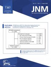


Jump to section
- Article
- EXECUTIVE SUMMARY
- INTRODUCTION
- METHODOLOGY
- SECTION 1: SYMPTOMATIC PATIENTS WITH SUSPECTED OR KNOWN CAD
- SECTION 2: ASYMPTOMATIC PATIENTS (WITHOUT SYMPTOMS OR ISCHEMIC EQUIVALENT)
- SECTION 3: DIAGNOSED HEART FAILURE (RESTING LV FUNCTION PREVIOUSLY ASSESSED BUT NO PRIOR CAD EVALUATION)
- SECTION 4: EVALUATION OF PATIENTS WITH KNOWN OR SUSPECTED CARDIAC SARCOIDOSIS
- SECTION 5: EVALUATION OF ARRHYTHMIAS WITHOUT ISCHEMIC EQUIVALENT (NO PRIOR CARDIAC EVALUATION)
- SECTION 6: SYNCOPE WITHOUT ISCHEMIC EQUIVALENT
- SECTION 7: ASSESSMENT OF CMD IN SYMPTOMATIC PATIENTS
- SECTION 8: SPECIFIC POPULATIONS
- SECTION 9: PRIOR TESTING OR PROCEDURES
- SECTION 10: PREOPERATIVE EVALUATION FOR NONCARDIAC SURGERY
- SECTION 11: DETERMINATION OF EXERCISE LEVEL BEFORE INITIATION OF EXERCISE PRESCRIPTION OR CARDIAC REHABILITATION
- BENEFITS AND HARMS OF IMPLEMENTING THE AUC GUIDANCE
- IMPLEMENTATION OF THE AUC GUIDANCE
- APPENDIX A: WORKGROUP MEMBERS AND EXTERNAL REVIEWERS
- APPENDIX B: DEFINITION OF TERMS AND ACRONYMS
- APPENDIX C: DISCLOSURES AND CONFLICTS OF INTEREST (COIs)
- APPENDIX D: PUBLIC COMMENTARY
- Acknowledgments
- REFERENCES
- Figures & Data
- Info & Metrics
Related Articles
Cited By...
- No citing articles found.