
Main menu

  • Home
  • Content
    • Current
    • Ahead of print
    • Past Issues
    • JNM Supplement
    • SNMMI Annual Meeting Abstracts
    • Continuing Education
    • JNM Podcasts
  • Subscriptions
    • Subscribers
    • Institutional and Non-member
    • Rates
    • Journal Claims
    • Corporate & Special Sales
  • Authors
    • Submit to JNM
    • Information for Authors
    • Assignment of Copyright
    • AQARA requirements
  • Info
    • Reviewers
    • Permissions
    • Advertisers
  • About
    • About Us
    • Editorial Board
    • Contact Information
  • More
    • Alerts
    • Feedback
    • Help
    • SNMMI Journals
  • SNMMI
    • JNM
    • JNMT
    • SNMMI Journals
    • SNMMI

User menu

  • Subscribe
  • My alerts
  • Log in
  • My Cart

Search

  • Advanced search
Journal of Nuclear Medicine
  • SNMMI
    • JNM
    • JNMT
    • SNMMI Journals
    • SNMMI
  • Subscribe
  • My alerts
  • Log in
  • My Cart
Journal of Nuclear Medicine

Advanced Search

  • Home
  • Content
    • Current
    • Ahead of print
    • Past Issues
    • JNM Supplement
    • SNMMI Annual Meeting Abstracts
    • Continuing Education
    • JNM Podcasts
  • Subscriptions
    • Subscribers
    • Institutional and Non-member
    • Rates
    • Journal Claims
    • Corporate & Special Sales
  • Authors
    • Submit to JNM
    • Information for Authors
    • Assignment of Copyright
    • AQARA requirements
  • Info
    • Reviewers
    • Permissions
    • Advertisers
  • About
    • About Us
    • Editorial Board
    • Contact Information
  • More
    • Alerts
    • Feedback
    • Help
    • SNMMI Journals
  • View or Listen to JNM Podcast
  • Visit JNM on Facebook
  • Join JNM on LinkedIn
  • Follow JNM on Twitter
  • Subscribe to our RSS feeds
Research ArticleGenitourinary

11C-PABA as a PET Radiotracer for Functional Renal Imaging: Preclinical and First-in-Human Study

Camilo A. Ruiz-Bedoya, Alvaro A. Ordonez, Rudolf A. Werner, Donika Plyku, Mariah H. Klunk, Jeff Leal, Wojciech G. Lesniak, Daniel P. Holt, Robert F. Dannals, Takahiro Higuchi, Steven P. Rowe and Sanjay K. Jain
Journal of Nuclear Medicine November 2020, 61 (11) 1665-1671; DOI: https://doi.org/10.2967/jnumed.119.239806
Camilo A. Ruiz-Bedoya
1Center for Infection and Inflammation Imaging Research, Johns Hopkins University School of Medicine, Baltimore, Maryland
2Department of Pediatrics, Johns Hopkins University School of Medicine, Baltimore, Maryland
  • Find this author on Google Scholar
  • Find this author on PubMed
  • Search for this author on this site
Alvaro A. Ordonez
1Center for Infection and Inflammation Imaging Research, Johns Hopkins University School of Medicine, Baltimore, Maryland
2Department of Pediatrics, Johns Hopkins University School of Medicine, Baltimore, Maryland
  • Find this author on Google Scholar
  • Find this author on PubMed
  • Search for this author on this site
Rudolf A. Werner
3Department of Nuclear Medicine, Hannover Medical School, Hannover, Germany
  • Find this author on Google Scholar
  • Find this author on PubMed
  • Search for this author on this site
Donika Plyku
4Russell H. Morgan Department of Radiology and Radiological Sciences, Johns Hopkins University School of Medicine, Baltimore, Maryland
  • Find this author on Google Scholar
  • Find this author on PubMed
  • Search for this author on this site
Mariah H. Klunk
1Center for Infection and Inflammation Imaging Research, Johns Hopkins University School of Medicine, Baltimore, Maryland
2Department of Pediatrics, Johns Hopkins University School of Medicine, Baltimore, Maryland
  • Find this author on Google Scholar
  • Find this author on PubMed
  • Search for this author on this site
Jeff Leal
4Russell H. Morgan Department of Radiology and Radiological Sciences, Johns Hopkins University School of Medicine, Baltimore, Maryland
  • Find this author on Google Scholar
  • Find this author on PubMed
  • Search for this author on this site
Wojciech G. Lesniak
4Russell H. Morgan Department of Radiology and Radiological Sciences, Johns Hopkins University School of Medicine, Baltimore, Maryland
  • Find this author on Google Scholar
  • Find this author on PubMed
  • Search for this author on this site
Daniel P. Holt
4Russell H. Morgan Department of Radiology and Radiological Sciences, Johns Hopkins University School of Medicine, Baltimore, Maryland
  • Find this author on Google Scholar
  • Find this author on PubMed
  • Search for this author on this site
Robert F. Dannals
4Russell H. Morgan Department of Radiology and Radiological Sciences, Johns Hopkins University School of Medicine, Baltimore, Maryland
  • Find this author on Google Scholar
  • Find this author on PubMed
  • Search for this author on this site
Takahiro Higuchi
5Okayama University Graduate School of Medicine, Dentistry and Pharmaceutical Sciences, Okayama, Japan; and
6Department of Nuclear Medicine, University Hospital, University of Würzburg, Würzburg, Germany
  • Find this author on Google Scholar
  • Find this author on PubMed
  • Search for this author on this site
Steven P. Rowe
4Russell H. Morgan Department of Radiology and Radiological Sciences, Johns Hopkins University School of Medicine, Baltimore, Maryland
  • Find this author on Google Scholar
  • Find this author on PubMed
  • Search for this author on this site
Sanjay K. Jain
1Center for Infection and Inflammation Imaging Research, Johns Hopkins University School of Medicine, Baltimore, Maryland
2Department of Pediatrics, Johns Hopkins University School of Medicine, Baltimore, Maryland
4Russell H. Morgan Department of Radiology and Radiological Sciences, Johns Hopkins University School of Medicine, Baltimore, Maryland
  • Find this author on Google Scholar
  • Find this author on PubMed
  • Search for this author on this site
  • Article
  • Figures & Data
  • Supplemental
  • Info & Metrics
  • PDF
Loading

Article Figures & Data

Figures

  • Tables
  • Additional Files
  • Figure1
    • Download figure
    • Open in new tab
    • Download powerpoint
  • FIGURE 1.
    • Download figure
    • Open in new tab
    • Download powerpoint
    FIGURE 1.

    11C-PABA PET/CT renal imaging in healthy rats. (A) Coronal dynamic 11C-PABA PET images of right kidney from representative rat show rapid cortical uptake after 2 min, followed by rapid clearance through pelvicalyceal system. (B) Maximum-intensity-projection 11C-PABA PET/CT images show abdominal aorta and iliac arteries 60 s after injection, as well as high activity in bladder (Bl) after 20 min, with low background signal in other tissues. (C) Average 11C-PABA time–activity curves for kidneys and bladder. Data are mean (solid lines) and SD (shaded areas) (n = 4). %ID = percentage injected dose.
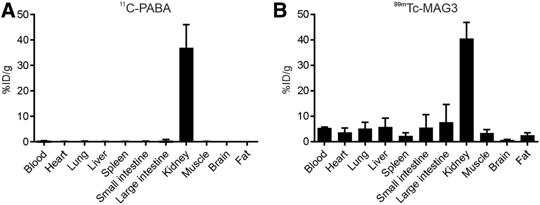
  • FIGURE 2.
    • Download figure
    • Open in new tab
    • Download powerpoint
    FIGURE 2.

    11C-PABA and 99mTc-MAG3 tissue biodistribution in rats. Healthy rats received simultaneous intravenous injections of 11C-PABA and 99mTc-MAG3 and were killed 30 min afterward. 99mTc-MAG3 counts were determined 2 h after 11C-PABA counts, when signal from 11C (physical half-life, 20 min) would have completely decayed. (A) Biodistribution of 11C-PABA was primarily within kidneys, with very low activity in all other measured organs. (B) Conversely, biodistribution of 99mTc-MAG3 had higher background activity. Data are mean and SD (n = 4).
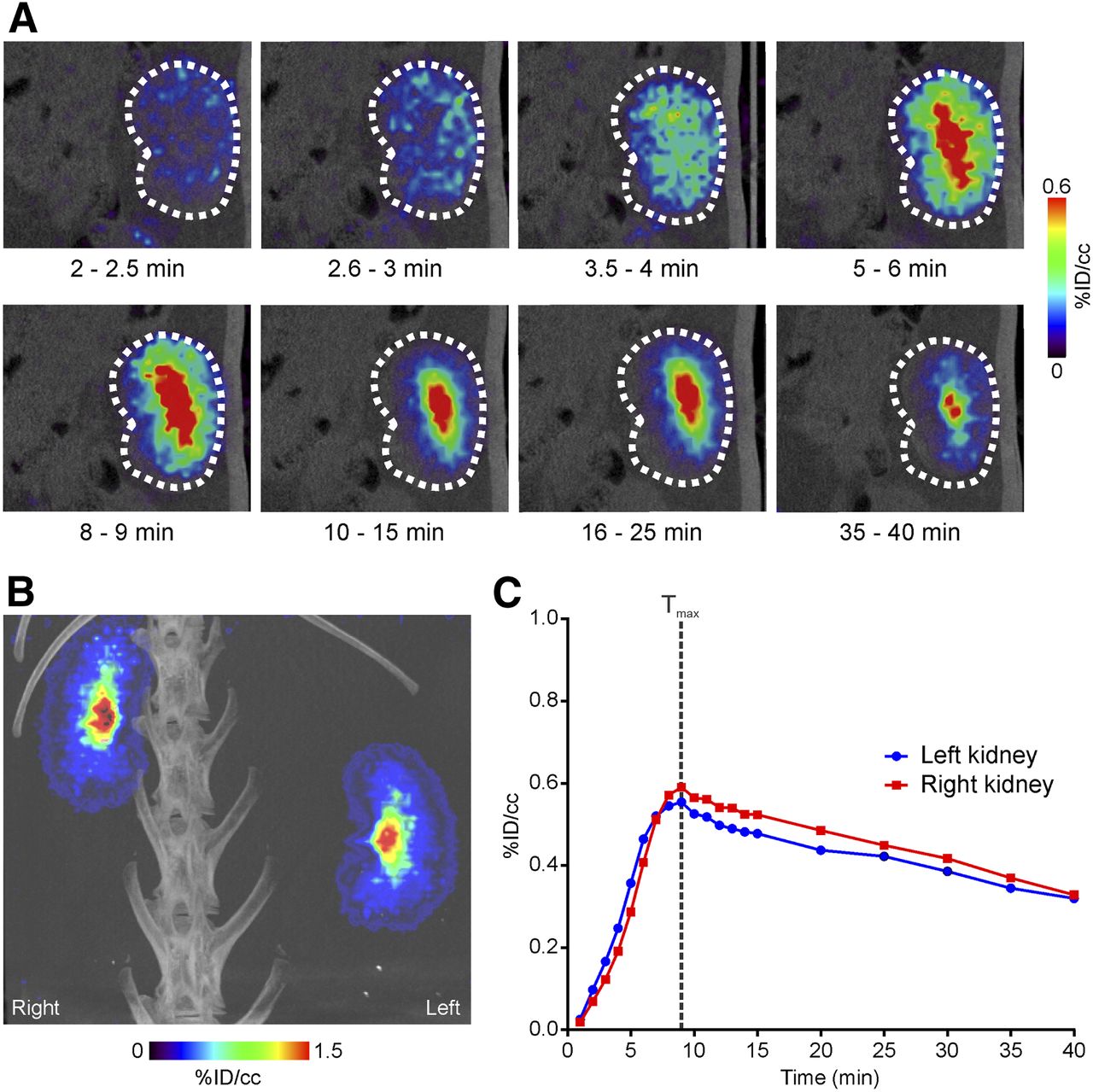
  • FIGURE 3.
    • Download figure
    • Open in new tab
    • Download powerpoint
    FIGURE 3.

    11C-PABA PET/CT renal imaging in healthy rabbits. (A) Coronal dynamic 11C-PABA PET/CT images of right kidney of representative rabbit show rapid delineation of renal cortex and then accumulation in pelvicalyceal system. (B) Maximum-intensity-projection 11C-PABA PET/CT image at 20 min shows similar uptake in left and right kidneys, with low background signal in other organs. (C) Average 11C-PABA time–activity curves for kidneys. Data are mean (n = 2). %ID = percentage injected dose.
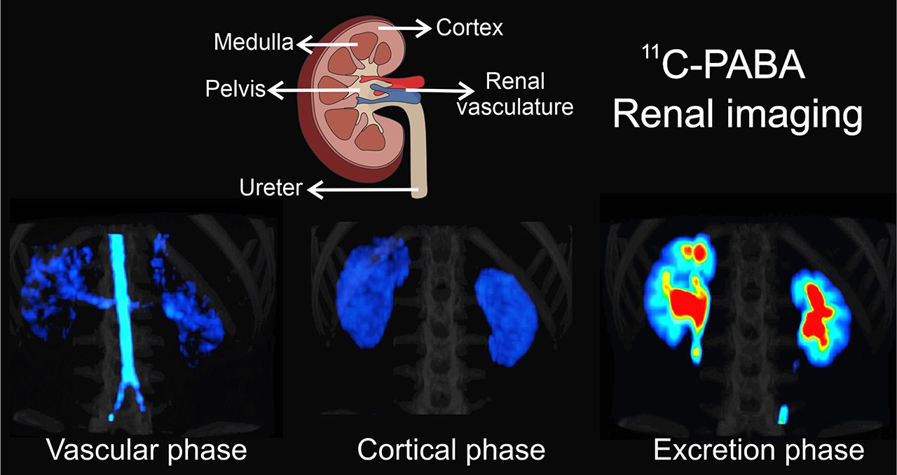
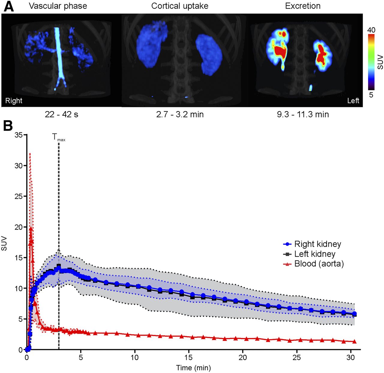
  • FIGURE 4.
    • Download figure
    • Open in new tab
    • Download powerpoint
    FIGURE 4.

    11C-PABA PET renal imaging in healthy human subjects. (A) Maximum-intensity-projection 11C-PABA PET/CT images of representative subject. Vascular phase is observed starting 22 s after injection, demonstrating renal perfusion. Cortical uptake phase is evident within 2.7 and 3.2 min, with maximal parenchymal uptake in renal cortex. Excretory phase is seen after 9 min, indicating maximal uptake in pelvicalyceal system. (B) Average 11C-PABA time–activity curves for kidneys and blood (aorta). Data are mean (solid lines) and SD (shaded areas) (n = 3).

Tables

  • Figures
  • Additional Files
    • View popup
    TABLE 1

    Characteristics of Human Subjects

    Subject no.SexAge (y)BMI (kg/m2)Creatinine (mg/dL)GFR (mL/min/1.73 m2)Albumin (g/dL)Injected 11C-PABA dose (MBq)
    1M23210.81544.9685.6
    2M25200.81434.8671.2
    3F30210.71344.8683.4
    • BMI = body mass index; GFR = glomerular filtration rate.

Additional Files

  • Figures
  • Tables
  • Supplemental Data

    Files in this Data Supplement:

    • Supplemental Data
    • Supplemental Video 1
PreviousNext
Back to top

In this issue

Journal of Nuclear Medicine: 61 (11)
Journal of Nuclear Medicine
Vol. 61, Issue 11
November 1, 2020
  • Table of Contents
  • Table of Contents (PDF)
  • About the Cover
  • Index by author
Print
Download PDF
Article Alerts
Sign In to Email Alerts with your Email Address
Email Article

Thank you for your interest in spreading the word on Journal of Nuclear Medicine.

NOTE: We only request your email address so that the person you are recommending the page to knows that you wanted them to see it, and that it is not junk mail. We do not capture any email address.

Enter multiple addresses on separate lines or separate them with commas.
11C-PABA as a PET Radiotracer for Functional Renal Imaging: Preclinical and First-in-Human Study
(Your Name) has sent you a message from Journal of Nuclear Medicine
(Your Name) thought you would like to see the Journal of Nuclear Medicine web site.
Citation Tools
11C-PABA as a PET Radiotracer for Functional Renal Imaging: Preclinical and First-in-Human Study
Camilo A. Ruiz-Bedoya, Alvaro A. Ordonez, Rudolf A. Werner, Donika Plyku, Mariah H. Klunk, Jeff Leal, Wojciech G. Lesniak, Daniel P. Holt, Robert F. Dannals, Takahiro Higuchi, Steven P. Rowe, Sanjay K. Jain
Journal of Nuclear Medicine Nov 2020, 61 (11) 1665-1671; DOI: 10.2967/jnumed.119.239806

Citation Manager Formats

  • BibTeX
  • Bookends
  • EasyBib
  • EndNote (tagged)
  • EndNote 8 (xml)
  • Medlars
  • Mendeley
  • Papers
  • RefWorks Tagged
  • Ref Manager
  • RIS
  • Zotero
Share
11C-PABA as a PET Radiotracer for Functional Renal Imaging: Preclinical and First-in-Human Study
Camilo A. Ruiz-Bedoya, Alvaro A. Ordonez, Rudolf A. Werner, Donika Plyku, Mariah H. Klunk, Jeff Leal, Wojciech G. Lesniak, Daniel P. Holt, Robert F. Dannals, Takahiro Higuchi, Steven P. Rowe, Sanjay K. Jain
Journal of Nuclear Medicine Nov 2020, 61 (11) 1665-1671; DOI: 10.2967/jnumed.119.239806
Twitter logo Facebook logo LinkedIn logo Mendeley logo
  • Tweet Widget
  • Facebook Like
  • Google Plus One
Bookmark this article

Jump to section

  • Article
    • Visual Abstract
    • Abstract
    • MATERIALS AND METHODS
    • RESULTS
    • DISCUSSION
    • CONCLUSION
    • DISCLOSURE
    • Acknowledgments
    • Footnotes
    • REFERENCES
  • Figures & Data
  • Supplemental
  • Info & Metrics
  • PDF

Related Articles

  • This Month in JNM
  • PubMed
  • Google Scholar

Cited By...

  • No citing articles found.
  • Google Scholar

More in this TOC Section

Genitourinary

  • Functional Renal Imaging with 2-Deoxy-2-18F-Fluorosorbitol PET in Rat Models of Renal Disorders
Show more Genitourinary

Translational

  • [99mTc]Tc-MY6349 Probe for Trop2-Targeted SPECT Imaging: From Preclinical to Pilot Clinical Study
  • Imaging Diverse Pathogenic Bacteria In Vivo with 18F-Fluoromannitol PET
  • Modeling Early Radiation DNA Damage Occurring During 177Lu-DOTATATE Radionuclide Therapy
Show more Translational

Similar Articles

Keywords

  • PABA
  • renal imaging
  • PET
  • renography
  • Translational science
SNMMI

© 2025 SNMMI

Powered by HighWire