


Abstract
Molecular imaging and targeted radiotherapy with radiolabeled cholecystokinin-2 receptor (CCK2R) targeting peptide probes holds high promise to improve the clinical management of patients with metastatic medullary thyroid carcinoma and other CCK2R-expressing malignancies. Low stability and suboptimal targeting of currently available radiolabeled peptide analogs has prompted us to seek new stabilization strategies. In this study, we present a new minigastrin analog with site-specific C-terminal modifications showing a highly optimized targeting profile. Methods: DOTA-D-Glu-Ala-Tyr-Gly-Trp-(N-Me)Nle-Asp-1-Nal-NH2 (DOTA-MGS5) radiolabeled with 111In, 68Ga, and 177Lu was evaluated in extensive in vitro stability studies. For 177Lu-DOTA-MGS5, additional metabolic studies were performed on BALB/c mice. Receptor affinity and cell uptake were studied in A431 human epidermoid carcinoma cells transfected with human CCK2R (A431-CCK2R), as well as the same cell line transfected with the empty vector (A431-mock). A431-CCK2R/A431-mock xenografted athymic BALB/c nude mice were used for biodistribution studies and small-animal SPECT/CT. Results: DOTA-MGS5 radiolabeled with 111In and 177Lu showed a highly increased stability against enzymatic degradation in different media up to 24 h of incubation. Similar results were observed for 68Ga-DOTA-MGS5 incubated up to 4 h. In the blood of mice injected with 177Lu-DOTA-MGS5, at least 70% intact radiopeptide was detected up to 1 h after injection. The unlabeled peptide and the complexes with the natural isotopes showed retained receptor affinity, and the radiopeptides showed unexpectedly high cell uptake in A431-CCK2R cells (>60% at 4 h). Regardless of the radiometal used for labeling, impressively high uptake in A431-CCK2R xenografts was found (∼20% injected activity/g 1–4 h after injection), whereas the uptake in A431-mock xenografts was negligible. Low background activity and favorable tumor-to-kidney ratios (4–6) allowed for high image contrast in small-animal SPECT/CT. Conclusion: The excellent targeting properties of DOTA-MGS5 support future clinical studies evaluating the diagnostic and therapeutic potential in patients with progressive or metastatic medullary thyroid carcinoma, as well as other advanced-stage CCK2R-expressing malignancies.
Cholecystokinin-2 receptors (CCK2R) are expressed at high incidence in medullary thyroid carcinomas (MTC, > 90%), small cell lung cancers (>50%), astrocytomas (>60%), insulinomas, stromal ovarian cancers, gastrointestinal stromal tumors, and more than 20% of gastroenteropancreatic tumors (1,2). CCK2R targeting, therefore, offers a highly personalized diagnostic and therapeutic approach for different cancers when safe and effective systemic therapies are lacking. This is especially the case for patients with unresectable, locally advanced, or metastatic MTC. Because of substantial side effects, targeted treatment with the multikinase inhibitors vandetanib and cabozantinib is restricted to patients with progressive and symptomatic disease (3). In patients with metastasized MTC, a higher tumor detection rate (>90%) was found for CCK2R imaging in comparison to somatostatin receptor imaging (40%) (4). Also, for about 20% of the patients with carcinoids and other neuroendocrine tumors, an additive value of CCK2R imaging was reported (5). Initial targeted radiotherapy in MTC patients showed encouraging therapeutic effects; nephrotoxicity was, however, found to be a major concern (6).
CCK2R-targeting peptide probes are based on the natural ligands cholecystokinin and gastrin. Gastrin analogs generally show higher receptor-specific uptake, but the kidney uptake is also high. Renal accumulation was successfully reduced by depletion of the penta-Glu sequence in minigastrin but was connected with reduced physiologic half-life and poor diagnostic efficacy (7). A high variety of synthetic modifications in the N-terminal part of the peptide sequence was investigated to increase enzymatic stability, reduce kidney retention, and improve tumor targeting (8,9). In the C-terminal sequence Trp-Met-Asp-Phe-NH2, known to be essential for receptor binding (10), only replacement of oxidation-sensitive Met allowed maintenance of receptor affinity (8,11).
For 2 novel radiopeptides, clinical trials have recently been initiated. In 4 MTC patients, 111In-labeled DOTA-(d-Glu)6-Ala-Tyr-Gly-Trp-Met-Asp-Phe-NH2 (CP04) visualized all known tumor lesions (12). Also, 177Lu-PP-F11N, derived from CP04 by replacement of Met with Nle, showed high CCK2R-specific tumor uptake in a patient with progressive, metastatic MTC (13). Despite physiologic uptake observed in stomach, colon, kidneys, and urinary bladder, the dosimetry studies indicate the feasibility of targeted radiotherapy.
A main issue remaining unsolved is the limited enzymatic stability of these linear peptide analogs in vivo. Rapid cleavage in the C-terminal receptor-specific sequence occurs, and no improvement in tumor uptake can be achieved by coadministration of protease inhibitors (13). We have recently described a new stabilization strategy based on site-specific amino acid modifications with unnatural aromatic amino acids and N-methylated amino acids preventing the metabolization of the C-terminal receptor-specific sequence. When applied to the truncated minigastrin analog DOTA-MG11 missing the penta-Glu motif (7), these modifications led to compounds with high CCK2R affinity, improved bioavailability, limited kidney retention, and highly increased tumor uptake (14).
In the present study, we have further refined our approach and present the new minigastrin analog DOTA-d-Glu-Ala-Tyr-Gly-Trp-(N-Me)Nle-Asp-1-Nal-NH2 (DOTA-MGS5) with an optimized targeting profile. Specific modifications were applied in the C-terminal receptor-specific sequence of the peptide, namely replacement of Met with Nle and of Phe with 1-naphtyl-alanine (1-Nal), as well as N-methylation of the peptide bond between Trp and Nle. A comprehensive preclinical evaluation of the DOTA-conjugated peptide analog radiolabeled with 111In, 68Ga, and 177Lu was performed. These studies included extensive in vitro and in vivo stability studies, receptor affinity and cell uptake studies, and tumor targeting and biodistribution studies in an animal tumor model, highlighting the specific features of this novel CCK2R-targeting peptide analog.
MATERIALS AND METHODS
Materials
All commercially obtained chemicals were of analytic grade and used without further purification. 111InCl3 was purchased from Mallinckrodt Medical. Non–carrier-added 177LuCl3 produced from highly enriched 176Yb was purchased from Isotope Technologies Garching. 68GaCl3 was obtained from a commercial 68Ge/68Ga generator with nominal activity of 1,850 MBq (Eckert and Ziegler Eurotope) eluted with 0.1N HCl solution (Rotem). DOTA-MG11 (DOTA-d-Glu-Ala-Tyr-Gly-Trp-Met-Asp-Phe-NH2) and CP04 used for comparative studies were purchased from piCHEM. The A431 human epidermoid carcinoma cell line stably transfected with human CCK2R (A431-CCK2R), as well as the same cell line transfected with the empty vector (A431-mock), was kindly provided by Dr. Luigi Aloj and cultured as described elsewhere (15).
Peptide Synthesis
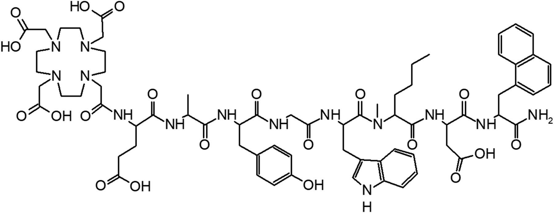
DOTA-MGS5 (DOTA-d-Glu-Ala-Tyr-Gly-Trp-(N-Me)Nle-Asp-1-Nal-NH2) was synthesized and analyzed following a previously described protocol (14). The chemical structure is displayed in Figure 1.

Chemical structure of DOTA-MGS5.
Radiolabeling and Characterization In Vitro
Standard protocols were used for labeling with the different radiometals (9,14,16). Radiochemical purity of the radiopeptides was analyzed using a high-performance liquid chromatography system described elsewhere (9) together with a water/acetonitrile/0.1% trifluoroacetic acid gradient with increasing concentrations of acetonitrile (111In and 177Lu: 0–3 min, 10%; 3–18 min, 10%–55%; 18–20 min, 55%–80%; 20–21 min, 80%–10%; 20–25 min, 10%; 68Ga: 0–2 min, 20%; 2–12 min, 20%–70%; 12–12.1 min, 70%–20%; 12.1–15 min, 20%).
The stability of 111In-, 68Ga- and 177Lu-labeled DOTA-MGS5 was investigated after incubation in phosphate-buffered saline (PBS; n = 1) and fresh human serum (1–2 nmol of peptide/mL, n = 2). A PBS sample of the radiolabeled peptides (∼0.05 nmol/mL) was used to determine the distribution coefficient (log D; n = 8). A serum sample (∼0.5 nmol/mL) was used to assess the protein binding by size-exclusion chromatography (n = 2). For comparison, the same assays were performed with 111In-CP04. The enzymatic degradation of radiolabeled DOTA-MGS5 (n = 2 for 111In; n = 1 for 68Ga and 177Lu) was further assessed in rat liver and kidney homogenates (∼0.5 nmol of peptide/mL). Previously published protocols were followed for this in vitro characterization (14,17).
Cell Uptake and Receptor Binding Studies
The receptor affinity of DOTA-MGS5 (n = 3), as well as CP04, DOTA-MG11, and pentagastrin (n = 1), was evaluated in competition assays against 125I-Tyr12-gastrin-I on A431-CCK2R cells. Binding assays, including also receptor binding studies with DOTA-MGS5 complexed with natIn, natLu and natGa (n = 1), were performed as described elsewhere (16). Half maximal inhibitory concentrations (IC50s) were calculated following nonlinear regression using Origin software (Microcal Origin, version 6.1).
Internalization and externalization experiments were performed on A431-CCK2R and A431-mock cells following previously published protocols (14,18). For internalization experiments, the cells were incubated with the radiopeptides (final peptide concentration of 0.4 nM) for up to 4 h, and the radioactivity of the lysed cells was determined in relation to the total radioactivity added (% internalized radioactivity). For externalization experiments, after 2 h of incubation the medium was replaced with 3 mL of fresh medium (unblocked) or medium containing 1 mM pentagastrin (blocked) and the efflux of radioactivity evaluated in relation to the cell uptake (% externalized radioactivity). Nonspecific binding was assessed in A431-mock cells. In an additional experiment, the cell uptake in dependence on the peptide concentration within the range of 0.04–160 nM was investigated. Furthermore, comparative internalization studies at 2 h of incubation were performed for DOTA-MGS5 and CP04 radiolabeled with different radiometals, including blocking of the receptor-specific uptake with 1 μM human minigastrin.
Animal Studies Evaluating the In Vivo Stability and Tumor Uptake
Metabolic and biodistribution studies were performed in accordance with the ethical standards of the institution and approved by the Austrian Ministry of Science (BMWFW-66.011/0075-WF/V/3b/2016).
The metabolic stability of 177Lu-DOTA-MGS5 (10, 30, and 60 min after injection; n = 1) and 177Lu-CP04 (30 min after injection; n = 1) was evaluated in 5- to 7-wk-old female BALB/c mice (Charles River). Mice were injected intravenously through a lateral tail vein with 20–45 MBq of the 177Lu-labeled peptide (≤2 nmol total peptide), and the percentage of intact radiopeptide in blood was analyzed as described elsewhere (14,18).
Biodistribution studies were performed on female athymic BALB/c nude mice (Charles River) injected subcutaneously with A431-CCK2R and A431-mock cells (2 × 10⁶ in 200 μL) at an age of 6–8 wk. After 7–11 d, when tumors reached a volume of about 0.3 mL (1 mouse did not develop the A431-mock xenograft), the mice were injected intravenously via a lateral tail vein with about 0.02 nmol of radiolabeled peptide (∼0.05 MBq for 111In, ∼0.8 MBq for 68Ga, ∼0.4 MBq for 177Lu) in groups of 4. After sacrifice at 1 or 4 h after injection, tumors and other tissues were removed and weighed, and the radioactivity was measured in the γ-counter (Perkin Elmer Life Sciences and Analytic Sciences) together with a standard. Results were expressed as percentage of injected activity per gram of tissue (%IA/g), and tumor-to-organ activity ratios were calculated for selected tissues. Statistical analysis (independent-samples t test, P < 0.05) was performed using Origin software.
The imaging properties of 111In-DOTA-MGS5 were evaluated by small-animal SPECT/CT. Three xenografted athymic BALB/c nude mice (Charles River) were injected with 10 MBq (0.3 nmol) of the radiolabeled peptide. Whole-body SPECT/CT images were obtained at 1 and 4 h after injection under inhaled anesthesia (1 L/min 1.5% isoflurane in O2) in the prone position on a heated bed using a small-animal SPECT/CT 4-head camera (Bioscan). SPECT images were acquired in helical scanning mode with 24 projections using 1.4-mm-pinhole collimators and a voxel size of 100 × 100 μm, and CT images were acquired with a 45-kVp x-ray source (180 projections over 6 min) (14). Region of interest analysis of the coregistered SPECT and CT images was performed using VivoQuant image analysis software (inviCRO LLC). After being scanned, the animals were sacrificed to determine the tracer uptake in tumors and other tissues.
RESULTS
Peptide Synthesis and Radiolabeling
DOTA-MGS5 was synthesized with moderate yield and high purity of at least 95%. Radiolabeling with the different radiometals generally resulted in radiochemical purity of at least 95% at a molar activity of no more than 35 GBq/μmol for 111In, no more than 40 GBq/μmol for 177Lu, and no more than 60 GBq/μmol for 68Ga.
Characterization of the Radiopeptides In Vitro
Table 1 summarizes the in vitro characteristics of radiolabeled DOTA-MGS5 in comparison with 111In-CP04. For 111In-DOTA-MGS5 and 177Lu-DOTA-MGS5, the stability in PBS and human serum was analyzed for up to 24 h after incubation. Because of the short half-life of 68Ga, 68Ga-DOTA-MGS5 was incubated only for up to 4 h. The radiolabeled complexes showed a high stability in aqueous solution. Also, in human serum more than 95% intact radiopeptide was found. Comparative studies with 111In-CP04 showed a high stability in PBS, whereas a lower stability (<90% intact radiopeptide) was found in serum. 68Ga-DOTA-MGS5, 177Lu-DOTA-MGS5, and 111In-DOTA-MGS5 showed a higher log D value than 111In-CP04, confirming a somewhat lower hydrophilicity. Protein binding was, however, comparable to 111In-CP04. A much higher degree of degradation, with less than 30% intact radiopeptide, after 2 h was found in rat liver homogenates than in human serum. 68Ga-DOTA-MGS5 showed a faster degradation over time than the 111In- and 177Lu-labeled radiopeptides. In rat kidney homogenates, all radiopeptides were almost completely degraded (Fig. 2).
In Vitro Characteristics of Radiolabeled DOTA-Peptides

Intact radiopeptide in rat homogenates of liver (A) and kidney (B).
Receptor Binding and Internalization Studies
A high CCK2R affinity with IC50s of less than 1 nM was observed for DOTA-MGS5 and for the complexes with the natural isotopes natIn, natLu, and natGa. A similar binding affinity was also found for pentagastrin, DOTA-MG11, and CP04 (Table 2).
IC50 as Calculated from Competition Assays Against 125I-Tyr12-Gastrin-I
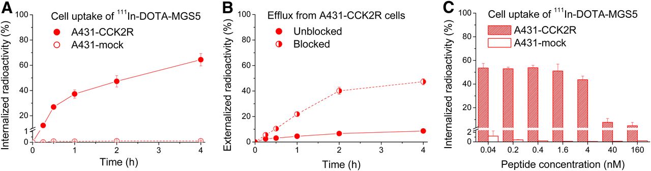
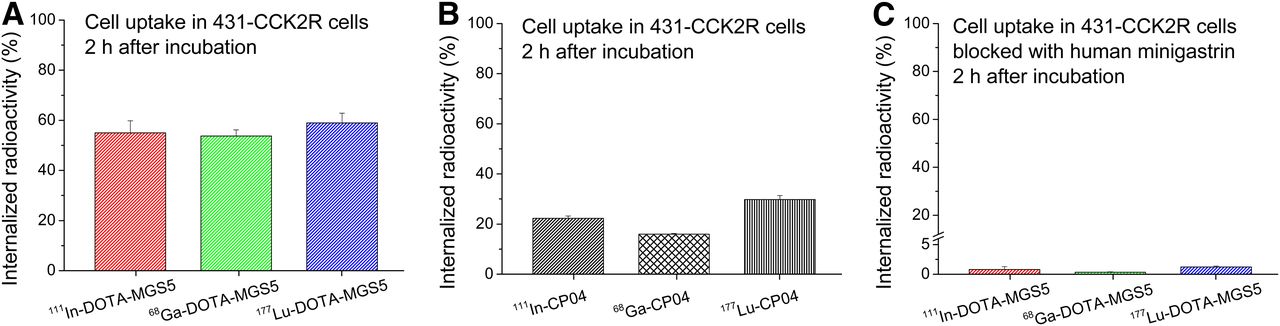
With 111In-DOTA-MGS5, remarkably high cell uptake values of more than 60% were reached in A431-CCK2R cells after 4 h of incubation (Fig. 3A). Efflux studies showed only a minor release of radioactivity (<10%) under unblocked assay conditions, whereas much higher values of more than 40% were reached when the reuptake was blocked by addition of pentagastrin (Fig. 3B). Stable uptake values of more than 50% were observed within a concentration range of 0.04–1.6 nM. At a concentration of 4 nM, the uptake was slightly reduced to approximately 40% and further dropped to 5%–10% at 40 nM and 160 nM (Fig. 3C). The nonspecific uptake in A431-mock cells was always below 1.5%. DOTA-MGS5 radiolabeled with 111In, 68Ga, and 177Lu showed a considerably increased cell uptake of 50%–60% in A431-CCK2R cells at 2 h after incubation (Fig. 4A) when compared with radiolabeled CP04 (15%–30%; Fig. 4B). Blocking studies with human minigastrin showed uptake values below 1.5%, confirming the receptor specificity (Fig. 4C).

Cell uptake of 111In-DOTA-MGS5. (A) Internalized activity in A431-CCK2R and A431-mock cells. (B) Efflux of radioactivity from A431-CCK2R cells. (C) Internalization in dependence on peptide concentration.

Cell internalization of DOTA-MGS5 (A) and CP04 (B), as well as blocking studies for DOTA-MGS5 (C).
Metabolic Studies and Biodistribution Studies In Vivo
In BALB/c mice injected with 177Lu-DOTA-MGS5, 86% intact radiopeptide was present in blood at 10 min after injection and still 77% and 70% intact radiopeptide could be detected at 30 and 60 min after injection, respectively. In comparative studies performed with 177Lu-CP04 at 30 min after injection, only 5% intact radiopeptide was observed in blood. Representative radiochromatograms at 30 min after injection are displayed in Figure 5.
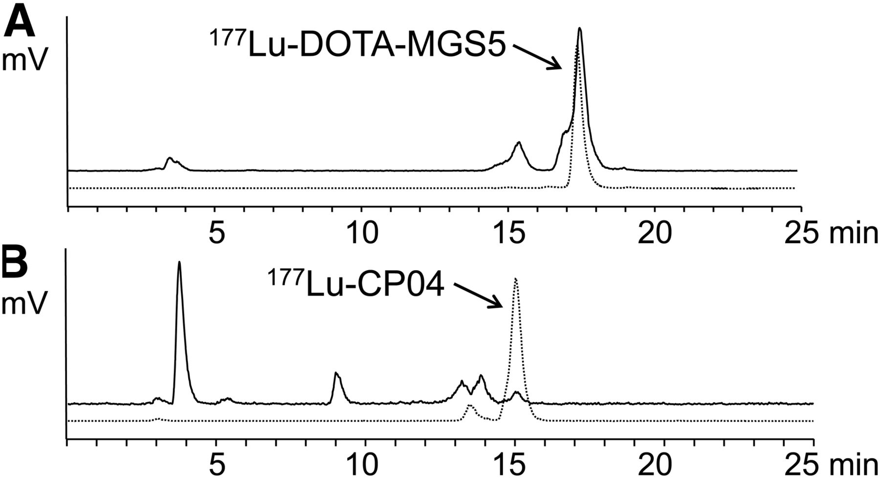
Radiometabolites as analyzed by radio–high-performance liquid chromatography from blood of BALB/c mice 30 min after injection with 177Lu-DOTA-MGS5 (A) and 177Lu-DOTA-CP04 (B). Dashed line shows radiochromatogram of radiopeptide before injection.
In the biodistribution studies, DOTA-MGS5 radiolabeled with different radiometals was rapidly cleared from the body, mainly through the kidneys, and a low nonspecific uptake generally occurred in most tissues (Fig. 6; Supplemental Table 1; supplemental materials are available at http://jnm.snmjournals.org). In blood, lung, heart, spleen, and liver, 68Ga-DOTA-MGS5 showed a slightly higher uptake than 111In-DOTA-MGS5 at 1 h after injection (P > 0.05). In CCK2R-expressing stomach and pancreas, the uptake of 68Ga-DOTA-MGS5 was lower but was significantly reduced only for stomach (P < 0.01). The uptake of 111In-DOTA-MGS5 significantly decreased over time in blood, lung, heart, spleen, liver, and kidney (P < 0.03). When 111In-DOTA-MGS5 and 177Lu-DOTA-MGS5 were compared at 4 h after injection, only in CCK2R-expressing pancreas (P < 0.02) was a significant difference found. The uptake in kidneys remained at less than 6 %IA/g for all radiopeptides and time points studied. Impressive tumor uptake and tumor retention were observed for 111In-DOTA-MGS5 in A431-CCK2R xenografts, with values of 19.53 ± 5.42 %IA/g at 1 h after injection and 23.49 ± 1.25 %IA/g at 4 h. A similar tumor uptake was also observed with 68Ga-DOTA-MGS5 (23.25 ± 4.70 %IA/g, 1 h after injection) and 177Lu-DOTA-MGS5 (24.45 ± 3.11 %IA/g, 4 h after injection), whereas only minimal uptake occurred in A431-mock xenografts (<1 %IA/g). When comparing the uptake of 111In-DOTA-MGS5 at 0.02 nmol (biodistribution, 4 h after injection) and 0.3 nmol (imaging, ∼5 h after injection), that in A431-CCK2R xenografts was reduced by 40% (P < 0.02) at the higher peptide amount. Also, the uptake in CCK2R-expressing pancreas (P < 0.01) and stomach (P < 0.02), as well as intestine (P < 0.01) and kidneys (P < 0.01), was significantly lower. The low activity levels in blood and normal tissue combined with strong uptake and prolonged retention in the tumor over time resulted in high tumor-to-organ activity ratios, especially at 4 h after injection, with mean values of 256–572 for blood, 6.11–6.53 for kidneys, and 3.08–4.74 for stomach (Supplemental Table 2).

Biodistribution of DOTA-MGS5 radiolabeled with different radiometals in A431-CCK2R/A431-mock xenograft–bearing BALB/c nude mice. Data are mean ± SD (n = 4). p.i. = after injection.
Small-animal SPECT/CT imaging with 111In-DOTA-MGS5 ultimately confirmed the favorable biodistribution profile (Fig. 7). On the maximum-intensity projections, a low background activity and high image contrast were observed. At 1 h after injection, the most prominent structure was the A431-CCK2R tumor. Excretion of radioactivity via the kidneys and bladder could also be seen. No uptake was observed in A431-mock tumors, which were visible only on CT. At 4 h after injection, the bladder was almost emptied and the kidneys could no longer be seen, whereas the tumor uptake remained comparatively high (0.41 and 0.36 MBq at 1 and 4 h, respectively). From the region-of-interest analysis performed on 2 mice, a tumor-to-kidney ratio of more than 2 and more than 4 was calculated for 1 and 4 h after injection, respectively.

Small-animal SPECT/CT images at 1 and 4 h after injection of BALB/c nude mice xenografted with A431-CCK2R (left) and A431-mock cells (right) injected with 111In-DOTA-MGS5. SPECT is in color scale ranging from 0.0015 to 0.011 kBq; CT is in gray scale.
DISCUSSION
Despite extensive radiopharmaceutical development, radiolabeled CCK2R-targeting peptide analogs have not yet been introduced into routine clinical practice. In the present study, we describe the novel minigastrin analog DOTA-MGS5 with site-specific stabilizations in the C-terminal receptor-specific sequence. It has recently been reported that the radiolabeled minigastrin analogs currently investigated in clinical trials are rapidly metabolized at this site by different proteases (13). The preclinical characterization of DOTA-MGS5 radiolabeled with 111In, 68Ga, and 177Lu shows that this new peptide analog fulfils important criteria for diagnostic and therapeutic use in nuclear medicine: excellent target-to-nontarget ratios, specific binding to tumor tissue, and high metabolic stability (19). To our knowledge, this is the first report on a radiolabeled CCK2R-targeting peptide probe with such an intense tumor uptake along with favorable tumor-to-kidney ratios.
Because most of the CCK2R-targeting radiopeptides previously developed have been studied in the A431-CCK2R/A431-mock mouse tumor model (9,14,20), this model was used for the preclinical evaluation of DOTA-MGS5. An impressively high and persistent tumor uptake (>20 %IA/g) was observed in A431-CCK2R xenografts together with considerably improved tumor-to-kidney ratios (>6). The uptake was clearly receptor-specific, as shown by the limited uptake in A431-mock xenografts (<1 %IA/g), and was confirmed in blocking studies on A431-CCK2R cells in vitro. The extraordinary targeting properties also become clear from the small-animal SPECT/CT imaging performed with 111In-DOTA-MGS5 at a 10-fold higher peptide amount, even though partial saturation effects occurred in A431-CCK2R tumors and in CCK2R-expressing tissues.
DOTA-MGS5 also seems highly promising for targeted radiotherapy, as the tumor uptake of 177Lu-DOTA-MGS5 is more than tripled in comparison with the radiopeptides currently used in clinical trials. 177Lu-CP04 and 177Lu-PP-F11N show a tumor uptake of 6.70 ± 0.56 and 6.94 ± 0.82 %IA/g in A431-mock xenografts when injected at a comparable peptide dose of 0.01 nmol. In xenografts based on human MTC cells (MZ-CRC-1), in line with a higher number of binding sites, a higher uptake (10–20 %IA/g) was observed for both radiopeptides. Still, tumor-to-kidney ratios with values of 3–4 were clearly inferior (13). In comparative cell uptake studies using CP04 and DOTA-MGS5 radiolabeled with different radiometals, we could confirm the highly increased cell internalization of DOTA-MGS5.
When analyzing our data, different parameters explain the optimized targeting profile of radiolabeled DOTA-MGS5.
Even though the C-terminal sequence Trp-X-Asp-Phe-NH2 (with X being Met or a Met-like substituent) (7) was postulated to be essential for receptor binding, DOTA-MGS5, with methylation of the peptide bond between Trp and Nle and the voluminous amino acid 1-Nal at the C terminus, showed high binding affinity. For CCK4 analogs, the exchange of Met with (N-Me)Nle resulted in increased CCK2R affinity (21), whereas the additional substitution of Phe with 1-Nal abolished this positive effect (22). As reported previously also for another minigastrin analog (23), we saw no metal-related changes on receptor affinity and cell uptake. We found evidence in the literature that the C terminus of gastrin assumes a well-defined β-turn that interacts with a hydrophobic pocket of the CCK2R (24). Especially, the aromaticity of Phe was found to increase polar receptor–ligand interactions (25). The amino acid substitutions applied in DOTA-MGS5 increase the lipophilicity of the radiopeptide, possibly promoting the receptor interaction.
We could show that the specific substitutions in the C-terminal sequence of DOTA-MGS5 successfully prevent the metabolic degradation in vivo. In the blood of BALB/c mice injected with 177Lu-DOTA-MGS5, 70% intact radiopeptide was still found at 1 h after injection. Such a stabilization could not be achieved with N-terminal modifications (13,26). This improved stability against enzymatic degradation in vivo was not an unexpected finding. Methylation of the peptide bond between Trp and Nle and introduction of voluminous amino acids at the C terminus were already investigated for CCK4 analogs, achieving compounds that are protected from degradation in rat brain homogenates (23). Higher bioavailability is likely to increment the amount of intact radiopeptide reaching the target site. In the efflux studies from A431-CCK2R cells, we found a much higher efflux of radioactivity under blocked assay conditions, suggesting that the intact radiopeptide is excreted from the cells and that only under unblocked assay conditions can reuptake occur.
Interestingly, DOTA-MGS5, because of its lower hydrophilicity, revealed a 3-fold increased protein binding in vitro compared with 111In-DOTA-MG11, with values of 41.0% versus 10.1% at 4 h (14). Also, 111In-CP04 showed an increased protein binding of 33.0%. The combination of increased enzymatic stability and increased protein binding enhancing the initial systemic circulation seems to be responsible for the reinforced specific CCK2R interaction at the tumor site. This hypothesis is supported by our recent findings with another 111In-labeled minigastrin analog showing C-terminal modifications. 111In-DOTA-MGS4 substituted with (N-Me)Nle and (N-Me)Phe (instead of 1-Nal) showed enzymatic stability similar to 111In-DOTA-MGS5 but a much lower protein binding. The blood levels of 111In-DOTA-MGS5 at 1 h after injection (1.19 ± 0.29 %IA/g) were more than 5 times higher than those of 111In-DOTA-MGS4 (0.2 ± 0.07 %IA/g). Consequently, 111In-DOTA-MGS5 showed a 2-fold higher tumor uptake in A431-CCK2R xenografts than did 111In-DOTA-MGS4 (14). At the later time point of 4 h after injection, the blood values of 111In-DOTA-MGS5 and 177Lu-DOTA-MGS5 declined to low levels of about 0.1 %IA/g, comparable to 111In-DOTA-MGS4 (0.08 ± 0.09 %IA/g), indicating that no prolonged systemic circulation occurred. Also, the 3-fold improvement in tumor uptake of 111In-CP04 when compared with 111In-DOTA-MG11 could be attributed to its higher protein binding rather than stabilization against metabolic degradation.
The impressive targeting properties become clear from the small-animal SPECT/CT imaging performed with 111In-DOTA-MGS5 and indicate that with this new CCK2R-targeting peptide analog a higher absorbed dose may be reached in the tumor also in targeted radiotherapy with 177Lu-DOTA-MGS5. The absorbed dose of the kidneys could potentially be reduced by the use of kidney protective agents such as Gelofusine (B. Braun) (27). Also, the higher uptake observed in the stomach when compared with 177Lu-CP04 and 177Lu-PP-F11N (13) seems not to be limiting for a therapeutic application, as it slowly decreases with time and in dependence on the injected peptide amount. In radiation therapy, doses of less than 45 Gy are generally accepted for the stomach without unacceptable complications (28). Besides improved molecular imaging, DOTA-MGS5 seems therefore especially promising for targeted radiotherapy in patients with advanced MTC and other CCK2R-expressing malignancies.
CONCLUSION
The high and persistent tumor uptake and favorable tumor to-background activity ratios, including kidneys, give high promise that new radiopharmaceuticals based on DOTA-MGS5 will be powerful peptide probes in the localization and treatment of patients with CCK2R-expressing tumors. DOTA-MGS5 radiolabeled with different radiometals—111In for SPECT, 68Ga for PET, or β-emitting radionuclides such as 177Lu—seems therefore most suited for future clinical studies evaluating the diagnostic and therapeutic potential in patients with progressive or metastatic MTC, as well as other advanced-stage CCK2R-expressing malignancies.
DISCLOSURE
This study was financially supported by the Austrian Science Fund (FWF) (project P 27844). Elisabeth von Guggenberg and Maximilian Klingler are inventors of the novel intellectual property presented in this article; the patent application submitted by the Medical University of Innsbruck is currently under evaluation. No other potential conflict of interest relevant to this article was reported.
Footnotes
Published online Dec. 7, 2018.
- © 2019 by the Society of Nuclear Medicine and Molecular Imaging.
REFERENCES
- Received for publication October 15, 2018.
- Accepted for publication November 13, 2018.


Jump to section
Related Articles
Cited By...
- Safety, Biodistribution, and Radiation Dosimetry of the 68Ga-Labeled Minigastrin Analog DOTA-MGS5 in Patients with Advanced Medullary Thyroid Cancer and Other Neuroendocrine Tumors
- Preclinical Evaluation of Minigastrin Analogs and Proof-of-Concept [68Ga]Ga-DOTA-CCK-66 PET/CT in 2 Patients with Medullary Thyroid Cancer
- Preliminary Clinical Experience with Cholecystokinin-2 Receptor PET/CT Using the 68Ga-Labeled Minigastrin Analog DOTA-MGS5 in Patients with Medullary Thyroid Cancer
- Cholecystokinin 2 Receptor Agonist 177Lu-PP-F11N for Radionuclide Therapy of Medullary Thyroid Carcinoma: Results of the Lumed Phase 0a Study